Abstract
The case of giant renal cyst measuring 30 cm, accompanied by abdominal swelling and erythrocytosis, is presented. A 45-year-old male presented with large abdominal mass, atrophic left kidney, hypertension, and erythrocytosis. The patient underwent multiple preoperative phlebotomies, open extirpation of the cyst, and nephrectomy. After the surgery, erythrocytosis ceased completely, blood pressure became normal without any medications whereas function of the remaining kidney was stable. The giant renal cysts measuring more than 15 cm are extremely rare. However, they can cause erythrocytosis and hypertension very frequently, especially in the case of cysts originating from the proximal tubule. To our knowledge, this is the largest renal cyst published in the literature that caused the above-mentioned complications.
INTRODUCTION
A simple renal cyst is an oval formation with a fibrous wall bordered by a single layer of flattened or cuboidal epithelium and filled with transudate-like clear fluid. Simple cysts differ very much in size, ranging from less than 1 cm to greater than 10 cm.Citation1 The cyst is not connected to any part of the nephron, although it may originate from a proximal or distal portion of the nephron.Citation2
Nowadays, the vast majority of cysts are discovered incidentally on ultrasonography, despite of the fact that cysts can produce an abdominal mass or pain, hematuria secondary to rupture into the pyelocalyceal system, and hypertension secondary to segmental ischemia.Citation3,Citation4 Cysts can cause calyceal or renal pelvic obstruction as well. Erythrocytosis has been reported in association with autosomal dominant polycystic kidney disease as well as in some cases of giant renal cysts.Citation5 The sources of excessive erythropoietin secretion are stroma cells of the cyst walls, that is, interstitial cells juxtaposed to proximal tubular cysts, so that erythrocytosis ceases after removal of the cyst in most cases.Citation6,Citation7
The treatment of benign simple cyst is performed only in the presence of symptoms, and it is usually surgical or minimally invasive. Unroofing of the cyst and percutaneous fluid aspiration followed with the injection of a sclerosing agent are the most frequent surgical approaches.Citation1,Citation8 Newer methods are percutaneous resection and intrarenal marsupialization and laparoscopic unroofing.Citation9,Citation10
CASE REPORT
A 45-year-old man visited the doctor due to epistaxis. Physical examination revealed a mass in the left side of the abdomen () and hypertension (160/95 mmHg) under regular antihypertensive medication of amlodipine (10 mg per day) and enalapril (20 mg BID), which he used during the last 2 years. Patient's blood analyses showed erythrocytosis with normal white blood cells and platelet count. Partial thromboplastin time was normal. Renal function and urine analysis were normal ().
Figure 1. A mass on the left side of the abdomen.
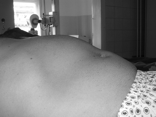
Table 1. Complete blood count and biochemical analysis before and after surgery
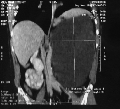
Ultrasound examination showed a large left renal cyst, and computed tomography proved the presence of the giant retroperitoneal cyst, sized 297 × 210 × 170 mm, filled with a clear fluid; small, atrophic left kidney dislocated cranially; and normal right kidney. Pancreas, stomach, aorta, vena cava, and small intestine were dislocated to the right side, whereas spleen was dislocated cranially ().
Figure 2. Coronal computed tomography section showing giant renal cyst filled with a clear fluid. Atrophic left kidney and the spleen are dislocated cranially. Pancreas, stomach, aorta, vena cava, and small intestine are dislocated to the right side.
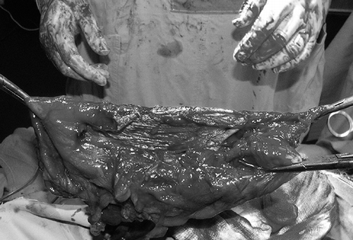
Due to the increased risk of thrombosis, three repeated phlebotomies with total evacuation of 1200 mL of blood were performed preoperatively, until the blood count was normal. After that, open surgical repair was performed. The cyst was dissected and opened and more than 5.5 L of clear yellowish liquid evacuated (). The cyst wall was resected and the nephrectomy was performed. The left kidney measured only about 4 cm. Pathological examination revealed simple renal cyst with typical epithelium, whereas the analysis of the cystic fluid showed no malignant cells. Two years after the surgery, the patient is in good health and his erythrocytosis ceased completely. Also, blood pressure is in normal range without any antihypertension medication, and the function of the remaining kidney is normal ().
Figure 3. Giant renal cyst with thick wall, after the opening and aspiration of the fluid.
DISCUSSION
Because simple renal cysts are progressively more common with age, they have been considered an acquired lesion, although some simple cysts are diagnosed in utero. The precursors of cysts are, most probably, ectasia and cystic dilatation of the tubules and ducts in older patients. Using computed tomography, Laucks and McLachlan demonstrated that the incidence of cysts was 20% by age 40 and 33% after age 60.Citation1
Giant renal cysts greater than 15 cm are rare and usually produce symptoms such as flank pain, hypertension, infection, hematuria, or erythrocytosis.Citation3 In the same time, polycythemia is one of the most common laboratory findings in patients with giant renal cyst originating from proximal tubule.Citation2,Citation6
Our patient did not have symptoms before epistaxis and did not have any regular work up before it. At that time, both diagnosis of the cyst and work up for erythrocytosis were performed. Therefore, late referral is the main reason for the late diagnosis of this cyst.
Nowadays, the treatment of symptomatic giant cysts should be minimally invasive, including percutaneous fluid aspiration with injection of sclerosing agent or laparoscopic unroofing. Open surgery should be restricted only for rare unclear cases requiring exploration or for cases in which some complications such as infection, recurrent cyst, or prolonged drainage could be expected.
In this case, open surgical procedure was performed due to the massive volume of the cyst and to enable surgical exploration. In the postoperative follow-up, the erythrocyte count remained normal and the preexisting hypertension ceased. The hypertension occurred most probably as the consequence of the cyst-related reduced renal blood flow and the activation of renin–angiotensin–aldosterone system. Also, absolute erythrocytosis might participate in the development of hypertension.
This case is unique in three reasons. First, the patient presented with no signs or symptoms, other than giant painless abdominal mass. Second, to our knowledge, this is the largest cyst published in the literature. Previously, Brown and Segura reported a 25-cm big left renal cyst.Citation3 Third, the hypertension ceased after the cyst removal and the patient did not require antihypertensive medication anymore.
Declaration of interest: The authors report no conflicts of interest. The authors alone are responsible for the content and writing of the paper.
REFERENCES
- Glassberg KI. Renal dysgenesis and cystic disease of the kidney. In: Walsh PC, Retik AB, Vaughan ED, Wein AJ, eds. Campbell's Urology. 7th ed., Philadelphia: Elsevier Science; 2003, electronical version.
- Franek E, Kokot F, Wiecek A, Erythropoietin concentration in cyst fluid in patients with simple renal cysts. Nephron. 1994;67:431–435.
- Brown JA, Segura JW, Blute ML. A giant left renal cyst presenting as obesity: A unique presentation. Arch Esp Urol. 1998;51:105–107.
- Giannakopoulos X, Charalabopoulos K, Charalabopoulos A, Golias CH, Peschos D, Sofikitis N. Giant simple renal cyst complicated with hypertension. Int J Clin Pract Suppl. 2005;147:69–71.
- Lezrek M, Fassi-Fehri H, Badet L, Marechal JM, Martin X. Remission of erythrocytosis and hypertension after treatment of a giant renal cyst. Urology. 2002;60:164.
- Eckardt KU, Mollmann M, Neumann R, Erythropoietin in polycystic kidneys. J Clin Invest. 1989;84:1160–1166.
- Poralla T, Arnold W, Eckhardt R, Hutteroth TH, Staritz M, Meyer zum Buschenfelde KH. Polycythemia in kidney cysts. A report on an unusual case. Dtsch Med Wochenschr. 1984;109:1364–1367.
- Bozkurt FB, Boyvat F, Tekin I, Aytekin C, Coskun M, Ozkardes H. Percutaneous sclerotherapy of a giant benign renal cyst with alcohol. Eur J Radiol. 2001;40:64–67.
- Hanash KA, Al-Othman K, Mokhtar A, Laparoscopic ablation of giant renal cyst. J Endourol. 2003;17:781–784.
- Singh I, Sharma D, Singh N. Retroperitoneoscopic deroofing of a giant renal cyst in a solitary functioning hydronephrotic kidney with a 3-port technique. Surg Laparosc Endosc Percutan Tech. 2003;13:404–408.