Abstract
Anastomotic pseudoaneurysms of transplanted kidneys are a very rare complication and encountered in less than 1% of such operations. They may be devastating and cause functional impairment and even loss of the graft. In this report, we present the first case of treatment of extrarenal pseudoaneurysm of arterial anastomosis in a renal transplant patient with endovascular coil embolization with the balloon remodeling technique. This method is mostly used in the treatment of wide-neck intracranial aneurysms.
INTRODUCTION
Anastomotic pseudoaneurysms of transplanted kidneys are a very rare complication and encountered in less than 1% of such operations. They may be devastating and cause functional impairment and even loss of the graft.Citation1–4 Treatment for this abnormality is necessary for the viability of the graft.
In this report, we present the first case of treatment of extrarenal pseudoaneurysm of arterial anastomosis in a renal transplant with endovascular embolization. Detachable coils were used with the balloon remodeling technique, which is mostly used in the treatment of wide-neck intracranial aneurysms ().
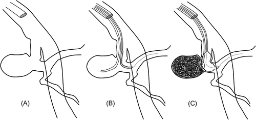
Figure 1. Drawings of the sequential technique used in balloon-assisted coiling. After the catheterization of the main vessel (A), balloon catheter is placed across the aneurysm neck and then the coil delivery microcatheter is placed inside the aneurysm (B). The balloon is inflated during coil insertion to prevent coil protrusion into the vessel lumen and microcatheter kick-out, and to provide sufficient coil packing (C).
CASE REPORT
A 55-year-old male patient with chronic glomerulonephritis received a renal transplant from a living related donor 14 years ago. The transplanted renal artery was anastomosed end to end to the left internal iliac artery. The patient was using cyclosporin 2 × 25 mg, prednisolone 1 × 1.5 mg, and acetylsalicylic acid 1 × 100 mg. He was hypertensive after transplantation, and metoprolol 1 × 100 mg and doxazosin 1 × 8 mg were started for treatment of hypertension. The patient did not have any complaints. The laboratory results showed no renal failure. Creatinine level was normal.
During routine follow-up examination of color Doppler ultrasonography (CDUS), a pseudoaneurysm with a diameter of 18 × 13 mm and an 8 mm neck was detected at the anastomotic site of the transplanted kidney in the left iliac fossa. CDUS performed 1 year before was reported normal. Abdominal magnetic resonance imaging (MRI) and magnetic resonance (MR) angiography confirmed the diagnosis, and a 90° angulation between the renal and the internal iliac arteries at the anastomotic site was also detected (). The patient was referred to the interventional radiology unit for endovascular treatment. One week before the procedure, clopidogrel 1 × 75 mg daily was started orally in addition to acetylsalicylic acid. Intravenous hydration was made with 1000 cc isotonic NaCl during 12 h before procedure to prevent or reduce the probable nephrotoxic side effects. Under local anesthesia, a centrolateral design 7F and 45 cm introducer sheath (Flexor Tuohy-Borst Sidearm, Balkin Up & Over Contralateral Design, Cook Medical, Inc., Bloomington, IN, USA) was placed through the right common femoral artery into the left common iliac artery. Then the left internal iliac artery was catheterized with a 7F guiding catheter (45° angled tip, Vista Brite Tip, Codman & Shurtleff, Inc., Raynham, MA, USA) to allow for introduction of both the microcatheter and the remodeling balloon. After digital subtraction angiography (DSA) (Advantx, General Electric Medical Systems, Milwaukee, WI, USA) was performed, the working projection of the aneurysm was defined. To seal the neck of the pseudoaneurysm during coil insertion, a compliant remodeling balloon, Scopernic20 (Balt, Montmorency, France), was placed across the pseudoaneurysm neck (). The Scopernic20 is an over-the-wire balloon with the provided 0.012 in. wire. Balloon inflation was accomplished with a 50:50 mixture of saline/contrast and a 1 mL syringe, a Cadence injection syringe (EV3, MTI, Irvine, CA, USA). This threaded syringe allows for slower, more calibrated balloon inflation, lowering the risk of vessel rupture. After insertion of the Scopernic20, the aneurysm was catheterized with a microcatheter, Excelsior1018 (Boston Scientific, Fremont, CA, USA). Then the aneurysm was packed with Guglielmi (Boston Scientific) and Micrus (Micrus Endovascular Corporation, San Jose, CA, USA) detachable bare coils. Before inserting each coil, the Scopernic20 was inflated to prevent both coil protrusion into the vessel lumen and microcatheter kick-out. After insertion of each coil, the balloon was deflated and we checked whether there was any coil protrusion. In the event of coil protrusion, the coil was partially retracted and the attempt was repeated until successful insertion was achieved with no coil protrusion. The procedure was continued until the pseudoaneurysm was totally embolized. The last angiography showed the total embolization of the pseudoaneurysm and the procedure was stopped. Then the balloon was inflated for the removal of the coil delivery microcatheter to prevent coil loops from being pulled into the vessel lumen by the microcatheter. The microcatheter was removed. Then the balloon was deflated and also removed. During the procedure, 5000 IU heparin was administered IV.
Figure 2. Magnetic resonance angiography shows a wide-neck pseudoaneurysm at the end-to-end arterial anastomotic site of the transplanted kidney on the left side. The anastomotic site angle is about 90°.
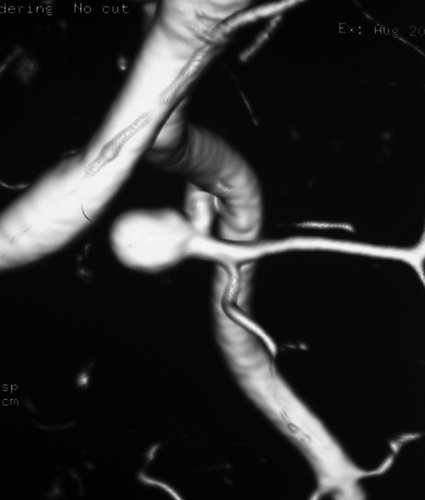
Figure 3. Digital subtraction angiography shows the wide-neck pseudoaneurysm (A). Unsubtracted (B) and subtracted (C) images show the balloon at the neck of the pseudoaneurysm, the coils, and the totally embolized aneurysm.

After the embolization, 1000 IU/h heparin IV infusion was continued for 1 day. Low molecular heparin (2 × 0.6 mL) was administered for 3 days subcutaneously after stopping the IV heparin. A plan was formulated to give acetylsalicylic acid (10 mg/day) orally for life-long use.
CDUS and conventional MRI were performed 1 day after the embolization. Follow-up CDUSs were performed 1, 3, and 12 months later. No recanalization of pseudoaneurysm, narrowing of the renal artery, or thromboembolic complication was seen. Serum creatinine levels were within normal values.
DISCUSSION
Vascular complications in renal transplant recipients are a significant cause of graft dysfunction related to high morbidity and mortality and seen in about 10% of transplantation patients. In the treatment of vascular complications including arterial stenoses, arteriovenous fistulas, pseudoaneurysms, and thrombosis, interventional radiologic methods play important roles. These complications can be detected and defined using CDUS, contrast-enhanced computed tomography, and MR angiography. But DSA is not just a gold standard imaging method; it also enables medical teams to perform endovascular treatment as we did in our case.Citation1,Citation5
Extrarenal, anastomotic pseudoaneurysm in a transplanted kidney is a rare complication and encountered in less than 1% of all patients. Intrarenal pseudoaneurysms are more common and they usually occur due to arterial injury during percutaneous procedures like needle biopsy. Extrarenal pseudoaneurysms are usually the result of defective vascular reconstruction or infection. So they are frequently seen at the anastomotic site and in the early weeks following surgery as a result of anastomotic defects.Citation3,Citation6 Late presentation, as in this case after 14 years, is a very rare condition. In the literature, late spontaneous renal artery pseudoaneurysms seen at the anastomotic sites were reported between 15 months and 20 years after surgery.Citation3,Citation7,Citation8
In addition to any degree of graft dysfunction, extrarenal pseudoaneurysms of transplanted kidneys may also cause the loss of the graft.Citation2–4 Rupture of the pseudoaneurysm is a probable serious complication and may cause death of the patient. Therefore, extrarenal pseudoaneurysms of transplanted kidneys must be treated. There are various options for treatment of extrarenal pseudoaneurysms of transplanted kidneys. Although surgical repair or transplant nephrectomy was most commonly used, Fujikata et al. reported a case of conservative treatment for mycotic aneurysm without rupture.Citation4,Citation6,Citation9,Citation10 In recent years, endovascular or percutaneous treatment of extrarenal pseudoaneurysms has been widely used. Although endovascular embolization is the most suitable technique in the treatment of intrarenal pseudoaneurysms and fistulas, there are limited endovascular techniques for treatment of the extrarenal pseudoaneurysms of transplanted kidneys.Citation5,Citation11
Fornaro et al. reported a case of extrarenal pseudoaneurysm treated with ultrasound-guided thrombin injection. This kind of treatment is suitable in a small number of cases because it allows pseudoaneurysms with narrow necks to be compressed and the flow of blood to be stopped by the ultrasound probe compression.Citation7 The stent-graft insertion at the neck of the pseudoaneurysm is the most suitable endovascular option in most cases. But if the anastomotic site is acutely angled, as in this case, the insertion of the stent-graft may be difficult and cause dissection or rupture of the artery.Citation12,Citation13 In our case, we did not prefer the stent-graft insertion due to the acute angle of the vessel at the site of the pseudoaneurysm. So we decided to treat the pseudoaneurysm with the balloon-assisted coil embolization technique most often used in the treatment of wide-neck intracranial aneurysms.Citation14,Citation15 The pseudoaneurysm was embolized with detachable coils without any complication with the help of the remodeling balloon, Scopernic20. In this case, recanalization of the embolized pseudoaneurysm may be seen as a late complication due to the wide neck of the pseudoaneurysm. If the recanalization occurs during follow-up, stent-assisted reembolization with coils can be performed.
In conclusion, endovascular treatment of extrarenal pseudoaneurysms at the anastomotic site of the transplanted kidneys can be performed with various interventional techniques such as balloon-assisted coil embolization as was done in our unique case.
Declaration of interest:
The authors report no conflicts of interest. The authors alone are responsible for the content and writing of the paper.
REFERENCES
- Dodd GD III, Tublin ME, Shah A, Zajko AB. Imaging of vascular complications associated with renal transplants. AJR Am J Roentgenol. 1991;157:449–459.
- Orlic P, Vukas D, Drescik I, Vascular complications after 725 kidney transplantations during 3 decades. Transplant Proc. 2003;35:1381–1384.
- Taghavi M, Shojaee Fard A, Mehrsai R, Shadman M. Late onset anastomotic pseudoaneurysm of renal allograft artery: Case report, diagnosis, and treatment. Transplant Proc. 2005;37:4297–4299.
- Asztalos L, Olvasztó S, Fedor R, Szabó L, Balázs G, Lukács G. Renal artery aneurysm at the anastomosis after kidney transplantation. Transplant Proc. 2006;38:2915–2918.
- Yagci AB, Parildar M, Oran I, Memis A. Interventional radiological management of vascular complications following renal transplantation. Diagn Interv Radiol. 2006; 12:206–210.
- Fujikata S, Tanji N, Iseda T, Ohoka H, Yokoyama M. Mycotic aneurysm of the renal transplant artery. Int J Urol. 2006;13:820–823.
- Fornaro J, Marincek B, Jungius KP. Pseudoaneurysm in the iliac fossa after renal transplantation – Treatment with ultrasound-guided thrombin injection. Abdom Imaging. 2007; 32:50–52.
- Düsünceli E, Atasoy C, Fitoz S, Yagci C. Late presentation of an extrarenal pseudoaneurysm in a renal transplant patient: CT and color Doppler US findings (case report). Tani Girisim Radyol. 2004;10:289–291 (Turkish).
- Renigers SA, Spigos DG. Pseudoaneurysm of the arterial anastomosis in a renal transplant. AJR Am J Roentgenol. 1978;131:525–526.
- Sharron JA, Esterl RM, Washburn WK, Abrahamian GA. Surgical treatment of an extrarenal pseudoaneurysm after kidney transplantation. Vasc Endovasc Surg. 2009; 43:317–321.
- Miyazaki T, Saitoh R, Doi T, Hayashida Y, Seishi I, Takahashi M. Embolization of a pseudoaneurysm in the transplanted kidney. AJR Am J Roentgenol. 1998; 171:1617–1618.
- Moosavi CA, Gujrathi SK, Friedman A, Fox D, Silberzweig JE. Endovascular repair of symptomatic renal transplant site pseudoaneurysm. Vasc Endovasc Surg. 2008–2009;42: 607–609.
- Poels JA, Riley PL. Extrarenal transplant artery pseudoaneurysm: A combined therapeutic approach. Cardiovasc Intervent Radiol. 2008;31:404–406.
- Baldi S, Mounayer C, Piotin M, Spelle L, Moret J. Balloon-assisted coil placement in wide-neck bifurcation aneurysms by use of a new, compliant balloon microcatheter. AJNR Am J Neuroradiol. 2003;24:1222–1225.
- Ross IB, Dhillon GS. Balloon assistance as a routine adjunct to the endovascular treatment of cerebral aneurysms. Surg Neurol. 2006;66:593–601; discussion 601–602.