Abstract
Solitary extramedullary plasmacytoma (EMP) is a rare plasma cell disorder mostly involving the upper airway; however, retroperitoneal infiltration is very rare. Kidney injury associated with EMP is exceptionally rare with only anecdotal reports. Herein we report a case of retroperitoneal EMP causing renal failure by the way of direct renal parenchymal infiltration. Renal parenchymal invasion should be considered in aggressive and refractory plasma cell dyscrasias with unexplained renal failure.
INTRODUCTION
Solitary extramedullary plasmacytomas (EMPs) are rare plasma cell disorders accounting for approximately 3% of all plasma cell neoplasms.Citation1 Most EMPs involve mucosa-associated lymphoid tissue of the upper airway; however, retroperitoneal infiltration is very rare.Citation2 Kidney injury associated with EMP is exceptionally rare with only anecdotal reports.Citation3,4 Herein we report a case of retroperitoneal EMP with direct parenchymal invasion of the kidney causing renal failure.
CASE REPORT
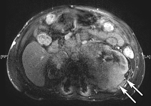
A 68-year-old male was admitted with increasing pretibial edema, exertional dyspnea, and low urinary output. About 14 years ago, a retroperitoneal mass lesion was detected and biopsy taken from the lesion revealed infiltration of the fat and connective tissue with mature plasma cells expressing monoclonal lambda light chain and immunoglobulin G (IgG) heavy chain compatible with the diagnosis of plasmacytoma. Repeated bone marrow biopsies showed normohypercellular marrow without clonal plasma cell infiltration. All together, the diagnosis of EMP was established. He had been followed up for about 14 years with various treatments, such as vincristine, adriablastin, dexamethasone, bortezomib, radiotherapy, and finally lenalidomide. He was refractory to all these treatments and the sizes of these intra-retroperitoneal mass lesions progressively increased. M-spike was not present in the protein electrophoresis, and serum and urinary immunoelectrophoresis were normal. Bence–Jones proteinuria was also absent. The right kidney was significantly smaller than the left one and had minimal or no contribution to creatinine clearance, as shown by renal scintigraphy. The first information about the asymmetry of the kidney sizes was taken in 2006, with smaller right kidney size of 100 × 60 mm while the left kidney was 136 × 15 mm. In 2008, right kidney was 96 × 55 mm in size. When the patient’s medical history was deepened, the patient was found to have three episodes of pyelonephritis between the years 2001 and 2002. Probably, chronic pyelonephritis was the responsible causative factor for the smaller right kidney. When the mass lesions increased in size, postrenal obstruction caused hydronephrosis of the already atrophied right kidney together with the normal-sized left kidney. Bilateral ureteral double J catheter insertion was performed and hydronephrosis was resolved. Baseline serum creatinine levels were stabilized to 1.4 mg/dL. After about 6 months, the patient developed oliguria and progressive renal failure with serum creatinine levels rising to 7 mg/dL necessitating hemodialysis treatment. In physical examination, abdomen was distended and multiple mass lesions were palpable. He had bilateral 2+ pretibial edema. Baseline biochemical parameters are presented in . There was no recent nephrotoxic chemical or drug use. In ultrasonography, significant hydronephrosis explaining this acute kidney failure could not be shown. For the treatment of a possible postrenal etiology, double J catheters were replaced with new metal stents; however, serum creatinine levels failed to decrease and urine output did not increase. In abdominal magnetic resonance (MR) imaging, multiple mass lesions with a maximum diameter of 90 mm were observed around the left renal hilus. Also a mass lesion of 70 mm indiscernible from the posterior region of the kidney was observed and it was considered to be the direct renal parenchymal infiltration by the plasmacytoma (A and B). The patient was evaluated to be refractory also to lenalidomide and thus lenalidomide was stopped. The patient was followed with hemodialysis treatment 3 times per week. He progressively lost weight and unfortunately died possibly because of high tumor burden.
Table 1. Baseline biochemical laboratory data.
Figure 1. MR image of the mass lesions indiscernible from the kidney around the left renal hilus and the posterior region of the kidney (arrows) (transverse view).
Note: MR, magnetic resonance.
DISCUSSION
Plasma cell dyscrasias are often associated with kidney disease. Mechanisms of kidney injury in plasma cell malignancies include cast nephropathy, amyloid light-chain (AL) amyloidosis, monoclonal Ig deposition disease, glomerulonephritis, tubulointerstitial nephritis, hyperviscosity syndrome, hypercalcemia, tumor lysis syndrome, and rarely direct parenchymal invasion by plasma cells.Citation5
EMPs are rare plasma cell proliferative disorders. Their diagnosis is based on the monoclonal plasma cell infiltration at a single disease site and the exclusion of systemic myeloma.Citation6 EMP occurs most commonly in the head and neck region, followed by gastrointestinal tract, central nervous system, thyroid, breast, parotid gland, testis, and lymph nodes.Citation7 Solitary EMP rarely occurs in the retroperitoneum. Cases of retroperitoneal EMP may have different clinical manifestations, such as renal failure,Citation3,4 flank pain and hematuria due to thrombosis of the renal vein,Citation8 obstructive jaundice,Citation9 and abdominal distention and pain.Citation10
Direct renal parenchymal infiltration may be the least common mechanism of renal failure due to plasma cell dyscrasias.Citation11–14 In the case reported by Kanoh et al.,Citation11 large soft-tissue mass emanating from the right kidney was observed to cause hydronephrotic changes. Treatment with radiotherapy alone was sufficient in this case. In another report,Citation12 a patient was presented with a right renal plasmacytoma and renal failure. Although the patient received multiple chemotherapies, he died 1 year after the diagnosis due to a rapidly progressing plasmatic cell dyscrasia. Igel et al.Citation13 reported a case of EMP arising in the kidney mimicking a renal cell carcinoma or a transitional cell carcinoma of the renal pelvis.
Radiotherapy is the treatment of choice in EMP.Citation7 Refractory patients may be treated with surgery and/or chemotherapy. In the literature, Kanoh et al.Citation11 reported that the treatment with radiotherapy alone was sufficient in their case. However, another patient died in spite of administration of multiple chemotherapeutics.Citation12 Our patient was also resistant to nearly all types of treatment modalities. Stem cell transplantation may be the choice of treatment in patients with high-risk plasmacytoma refractory to therapies.Citation15
In conclusion, direct renal parenchymal infiltration is a very rare cause of renal failure due to retroperitoneal EMP. It should be considered in aggressive and refractory plasma cell dyscrasias with unexplained renal failure.
Declaration of interest: The authors report no conflicts of interest. The authors alone are responsible for the content and writing of the paper.
REFERENCES
- Dimopoulos MA, Kiamouris C, Moulopoulos LA. Solitary plasmacytoma of bone and extramedullary plasmacytoma. Hematol Oncol Clin North Am. 1999;13:1249–1257.
- Ooi GC, Chim JC, Au WY, Khong PL. Radiologic manifestations of primary solitary extramedullary and multiple solitary plasmacytomas. AJR Am J Roentgenol. 2006;186:821–827.
- Marks ES, Lee KM. Acute renal failure secondary to vascular occlusion by a retroperitoneal plasmacytoma. Cancer. 1984;53:1228–1229.
- Gionanlis LC, Bamichas GI, Smias CV, Tzarou VN, Sombolos KI. Retroperitoneal plasmacytoma causing renal obstruction. Int Urol Nephrol. 2008;40(2):555–556.
- Heher EC, Goes NB, Spitzer TR, . Kidney disease associated with plasma cell dyscrasias. Blood. 2010;116(9):1397–1404.
- Dimopoulos MA, Hamilos G. Solitary bone plasmacytoma and extramedullary plasmacytoma. Curr Treat Options Oncol. 2002;3:255–259.
- Weber DM. Solitary bone and extramedullary plasmacytoma. Hematology Am Soc Hematol Educ Program. 2005;373–376.
- Kobayashi H, Itoh T, Murata R, Tanabe M. Primary retroperitoneal plasmacytoma with tumor thrombus within the renal vein. J Urol. 1992;147:452–454.
- Chen TC, Wu JH, Ng KF, Lien JM, Hung CF. Solitary extramedullary plasmacytoma in the retroperitoneum. Am J Hematol. 1998;58:235–238.
- Sered S, Nikolaidis P. CT findings of perirenal plasmacytoma. AJR Am J Roentgenol. 2003;181:888.
- Kanoh T, Katoh H, Izumi T, Tsuji M, Okuma M. Renal plasmacytoma. Rinsho Ketsueki. 1993;34(11):1470–1473.
- Tejido Sánchez A, Hernández Martínez E, Ortíz MC, . Renal plasmacytoma. Report of a new case. Arch Esp Urol. 2001;54(7):718–722.
- Igel TC, Engen DE, Banks PM, Keeney GL. Renal plasmacytoma: Mayo clinic experience and review of the literature. Urology. 1991; 37(4):385–389.
- Kandel LB, Harrison LH, Woodruff RD, Williams CD, Ahl Jr. ET, Renal plasmacytoma: A case report and summary of reported cases. J Urol. 1984;132(6):1167–1169.
- Jantunen E, Koivunen E, Putkonen M, Siitonen T, Juvonen E, Nousiainen T. Autologous stem cell transplantation in patients with high-risk plasmacytoma. Eur J Hematol. 2005;74(5):402–406.