Abstract
Malpositioning of long-term hemodialysis catheter is a known complication that may lead to a gradual decline in blood flow. We report two rare catheter malpositions in the hepatic veins in two patients with end-stage renal disease in whom a dialysis catheter was inserted through the left external jugular vein and the right external jugular vein. Because of gradual reduction of catheters’ blood flow, an angiography was performed, which confirmed catheters’ tip positioning into the hepatic veins. The catheters were replaced in the correct site.
INTRODUCTION
Malposition of a central venous catheter is a common complication (4%) and requires appropriate management (correction, replacement, or removing).Citation1 The insertion of the hemodialysis catheter’s tip into the hepatic veins is a very rare condition and only one case is referred in literature.Citation2 We present two cases of hemodialysis catheter dysfunction due to the catheters’ tip insertion into the hepatic veins.
REPORT OF CASES
First Case
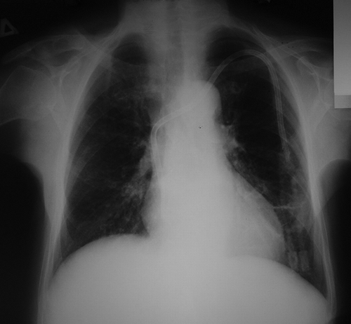
An 85-year-old female patient, with end-stage renal disease (unknown primary disease) in hemodialysis therapy three times per week for 5 years, was admitted for management of a dysfunctioning hemodialysis catheter. Her medical history included arterial hypertension for the last 30 years and left cardiac failure of diastolic type with episodes of acute pulmonary edema on the basis of hypervolemia because of the chronic renal disease. Also from clinical and laboratory testing (low parathormone and changing levels of calcium), a dynamic renal bone disease is referred. In the beginning, a temporal dual lumen hemodialysis catheter was inserted via the right jugular vein and a distal radial-cephalic arteriovenous anastomosis was created at the left forearm, which was in function for 10 months. After that, because of native vascular access thrombosis and left cardiac failure, a long-term, dual-lumen, cuffed, hemodialysis catheter (diameter 14, 5 Fr, cuff to tip 19 cm) was inserted through the left external jugular vein with the tip position at the junction of the superior vena cava to the right atrium. The rate of blood flow was found to be more than 500 mL/min. This catheter was in function for 3 years and 7 months. Because of gradual blood flow reduction and poor response to thrombolytic therapy, the catheter was replaced by the Seldinger method with a longer one (diameter 14, 5 Fr, cuff to tip 23 cm), through the same route but with a different subcutaneous tunnel. The catheter’s tip position into the right atrium was confirmed via fluoroscopy and the rate of blood flow was found to be more than 500 mL/min. Two months later, the catheter’s blood flow gradually reduced and the patient underwent angiography with a contrast agent injected separately from the two lumens of the catheter. The angiography from the venous limb of the catheter showed tip positioning into the hepatic veins (). The catheter was replaced with a same kind of catheter by the Seldinger method, through the same route but with a little longer subcutaneous tunnel, and its tip positioning into the right atrium was confirmed by fluoroscopy (); the rate of blood flow was found to be more than 500 mL/min.
Figure 1. Angiography of first case: hemodialysis catheter’s tip positioning into the hepatic veins.
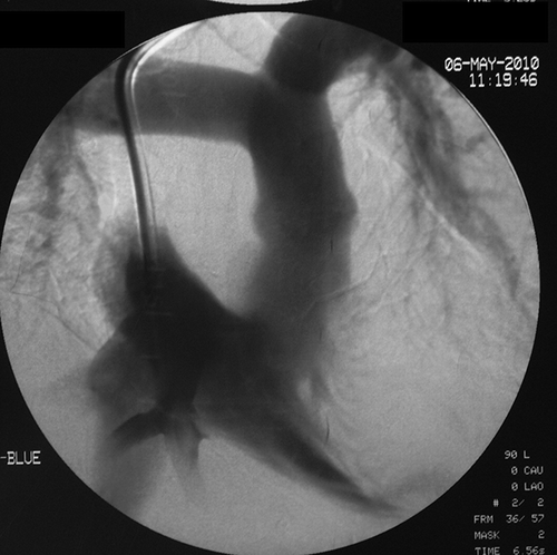
Figure 2. Chest radiography: hemodialysis catheter’s tip positioning into the right atrium.
Second Case
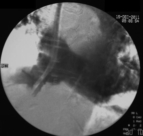
A 75-year-old female patient, with end stage renal disease (primary disease diabetes mellitus) in hemodialysis therapy three times per week for 3 years, was admitted for management of a dysfunctioning hemodialysis catheter. Her medical history included arterial hypertension, diabetes mellitus, and mitral valve insufficiency. In the beginning, a long-term, dual-lumen, cuffed, hemodialysis catheter (diameter 14, 5 Fr, cuff to tip 23 cm) was inserted through the right external jugular vein providing blood flow more than 500 mL/min. The catheter’s tip position into the right atrium was confirmed via fluoroscopy at the time of insertion. This catheter was in function for 3 years. The patient was hemodynamically stable during the dialysis. Over the last 3 months, the rate of blood flow from the arterial limb of the catheter decreased with a poor response to thrombolytic therapy. The angiography with a contrast agent injected separately from the two lumens of the catheter showed the catheter’s tip positioning into the hepatic veins (). The catheter was replaced by the Seldinger method with another catheter, through the same route but with a different subcutaneous tunnel, checking fluoroscopically its tip positioning into the right atrium, and the rate of blood flow was found to be more than 500 mL/min.
Figure 3. Angiography of second case: hemodialysis catheter’s tip positioning into the hepatic veins.
DISCUSSION
The use of permanent central vein catheters for hemodialysis is a solution to the problem of hemodialysis access in patients suffering from end-stage renal disease in cases where there are no alternative possibilities for the creation of a vascular accessCitation3 or where the creation of a vascular access for hemodialysis is not recommended.
Aberrant placement of a permanent central vein catheter for hemodialysis is a well-recognized condition that is related to the catheter’s tip position outside the major vein trunks or the right atrium.Citation4
Sometimes, even if the permanent central vein catheter for hemodialysis is inserted at a correct anatomical position (major vein trunk or right atrium), because of pericatheter sheath formation, the blood flows in via the collateral vein set. This leads to a decrease in the rate of blood flow through the catheter.Citation5
The site of permanent central vein catheters for hemodialysis is checked by fluoroscopy or roentgenography.Citation6 In all cases of displacement or dysfunction of permanent central vein catheters for hemodialysis, an X-ray control by contrast medium injection in both of the catheter lumens, separately, reveals the catheter’s tip positioning as well as the thrombosis of main vein trunks and the collateral vein set.Citation1,7 In both the cases in our study, the angiography showed the site of the catheter’s tip and blood supply from the hepatic veins.
In cases of insertion of a longer catheter, it is possible that the catheter’s tip enters through the inferior vena cava into the hepatic veins. This may be harmful to the liver, requiring immediate action,Citation2 or may contribute to catheter malfunctioning as happened with both cases in our study. Over time, the fibrosis of the venous endothelium due to the contact with the catheter and the sheath formation decreases the rate of blood flow in the catheter.Citation8 Additionally, the tunnelled hemodialysis catheter is in constant motion due to the movement of the heart as well as the upper body.Citation9 For this reason, the catheter’s tip movement may cause damage to the wall of the vessel or the atrium, resulting in mural thrombus formation at the point of contact.Citation10
Catheter replacement over a guidewire has become an accepted technique for replacing a malfunctioning catheter,Citation11 a technique that we have used in both cases in our study. Catheter insertion over a guidewire is associated with less discomfort and a significantly lower rate of mechanical complications than those inserted percutaneously at a new site.Citation12
CONCLUSION
In cases of the hemodialysis catheter insertion into the right atrium, its tip can be moved or erroneously inserted through the inferior vena cava into the hepatic veins. The correct positioning of a hemodialysis catheter must be checked fluoroscopically while its malfunction must be checked by X-ray control by contrast medium injection in both of the lumens, separately, for repositioning or replacing the catheter.
Declaration of interest: The authors report no conflicts of interest. The authors alone are responsible for the content and writing of the article.
REFERENCES
- Skandalos I, Hatzibaloglou A, Evagelou I, . Deviations of placement/function of permanent central vein catheters for hemodialysis. Int J Artif Organs. 2005;28(6):583–590.
- Lindner U, Haas CS. Malposition of a hemodialysis catheter in a hepatic vein. Scand J Urol Nephrol. 2010;44(5):364–365.
- Cetinkaya R, Odabas AR, Unlu Y, Selcuk Y, Ates A, Ceviz M. Using cuffed and tunnelled central venous catheters as permanent vascular access for hemodialysis: a prospective study. Ren Fail. 2003;25(3):431–438.
- Messina M, Morale W, Viglianesi A, . A typical placement of hemodialysis catheters in patients with complete and irreversible obstruction of central venous vessels. J Vasc Access. 2011; 12(1):21–27.
- Suhocki PV, Conlon PJ Jr, Knelson MH, Harland R, Schwab SJ. Silastic cuffed catheters for hemodialysis vascular access: thrombolytic and mechanical correction of malfunction. Am J Kidney Dis. 1996;28(3):379–386.
- Sotirakopoulos N, Skandalos L, Tsitsios T, Stambolidou M, Karamoschos K, Mavromatidis K. The incorrect placement of hemodialysis catheters in veins. The necessity for urgent X-ray evaluation for its position. Ren Fail. 2001;23(1):127–133.
- Rasmussen RL. The catheter-challenged patient and the need to recognize the recurrently dysfunctional tunneled dialysis catheter. Semin Dial. 2010;23(6):648–652.
- Forauer AR, Theoharis C. Histologic changes in the human vein wall adjacent to indwelling central venous catheters. J Vasc Interv Radiol. 2003;14(9):1163–1168.
- Nazarian GK, Bjarnason H, Dietz CA Jr, Bernadas CA, Hunter DW. Changes in tunneled catheter tip position when a patient is upright. J Vasc Interv Radiol. 1997;8(3):437–441.
- Bayón J, Martín M, García-Ruíz JM, Rodríguez C. “We have a tenant” a right atrial thrombus related to a central catheter. Int J Cardiovasc Imaging. 2011;27(1):5–6.
- Wong JK, Sadler DJ, McCarthy M, Saliken JC, So CB, Gray RR. Analysis of early failure of tunneled hemodialysis catheters. AJR Am J Roentgenol. 2002;179(2):357–363.
- Cobb DK, High KP, Sawyer RG, . A controlled trial of scheduled replacement of central venous and pulmonary-artery catheters. N Engl J Med. 1992;327(15):1062–1068.