Abstract
Purpose: The aim of this study is to determine the optimal angle of needle entry in the sagittal plane for internal jugular vein (IJV) catheterization with the central approach while the head is in the neutral position. Methods: The contrast–enhanced carotid artery computed tomography angiographies of 123 consecutive patients were retrospectively reviewed. The point of merger between the sternal and clavicular heads of the sternocleidomastoid muscle was assumed as a clinical entry (CE) point. The angle between CE point and the center of the IJV, the depth, diameter of the vessels and the degree of overlap between the IJV and carotid artery (CA) were measured. Results: The angles between the CE point and the center of the IJVs were similar, 7° ± 13° medial and 8° ± 12° medial on the right and the left side, respectively. The center of IJVs from the CE point was between 0° and 16° toward the medial in 79.8% on the right side and 89.9% on the left side of patients. The diameters of the right IJVs were greater than the left IJVs (p = 0.001). The depth from the skin and overlap between IJV and CA did not vary between the two sides. Conclusions: When a central approach is used for right internal jugular vein (RIJV) cannulation with a neutral head position, the orientation of the angle of needle entry (i.e., 16°) medially in the sagittal plane may quadruple the success rate of RIJV catheterization compared to the success rate of a laterally oriented angle of entry as recommended by the classic method.
INTRODUCTION
Internal jugular venous (IJV) catheters for hemodialysis are a commonly employed temporary vascular access for hemodialysis.Citation1 This procedure has been performed by a blind method using only anatomical landmarks for many years. Although success rates are high while the blind method is used, inadvertent carotid artery (CA) puncture is seen in up to 21% during IJV catheterization and may result in serious neurological damage and even death in high-risk patients.Citation2–Citation4 CA puncture poses greater risk for patients with uremia, arteriosclerosis, and coagulopathy.Citation5 It has been recommended that this procedure be performed under guidance by ultrasound (US) due to greater success rates and fewer complications.Citation1 However, in the event that no US is available or there is no physician experienced in placing a central venous catheter guided by US, physicians should develop sufficient skill to achieve successful central venous catheterization using the blind method.Citation6
IJV catheterization can be achieved by an anterior, central, or posterior approach, using the meeting point of the sternal and clavicular ends of the sternocleidomastoid (SCM) muscle in alignment with the cricoids cartilage as anatomical landmarks.Citation7 CA pulsations can also being used as an auxiliary method, depending on the physicians’ preference. Although no superior qualities have been reported among the mentioned approaches,Citation8 the central approach is most commonly used.Citation3,Citation4,Citation8,Citation9 Within the framework of the central approach, the head is often rotated toward the opposite side and the needle enters at the meeting point of the two ends of the SCM muscle in alignment with the cricoids cartilage and is moved toward the nipple on the same side.Citation3,Citation4 When the head is rotated toward the opposite side, the landmarks become more clear, cannulation becomes easierCitation3,Citation10; however, the probability of overlap between the jugular vein and anatomical landmarks is increased.Citation11 Conversely, anatomical studies using US have reported that the probability of overlap between the CA and the jugular vein is increased by turning the head toward the opposite side; thus, it would actually be more advantageous to keep the head in a neutral position to decrease the risk of CA puncture.Citation3,Citation10–Citation12 In addition, maintaining the head in the neutral position may be necessary in cases of cervical trauma or fusion. Keeping the head straight, however, may change the relationship between the anatomical landmarks and the jugular vein and decrease the probability of successful cannulation.Citation3,Citation13 Therefore, knowledge of the optimal needle entry angle for IJV catheterization while the head is in the neutral position is important for physicians.
In a randomized prospective study by Apiliogullari et al.,Citation3 the authors performed IJV cannulation through the central approach with the head in a neutral position and with the head rotated toward the opposite side, directing the needle toward the ipsilateral nipple, and reported that the success rate with the neutral head position was significantly low with the 90% study power. The findings reported by Apiliogullari et al.Citation3 provoked the following question: when IJV cannulation is performed through the central approach while the head is in the neutral position, to which side and at what angle the needle should be directed in order to reach the jugular vein.
The primary goal of this retrospective computerized tomography (CT) study is to determine the optimal needle angle of entry in the sagittal plane for IJV catheterization when the central approach is used while the head is in the neutral position.
MATERIALS AND METHODS
The study protocol was approved by the Institutional Review Board of the Medical Faculty Hospital, Selcuk University. We retrospectively reviewed the contrast–enhanced CA CT angiography of 123 consecutive patients aged from 18 to 65 years between July and September 2012. The purpose of these carotid CT angiograms was to evaluate CA diseases which are usually created by atherosclerosis. CT scans were excluded from the study if any of the following conditions were observed: inadequate vessel opacification, inappropriate CT scan sections, thrombosis of IJV, anatomical neck abnormalities, neck and upper thoracic masses, goiters, and lymphadenopathies
CT Technique
All images were captured with the patient in a supine position with the legs extended, and the subjects were asked to hold their breath. The CT scans were obtained with a four-detector-row CT scanner (Aquillon; Toshiba Medical Systems, Tokyo, Japan), and the images were reconstructed at 3.0-mm intervals. The required dose of noniodinated contrast material, determined according to the patient’s weight, was administered using a power injector.
Study Measurement
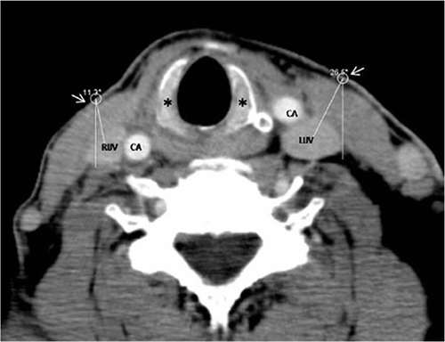
All measurements were performed separately for both the right internal jugular vein (RIJV) and the left internal jugular vein (LIJV). The point of merger between the sternal and clavicular heads of the SCM at the cross-sectional level of the cricoid cartilage was assumed as a central landmark. Vertical projection of this anatomical region from the skin was accepted as the clinical entry (CE) point. The angle between this point and the center of the IJV was automatically calculated on workstation (Vitrea Workstation, Toshiba Medical Systems, Tokyo, Japan) ().
The depth of the vessels below the skin was measured from the skin to the margin of the vein closest to the skin’s surface. The diameter of the vessels was measured between the furthest two points of the vein wall in the transverse plane. Measurements of IJVs ≤7 mm were accepted as potentially difficult catheterization and were recorded.Citation14,Citation15
Figure 1. Axial CT image shows the automatically calculated angles between the clinical entry point (arrows) and the center of the IJVs at the cross-sectional level of the cricoid cartilage (*). CA, carotid artery; RIJV, right internal jugular vein; LIJV, left internal jugular vein.
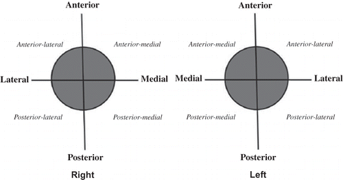
Both common CAs were taken as reference points for measuring the position of the IJV (). The degree of overlap between the IJV and CA was defined as follows: (1) the IJV overlapped ≤25% of the diameter of the CA; (2) the IJV overlapped 26–50% of the diameter of the CA; or (3) the IJV overlapped >50% of the diameter of the CA.
Figure 2. Common carotid arteries were taken as reference points and perpendicular axes were used to demarcate the right and left internal jugular veins positions. O: common carotid arteries.
The sample size was based on that used in a previous retrospective CT study.Citation16 Data were tested for normality using the Kolmogorov–Smirnov normality test, and the results were expressed as mean ± SD or number of patients. Categorical data were analyzed using the chi-square test, and the Student’s t test was used for normally distributed data. A p-value of <0.05 was considered statistically significant.
RESULTS
We retrospectively evaluated 123 consecutive cases of contrast-enhanced CA CT angiography studies. Thirty-four patients were excluded from the analysis due to inadequate vessel opacification (n = 6), inappropriate CT scan sections (n = 7), thrombosis of IJV (n = 2), and anatomical neck abnormalities (previous neck surgery, neck masses, goiters, and lymphadenopathies; n = 19). Thus, a total of 178 IJVs from 89 study participants (42 females and 47 males, mean age 46 ± 11.9 years, range: 18–65 years) were evaluated.
The results of the CT scan analyses are presented in and 2. The angles between the CE point and the center of the IJVs were similar in measure as 7° ± 13° medial (ranged from 74° medial to 20° lateral) and 8° ± 12° medial (ranged from 74° medial to 12° lateral) on the right and the left side, respectively (). Central of IJVs from the CE point was between 0° and 16° toward the medial in 79.8% on the right side and 89.9% on the left side of the patients (). The diameters of the RIJVs were significantly greater than those of the LIJVs (p = 0.001). Number of patients with ≤7 mm were 4 and 19 in the RIJV and LIJV, respectively (p = 0.001). The depth from the skin, position, and overlap between IJV and CA did not vary significantly between the right and left sides ().
Table 1. Angles between the clinical entry point and the center of the internal jugular veins.
Table 2. Results of the analysis of the computerized tomography scans.
DISCUSSION
Although central vein catheterization has been used for many years, modern technology has provided the capability to perform this procedure under safer conditions. According to the current understanding, several factors can increase success rates and decrease complication rates for IJV catheterization, including vein superficiality, diameter, and the distance from both CA and pleura. Efforts to achieve safer IJV catheterization frequently aim at increasing the diameter of the IJV or decreasing the possibility of CA puncture. The Trendelenburg position, Valsalva maneuver, humming, and positive end expiratory pressure (PEEP) application are known to increase IJV diameter.Citation17,Citation18 When the head is in the neutral position, the degree of overlap decreases, especially between the right jugular vein and the CA, and therefore the risk of CA puncture is decreased.Citation10 In order to verify this information, Lamperti et al.Citation10 studied the effects of US-guided IJV catheterization with the head in a neutral position and turning it to the side on the rates of success and complications in1332 adult patients. The authors reported no difference between the two groups with respect to success rates or complications. The most substantial limitation of the study was likely related to the use of US, because skilled US by an experienced technician should increase success rates regardless of head position .5 [TS: Set “5” in superscript position].
A recently published US study conducted by Park et al.Citation13 reported that the central point of the IJV only 0.28 ± 0.78 cm lateral to the apex of the triangle (distance ranged from 1 cm medial to 2.5 cm lateral) in 100 adult patients with the head in the neutral position. Based on the results of present study, angles between the CE point and the center of the IJVs were similar: 7° ± 13° medial and 8° ± 12° medial on the right and the left sides, respectively. These results may explain the high failure rate reported by Apiliogullari et al.Citation3 where the needle was directed laterally using central landmarks.
The results also indicate that the distances of the right and left IJVs from the skin are similar (16.8 and 16.3 mm, respectively). This finding is inconsistent with a previous report suggesting distances of 17.4 mm on the right and 18.7 mm on the left with statistical difference (p < 0.0001).19 Although the distance from the skin may not provide a basis for preference between the right and left sides, the larger diameter of the RIJV, and number of patients with ≤7 mm diameter were high in left side consistent with reports of previous studies, supports a preference of the right side.Citation14–Citation16,Citation19
Sulek et al.Citation20 stated that there was no difference in overlap between the right and left IJV in the neutral position based on their US study, while overlap increased when the head was rotated toward the opposite side at an 80° angle. The results of our retrospective evaluation, consistent with previous studies, revealed no difference between the right and the left sides with respect to overlap with the CA.Citation11,Citation20
The present study has some limitations that must be considered: the lack of different approach groups and different age groups. Further studies are needed to determine the optimal needle entry angle for IJV catheterization when the different approaches (e.g., anterior, posterior, and precarotid approach) were used in different age groups.
Based on the results of present retrospective evaluation of CT scans, when a central approach is used for RIJV catheterization with a neutral head position, the orientation of the angle of needle entry 16° medially in the sagittal plane may quadruple the success rate of RIJV cannulation compared to the success rate of an laterally oriented angle of entry as recommended by the classic method. Our assessments are based on two-dimensional CT scan images; therefore, clinical studies are needed to verify the applicability of our conclusion in daily practice.
Declaration of interest: The authors report no conflicts of interest.
REFERENCES
- Bansal R, Agarwal SK, Tiwari SC, Dash SC. A prospective randomized study to compare ultrasound-guided with non ultrasound-guided double lumen internal jugular catheter insertion as a temporary hemodialysis access. Ren Fail. 2005;27:561–564.
- Feller-Kopman D. Ultrasound-guided internal jugular access: a proposed standardized approach and implications for training and practice. Chest. 2007;132:302–309.
- Apiliogullari B, Kara I, Apiliogullari S, Arun O, Saltali A, Celik JB. Is a neutral head position as effective as head rotation during landmark-guided internal jugular vein cannulation? Results of a randomized controlled clinical trial. J Cardiothorac Vasc Anesth. 2012; 26:985–988. doi:10.1053/j.jvca.2012.07.005.
- Kocum A, Sener M, Calıskan E, Bozdogan N, Atalay H, Aribogan A. An alternative central venous route for cardiac surgery: supraclavicular subclavian vein catheterization. J Cardiothorac Vasc Anesth. 2011;25:1018–1023.
- Kara I, Ozbek S, Apiliogullari S, Duman A, Celik JB, Ozbek S. Seeing is believing: ultrasound-guided internal jugular vein cannulation. Anesth Analg. 2012;115:1471.
- National Institute for Clinical Excellence. Guidance on the use of ultrasound locating devices for placing central venous catheters. Technology Appraisal Guidance No. 49, September 2002. Available at:http://www.nice.org.uk. Accessed February, 2012.
- Sznajder JI, Zveibil FR, Bitterman H, Weiner P, Bursztein S. Central vein catheterization: failure and complication rates by three percutaneous approaches. Arch Intern Med. 1986;146:259–261.
- Hessel EA 2nd. Landmark-guided internal jugular vein cannulation: is there still a role and, if so, what should we do about it? J Cardiothorac Vasc Anesth. 2012; 26(6):979–981. doi:10.1053/j.jvca.2012.08.002.
- Suarez T, Baerwald JP, Kraus C. Central venous access: the effects of approach, position, and head rotation on internal jugular vein cross-sectional area. Anesth Analg. 2002;95:1519–1524.
- Lamperti M, Subert M, Cortellazi P, et al. Is a neutral head position safer than 45-degree neck rotation during ultrasound-guided internal jugular vein cannulation? Results of a randomized controlled clinical trial. Anesth Analg. 2012;114:777–784.
- Troianos CA, Kuwik RJ, Pasqual JR, Lim AJ, Odasso DP. Internal jugular vein and carotid artery anatomic relation as determined by ultrasonography. Anesthesiology. 1996;85:43–48.
- Lieberman JA, Williams KA, Rosenberg AL. Optimal head rotation for internal jugular vein cannulation when relying on external landmarks. Anesth Analg. 2004;99:982–988.
- Park SY, Kim MJ, Kim MG, et al. Changes in the relationship between the right internal jugular vein and an anatomical landmark after head rotation. Korean J Anesthesiol. 2011;61:107–111.
- Mey U, Glasmacher A, Hahn C, et al. Evaluation of an ultrasound-guided technique for central venous access via the internal jugular vein in 493 patients. Support Care Cancer. 2003;11:148–155.
- Lorchirachoonkul T, Ti LK, Manohara S, et al. Anatomical variations of the internal jugular vein: implications for successful cannulation and risk of carotid artery puncture. Singapore Med J. 2012;53:325–328.
- Lim CL, Keshava SN, Lea M. Anatomical variations of the internal jugular veins and their relationship to the carotid arteries: a CT evaluation. Australas Radiol. 2006;50:314–318.
- Lewin MR, Stein J, Wang R, et al. Humming is as effective as Valsalva’s maneuver and Trendelenburg’s position for ultrasonographic visualization of the jugular venous system and common femoral veins. Ann Emerg Med. 2007;50: 73–77.
- Lee SC, Han SS, Shin SY, Lim YJ, Kim JT, Kim YH. Relationship between positive end-expiratory pressure and internal jugular vein cross-sectional area. Acta Anaesthesiol Scand. 2012;56:840–845.
- Ishizuka M, Nagata H, Takagi K, Kubota K. Right internal jugular vein is recommended for central venous catheterization. J Invest Surg. 2010;23:110–114.
- Sulek CA, Gravenstein N, Blackshear RH, Weiss L. Head rotation during internal jugular cannulation and the risk of carotid artery puncture. Anesth Analg. 1996;82:125–128.