Abstract
Contrast-induced nephropathy (CIN) is a common cause of hospital-acquired acute kidney injury (AKI) and a source of significantly increased short- and long-term mortality. Studies of large cohorts have revealed that more than half of these cases are in subjects undergoing cardiac catheterization and intra-arterial coronary angiography, and nearly a third follow computed tomography (CT) scans. Neutrophil gelatinase-associated lipocalin (NGAL) represents an early predictive troponin-like biomarker for AKI. Its role in the timely diagnosis of CIN has already been examined in adults and children undergoing coronary angiography and a meta-analysis revealed a very good performance of plasma or urine NGAL in the prediction of CIN. Much of these data have been extrapolated to patients receiving intravenous (IV) contrast agent for CT scans, although major differences in patient populations, contrast volume administered and intra-procedural complications between the two settings exist. In this context, a recent prospective study by our group evaluated plasma NGAL, measured using standardized Τriage® NGAL test (Biosite Incorporated, San Diego, CA) at baseline and 6-h post-procedure, for early detection of CIN among hospitalized patients undergoing elective contrast-enhanced CT. CIN, defined as an increase in serum creatinine (SCr) of >25% or >0.5 mg/dL from baseline within 48-h post-procedure, was found in 8.51% of subjects. In contrast, significant elevation of plasma NGAL was found at 6-h post-procedure with excellent performance characteristics. This review presents the current status of NGAL in the prediction of CIN after IV contrast administration among hospitalized patients undergoing elective contrast-enhanced CT.
Introduction
Contrast-induced nephropathy (CIN) is a common cause of hospital-acquired acute kidney injury (AKI), accounting for up to 12% of cases, and is associated with an average in-hospital mortality of 6%.Citation1 The exact risk for CIN is difficult to determine. The reported incidence of CIN varies widely depending on the presence or absence of risk factors, in particular pre-existing chronic kidney disease (CKD), the amount and type of administered contrast agent and the nature of the radiologic procedure.Citation1–3 However, despite the widespread use of newer, less nephrotoxic contrast agents and the implementation of preventive strategies over the last decades, the risk of CIN continues to be considerable, especially in the in-patient setting.Citation3 Studies of large cohorts have revealed that more than half of CIN cases complicate cardiac catheterization and intra-arterial coronary angiography, and nearly a third follow computed tomography (CT) scans.Citation2 Advances in diagnostic and interventional imaging techniques have contributed to a continuously increasing number of patients exposed to iodinated contrast media. Indeed, the number of contrast-enhanced CT scans has been increasing by approximately 2 million per year in the United States over the last decade.Citation3
CIN is an acute decline in renal function that occurs 48–72 h after intravascular injection of contrast medium. The most commonly used clinical definition is a relative rise in serum creatinine (SCr) of at least 25% from the baseline value or an absolute increase in SCr by at least 0.5 mg/dL (44 μmol/L) within 48–72 h after contrast administration, in the absence of other obvious causes.Citation1–3 However, other definitions are also in use. Indeed, the Acute Kidney Injury Network proposed using the same standardized definition in all cases of AKI: increase in SCr of at least 0.3 mg/dL or at least 50% from baseline or reduction in urine output (<0.5 mL/kg/h for >6 h) within 48 h after contrast exposure.Citation3 This definition, however, has not been widely adopted as yet. Furthermore, the criterion of oliguria does not apply for most cases of CIN.
AKI due to CIN is largely asymptomatic and establishing the diagnosis is currently based on functional biomarkers such as serial SCr measurements. Unfortunately, SCr is a delayed and not always a reliable indicator of AKI.Citation4 Over 50% of kidney function must be lost before SCr begins to rise. Animal studies have identified several interventions that can prevent and/or treat AKI if instituted early in the disease course.Citation5 The lack of early biomarkers has hampered our ability to translate these promising therapies to human AKI. Therefore, the pursuit of biomarkers with improved performance in early and accurate diagnosis is an area of intense contemporary research with the ultimate goal to implement currently available therapeutic interventions in a timely manner and, consequently, reduce the unacceptably high morbidity and mortality rates associated with AKI.Citation6
Over the last decade, the application of innovative technologies has identified, in urine and plasma or serum, several candidates that are emerging as biomarkers for the early detection of AKI ().Citation4,Citation6,Citation7 Neutrophil gelatinase-associated lipocalin (NGAL) is the most promising novel AKI biomarker.Citation7 The role of NGAL in the early diagnosis of CIN has already been established in adults and children undergoing coronary angiography, whereas its performance in patients undergoing contrast-enhanced CT is unclear and most of the data come from extrapolation. The role of NGAL as an AKI biomarker and the current status of NGAL in the prediction of AKI due to CIN and, in particular, after IV contrast administration for CT scan are reviewed in this article.
Table 1. Principal biomarkers for early detection of AKI in humans.
NGAL biology
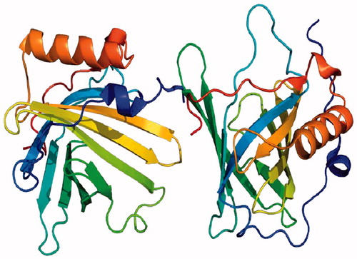
NGAL is also known as lipocalin-2 (LCN2) and was originally identified as a 25-kDa protein covalently bound to gelatinase from secondary granules of human neutrophils.Citation8 Mature peripheral neutrophils lack NGAL mRNA expression, and NGAL protein is synthesized at the early-myelocyte stage of granulopoiesis during the formation of secondary granules. NGAL mRNA is normally expressed in a variety of adult human tissues, including bone marrow, uterus, prostate, salivary gland, stomach, colon, trachea, lung, liver and kidney.Citation9 Several of these tissues are prone to exposure to micro-organisms, and constitutively express the NGAL protein at low levels. The promoter region of the NGAL gene contains binding sites for a number of transcription factors, including nuclear factor (NF)-κB.Citation9 This could explain the constitutive, as well as inducible, expression of NGAL in several of the non-hematopoietic tissues. Like other lipocalins, NGAL forms a barrel-shaped tertiary structure with a hydrophobic calyx that binds small lipophilic molecules.Citation9 NGAL structure is depicted in .
Figure 1. NGAL structure. Note: Permission is granted to copy, distribute and/or modify this document under the terms of the GNU Free Documentation License, Version 1.2 or any later version published by the Free Software Foundation; with no Invariant Sections, no Front-Cover Texts, and no Back-Cover Texts. A copy of the license is included in the section entitled GNU Free Documentation License. (From Wikipedia, the free encyclopedia.)

Functional roles of NGAL
The major ligands for NGAL are siderophores, small iron-binding molecules. Siderophores are synthesized by bacteria to acquire iron, and NGAL exerts a bacteriostatic effect by depleting siderophores. Therefore, NGAL is considered a critical component of innate immunity to bacterial infection. Experimental evidence for this role is derived from mice that are genetically modified to lack the NGAL gene, which renders them more susceptible to Gram-negative bacterial infections and death from sepsis.Citation7 On the other hand, siderophores produced by eukaryotes participate in NGAL-mediated iron shuttling that is critical to various cellular responses, such as proliferation and differentiation.Citation7–9 This property provides a potential molecular mechanism for the documented role of NGAL in enhancing the epithelial phenotype. During kidney development, NGAL promotes epithelial differentiation of the mesenchymal progenitors, leading to the generation of glomeruli, proximal tubules, Henle’s loop and distal tubules. Furthermore, although NGAL is expressed only at very low levels in several human tissues, it is markedly induced in injured epithelial cells, including the kidney. This is likely mediated via NF-κB, which is known to be rapidly activated in kidney tubule cells after acute injuries, and plays a central role in controlling cell survival and proliferation.Citation10 NGAL induction in the context of AKI leads to marked preservation of function, attenuation of apoptosis and an enhanced proliferative response. This protective effect is dependent on the chelation of toxic iron from extracellular environment and the regulated delivery of siderophore and iron to intracellular sites.Citation10
Additionally, NGAL is markedly induced in a number of human cancers, where it often represents a predictor of poor prognosis.Citation7 The NGAL gene is known to be induced by a number of tumor-promoting agents, including SV40 and polyoma virus, phorbol esters, hepatocyte growth factor, retinoic acid, glucocorticoids and NF-κB.Citation11 The overexpressed NGAL protein binds to matrix metalloproteinase (MMP)-9, thereby preventing MMP-9 degradation and increasing MMP-9 enzyme activity. In turn, MMP-9 activity promotes cancer progression by degrading the basement membranes and extracellular matrix, liberating VEGF, and thus enabling angiogenesis, invasion and metastasis.Citation11
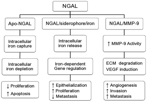
A potentially unifying hypothesis to reconcile these seemingly contradictory roles of NGAL in human biology is offered in . The mechanisms for the intracellular uptake of NGAL via receptors such as megalin, and for intracellular trafficking via endosomes have been elucidated.Citation7 The subsequent molecular pathway taken by NGAL may be largely dependent on the type of molecule it is complexed with. Indeed, NGAL that is devoid of siderophore and iron (Apo-NGAL) rapidly scavenges intracellular iron. The resultant intra-cellular iron depletion results in a decrease in the mammalian cell’s proliferative ability and induction of apoptosis. On the other hand, when NGAL is bound to siderophore and iron, there is a rapid release of iron with regulation of iron-dependent molecular pathways, and downstream induction of proliferation and epithelial transformation. Finally, when NGAL is complexed with MMP-9 instead, there is enhancement of the active MMP-9 pool with resultant upregulation of MMP-9’s well known proangiogenic and proinvasive properties.
Sources of urinary and plasma NGAL
Although plasma NGAL is freely filtered by the glomerulus, it is almost totally reabsorbed in the proximal tubules by efficient megalin-dependent endocytosis.Citation10 Thus, any urinary excretion of NGAL is likely only when there is concomitant proximal renal tubular injury that precludes NGAL reabsorption and/or increases de novo NGAL synthesis. However, gene expression studies in AKI have demonstrated a rapid and massive (1000-fold) upregulation of NGAL mRNA in the thick ascending limb of Henle’s loop and the collecting ducts.Citation6,Citation10 The resultant synthesis of NGAL protein in the distal nephron and secretion into the urine appears to comprise the major fraction of urinary NGAL. The latter is a monomeric molecule, whereas urinary NGAL originating from neutrophils is dimeric. The over-expression of NGAL in the distal tubule and rapid secretion into the lower urinary tract is in accord with its teleological function as an antimicrobial strategy. It is also consistent with the proposed role for NGAL in promoting cell survival and proliferation, given the recent documentation of abundant apoptotic cell death in distal nephron segments in several animal and human models of AKI.Citation6,Citation10
Regarding plasma NGAL in AKI, the kidney itself does not appear to be a major source, since direct ipsilateral renal vein sampling after unilateral ischemia indicates that the NGAL synthesized in the kidney is not introduced efficiently into the circulation, but is abundantly present in the ipsilateral ureter.Citation10 However, it is now well known that AKI results in a dramatically increased NGAL mRNA expression in distant organs,Citation6,Citation10 especially the liver and lungs, and the over-expressed NGAL protein released into the circulation may constitute a distinct systemic pool. Additional contributions to the systemic pool in AKI may derive from the fact that NGAL is an acute phase reactant and may be released from neutrophils, macrophages and other immune cells.Citation6 Furthermore, any decrease in glomerular filtration rate (GFR) resulting from AKI would be expected to decrease the renal clearance of NGAL, with subsequent accumulation in the systemic circulation. The relative contribution of these mechanisms to the rise in plasma NGAL after AKI remains to be determined.
NGAL in the prediction of AKI
Preclinical transcriptome profiling studies identified NGAL to be one of the most upregulated genes in the kidney very early after acute injury in animal models.Citation12 Downstream proteomic analyses also revealed NGAL to be one of the most highly induced proteins in the kidney after ischemic or nephrotoxic AKI in animal models.Citation13 The incidental finding that NGAL protein was easily detected in the urine soon after AKI in animal studies has initiated a number of translational studies to evaluate NGAL as a non-invasive biomarker in human AKI. In a cross-sectional study of adults with established AKI (doubling of serum creatinine) from varying etiologies, a marked increase in urine and serum NGAL was documented by western blotting when compared with normal controls.Citation14 Urine and serum NGAL levels correlated with serum creatinine and kidney biopsies in subjects with AKI who demonstrated intense accumulation of immunoreactive NGAL in cortical tubules, confirming NGAL as a sensitive index of established AKI in humans.Citation7 A number of subsequent studies have now implicated NGAL as a promising, non-invasive, troponin-like, early diagnostic biomarker for AKI in various common clinical settings.Citation5–7 NGAL appears to fulfill many characteristics of an ideal AKI biomarker as demonstrated in .
Table 2. NGAL as an AKI biomarker.
NGAL in CIN
Several investigators have examined the role of plasma and urine NGAL as a predictive biomarker of CIN in adults and children undergoing coronary angiography with intra-arterial contrast administration and revealed good diagnostic and prognostic performance.Citation15–20 In a prospective study of 91 children with congenital heart disease undergoing elective cardiac catheterization and angiography with contrast administration, both urine and plasma NGAL measured by using a research-based enzyme-linked immunosorbent assay (ELISA) at 2-h post-procedure predicted CIN, defined as a 50% increase in SCr from baseline, with an area under the receiver-operating characteristic curve (AUC-ROC) of 0.91–0.92.Citation18 Furthermore, pilot studies in adults with normal SCr receiving contrast for coronary angiography and intervention detected a significant rise in both urine and serum NGAL 2–4 h post-procedure, whereas serum cystatin C levels increased significantly only 24 h after contrast exposure.Citation15 However, none of these subjects developed CIN, as SCr remained unchanged 48-h post-procedure.Citation15 An extension of the same study in 100 patients confirmed these results and revealed CIN incidence of 11%.Citation16 A study from China detected CIN in 8.7% of adults with normal renal function undergoing coronary angiography and found that urinary NGAL measured by a research-based assay at 24-h post-procedure increased significantly in the CIN group, but not in the non-CIN group.Citation17 ROC curve analysis demonstrated that urinary NGAL at 24 h showed a good performance in early diagnosis of CIN as compared to SCr (AUC-ROC 0.73). Another study from Egypt showed a significant increase in serum NGAL 4 and 24 h after coronary interventions among adults with normal SCr.Citation19 CIN, defined as a 25% increase in SCr from baseline, was detected in 6.7% of the participants. Finally, a meta-analysis revealed an overall AUC-ROC of 0.894 for CIN prediction, when plasma or urine NGAL was measured within 6 h after contrast administration for coronary procedures.Citation20 A comparative presentation of the above-mentioned studies is provided, among others, in .
Table 3. Studies evaluating performance of NGAL and other biomarkers in CIN.
Much of these above-mentioned data have been arbitrarily extrapolated to patients receiving intravenous (IV) contrast for CT scans despite major differences in patient populations, contrast volume administered and intra-procedural complications between the two settings.Citation21 It is currently unclear how far the conclusions of such studies can be extrapolated to patients receiving IV contrast for CT scans. Furthermore, although the number of coronary and conventional angiograms has been relatively stable, the number of contrast-enhanced CT scans has been increasing by approximately 2 million per year in the United States over the last decade.Citation22 Of note, 90% of contrast media are used for CT imaging.Citation22,Citation23 Therefore, the widespread use of contrast-enhanced CT necessitates studies focusing on NGAL performance as an early and accurate AKI biomarker in this setting.Citation23
NGAL in CIN after IV contrast for CT scan
Our group designed a study to prospectively evaluate plasma NGAL measured by using standardized, point-of-care Τriage® NGAL device (Biosite Incorporated, San Diego, CA)Citation24 for early CIN detection among hospitalized patients undergoing elective contrast-enhanced CT.Citation25 CIN, defined by an increase in SCr of >25% or >0.5 mg/dL from baseline within 48 h after contrast exposure, was found in 8.5% of subjects, but detection by SCr was possible 24–48 h post-procedure. In contrast, significant elevation of plasma NGAL by nearly 10-fold was found at 6-h post-procedure in those with versus those without CIN, with excellent performance characteristics of 6-h plasma NGAL for CIN prediction (area under the ROC curve of 1.00 with 95% CI of 0.92–1.00 at the cut-off value of 200 ng/mL).
In summary, our study demonstrated that plasma NGAL 6 h after contrast administration measured by the rapid, point-of-care Τriage® NGAL test appeared to be a clinically useful biomarker in the early prediction of CIN in hospitalized patients undergoing elective conventional contrast-enhanced CT. Therefore, plasma NGAL appears to be a powerful early biomarker of CIN that precedes the increase in SCr by several hours. Furthermore, the relatively high CIN incidence detected in this well-controlled population underlines the importance of early diagnosis by an adequate and simple procedure as the 6-h plasma Τriage® NGAL test that was evaluated in our study.
Our study is the first one that evaluates plasma NGAL performance for early CIN detection specifically after IV contrast administration. Our population consisted of subjects with well-preserved renal function and, thus, at low-to-medium risk of CIN. The predictive performance of NGAL varies with baseline renal function, and appears to be optimal in subjects with normal pre-procedure renal function.Citation26 Furthermore, our design involved only elective CT scans, excluding emergent ones usually performed in subjects with major comorbidities and potentially at higher risk of CIN.Citation27,Citation28 The magnitude of rise by nearly 10-fold from baseline supports previous literature data that NGAL is a highly discriminatory biomarker with a wide dynamic range and cut-off values that allow for risk assessment and stratification.Citation5,Citation6
The reported incidence of CIN varies widely depending on the presence or absence of risk factors, in particular pre-existing CKD, the amount and type of agent administered and the exact radiologic procedure.Citation28 Current data coming mainly from patients undergoing coronary angiography with intra-arterial contrast administration indicated that CIN occurs in 4–20% of cases.Citation27,Citation28 By contrast to that associated with angiography, the risk of CIN associated with contrast-enhanced CT scans is quite low, even among patients with CKD.Citation23,Citation29 Therefore, in a study of in- and out-patients with eGFR less than 60 mL/min/1.73 m2 undergoing elective contrast-enhanced CT, only 3.5% had an increase in SCr of greater than 0.5 mg/dL (44 μmol/L) within 48 to 96 h post-procedure.Citation29 In addition, three recent prospective trials involving contrast-enhanced CT in patients with eGFR less than 60 mL/min/1.73 m2 found an overall incidence of CIN, defined as a ≥25% increase in SCr, of approximately 5%.Citation30–32 In view of these data, CIN incidence of 8.5% reported in our study and others appears to be relatively high. However, above-mentioned differences in study populations and variations in study design and methodology (e.g., assay used for NGAL measurement) should be taken into account in the interpretation of the results.
Previous studies on NGAL performance as predictive biomarker of CIN used mainly research-based ELISA assays for plasma NGAL measurement,Citation15–20 which are not practical in the clinical setting. In this regard, a major advance has been the development of a standardized, point-of-care Τriage® NGAL test (Biosite Incorporated, San Diego, CA) for plasma NGAL measurement.Citation24 A potential limitation is the suboptimal assay performance in the lower-range values. However, a meta-analysis on the accuracy of NGAL in the diagnosis of AKI revealed better performance of standardized assays compared with individually developed research-based ones.Citation20 Τriage® NGAL device, although validated in several clinical settings with favorable results,Citation24 had not been evaluated so far in subjects undergoing contrast-enhanced CT scans. Our studyCitation25 supports its use in the latter clinical setting as well.
With respect to the sample source, the majority of biomarkers for AKI described to date have been determined in the urine.Citation5,Citation6 NGAL urinary diagnostics do have several advantages, but also some important limitations compared to plasma/serum NGAL (). Indeed, urine NGAL levels were correlated with GFR in subjects with CKD due to glomerulonephritis, as well as in those with autosomal dominant polycystic kidney disease.Citation6,Citation33 Furthermore, urinary NGAL may be increased in urinary tract infections.Citation6 Simultaneously, plasma or serum NGAL measurements may be influenced by a number of co-existing variables such as CKD, hypertension, systemic infections, inflammatory conditions and malignancies.Citation4–7 In case of intra-arterial coronary angiography and intervention, NGAL might be released from atherosclerotic plaques and, subsequently, increase plasma pool, in the absence of co-existing AKI.Citation6,Citation7,Citation21,Citation22 However, the increase in both plasma/serum and urine NGAL in the above-mentioned situations is generally much less than that typically encountered in AKI. In any case, plasma-based diagnostics have revolutionized many aspects of clinical medicine, as exemplified by the use of troponins for the early diagnosis of acute myocardial infarction and the value of B-type natriuretic peptide for prognostication in acute coronary syndromes.Citation5,Citation6,Citation34
Table 4. Urinary versus plasma/serum NGAL diagnostics.
Another study on NGAL performance in CIN after exposure to IV contrast material has been published almost simultaneously to ours.Citation35 Both serum and urine NGAL showed excellent performance in predicting CIN, 8 h after iodinated contrast administration (AUC-ROC 0.995 and 0.992 for serum and urine NGAL, respectively), while SCr level changes occurred 24-h post-procedure. Despite differences in study design and participants, these results appear to be in line with our own findings, providing further support to plasma/serum NGAL role as an early predictive biomarker of CIN after IV contrast administration. A comparative presentation of studies evaluating NGAL performance in CIN is provided in .
Apart from NGAL, several other biomarkers, such as cystatin C, Interleukin 18 (IL-18) and liver-type fatty acid-binding protein (L-FABP), have been evaluated in the prediction of CIN, with inconsistent results due, mainly, to differences in study populations, design and clinical settings.Citation36–43 NGAL, both in plasma and urine, shows superior performance as early CIN biomarker.Citation36,Citation37 Indeed, it was pronounced by experts in the field as the “renal troponin”.Citation6 A brief comparative presentation of studies on biomarkers for the early prediction of CIN is shown in . However, results on biomarkers’ performance should always be interpreted with caution, in particular, when validated in heterogenic populations with co-morbid conditions, such as diabetes mellitus, vascular disease and CKD.
Summary
Over the last decade there has been considerable progress in the discovery and development of biomarkers of AKI, and several has now been evaluated in various clinical settings. NGAL as a promising AKI biomarker has successfully passed through the pre-clinical, assay development and initial clinical testing stages of the biomarker development process. It has now entered the prospective screening stage, facilitated by the development of standardized laboratory platforms, in an attempt to identify the best biomarkers for each purpose (risk assessment, diagnosis, determination of cause for differential diagnosis and prognosis).
CIN is a common cause of hospital-acquired AKI and a source of substantial morbidity and mortality. Several biomarkers have been evaluated in the early prediction of CIN, not always with robust results. The application of such biomarkers in clinical practice remains controversial, especially for populations with major co-morbidities. However, NGAL, both in plasma/serum and urine, appears to fulfill many characteristics of an ideal early AKI biomarker. Its role in the timely diagnosis of CIN has already been examined in adults and children undergoing coronary angiography and a meta-analysis revealed a very good performance of plasma or urine NGAL, measured within 6 h after contrast administration, for CIN early diagnosis. However, limited information exists on NGAL performance in CIN after IV contrast administration for CT scans. A recently published study from our group offered some data on this understudied area. Therefore, plasma NGAL measured by a rapid, standardized assay appears to be a powerful early predictive biomarker for CIN among the important group of hospitalized patients undergoing elective contrast-enhanced CT. Our results could serve as a useful working hypothesis for further studies with a larger number of patients to demonstrate the potential role of NGAL for CIN prediction in this population.
Although growing body of evidence suggests that the incidence of CIN may be less with IV contrast administration than that reported in coronary diagnostic procedures, the widespread use of contrast-enhanced CT examinations still leaves a sizeable population vulnerable to this event. An important incidence of CIN was confirmed in the well-controlled population of our study making an early diagnostic tool even more desirable. Indeed, an increase in plasma NGAL would potentially trigger clinicians to monitor patients more closely, avoid treatment with additional nephrotoxins, and optimize hydration and renal perfusion to prevent further injury, limit re-hospitalization and, subsequently, reduce CT-associated morbidity and cost. However, our ability of early intervention in the evolution of CIN was limited up to now by the lack of an early, appropriately sensitive and specific marker of kidney injury. Therefore, timely detection of imminent CIN by an accurate and easy-to-perform procedure as the standardized, point-of-care plasma Τriage® NGAL test might enable early initiation of interventions or, at least, promote increased vigilance to improve the mid-term adverse outcomes associated with this rather common clinical complication.
Declaration of interest
The authors report no conflicts of interest. The authors alone are responsible for the content and writing of the paper.
References
- Pannu N, Wiebe N, Tonelli M. Prophylaxis strategies for contrast-induced nephropathy. JAMA. 2006;295:2765–2779
- Nash K, Hafeez A, Hou S. Hospital-acquired renal insufficiency. Am J Kidney Dis. 2002;39:930–936
- Solomon R. Contrast media nephropathy – how to diagnose and how to prevent? Nephrol Dial Transplant. 2007;22:1812–1815
- Nickolas TL, Barasch J, Devarajan P. Biomarkers in acute and chronic kidney disease. Curr Opin Nephrol Hypertens. 2008;17:127–132
- Devarajan P. Update on mechanisms of ischemic acute kidney injury. J Am Soc Nephrol. 2006;17:1503–1520
- Devarajan P. Neutrophil gelatinase-associated lipocalin – an emerging troponin for kidney injury. Nephrol Dial Transplant. 2008;23:3737–3743
- Devarajan P. Neutrophil gelatinase-associated lipocalin: a promising biomarker for human acute kidney injury. Biomark Med. 2010;4:265–280
- Cowland JB, Sorensen OE, Schested M, Borregaard N. Neutrophil gelatinase-associated lipocalin is up-regulated in human epithelial cells by IL-1β but not by TNF-α. J Immunol. 2003;171:6630–6639
- Mishra J, Mori K, Ma Q, et al. Amelioration of ischemic acute renal injury by neutrophil gelatinase-associated lipocalin. J Am Soc Nephrol. 2004;15:3073–3082
- Schmidt-Ott KM, Mori K, Li JY, et al. Dual action of neutrophil gelatinase-associated lipocalin. J Am Soc Nephrol. 2007;18:407–413
- Devarajan P. Neutrophil gelatinase-associated lipocalin: new paths for an old shuttle. Cancer Ther. 2007;5(B):463–470
- Yuen PST, Jo S-K, Holly MK, Hu X, Star RA. Ischemic and nephrotoxic acute renal failure are distinguished by their broad transcriptomic responses. Physiol Genomics. 2006;25:375–386
- Mishra J, Ma Q, Prada A, et al. Identification of neutrophil gelatinase-associated lipocalin as a novel urinary biomarker for ischemic injury. J Am Soc Nephrol. 2003;4:2534–2543
- Mori K, Lee HT, Rapoport D, et al. Endocytic delivery of lipocalin-siderophore-iron complex rescues the kidney from ischemia-reperfusion injury. J Clin Invest. 2005;115:610–621
- Bachorzewska-Gajewska H, Malyszko J, Sitniewska E, Malyszko JS, Dobrzycki S. Neutrophil-gelatinase-associated lipocalin and renal function after percutaneous coronary interventions. Am J Nephrol. 2006;26:287–292
- Bachorzewska-Gajewska H, Malyszko J, Sitniewska E, et al. Could neutrophil gelatinase-associated lipocalin (NGAL) and cystatin C predict the development of contrast-induced nephropathy after percutaneous coronary interventions in patients with stable angina and normal serum creatinine values? Kidney Blood Press Res. 2007;30:408–415
- Ling W, Zhaohui N, Ben H, et al. Urinary IL-18 and NGAL as early predictive biomarkers in contrast-induced nephropathy after coronary angiography. Nephron Clin Pract. 2008;108:c176–c181
- Hirsch R, Dent C, Pfriem H, et al. NGAL is an early predictive biomarker of contrast-induced nephropathy in children. Pediatr Nephrol. 2007;22:2089–2095
- Shaker OG, El-Shehaby A, El-Khatib M. Early diagnostic markers for contrast nephropathy in patients undergoing coronary angiography. Angiology. 2010;61:731–736
- Haase M, Bellomo R, Devarajan P, Schlattman P, Haase-Fielitz A, the NGAL Meta-analysis Investigator Group. Accuracy of neutrophil gelatinase-associated lipocalin (NGAL) in diagnosis and prognosis in acute kidney injury: a systematic review and meta-analysis. Am J Kidney Dis. 2009;54:1012–1024
- Rao QA, Newhouse JH. Risk of nephropathy after intravenous administration of contrast material: a critical literature analysis. Radiology. 2006;239:392–397
- Rudnick MR, Goldfarb S, Tumlin J. Contrast-induced nephropathy: is the picture any clearer? Clin J Am Soc Nephrol. 2008;3:261–262
- Solomon R. Contrast-induced acute kidney injury: is there a risk after intravenous contrast? Clin J Am Soc Nephrol. 2008;3:1242–1243
- Dent CL, Ma Q, Dastrala S, et al. Plasma NGAL predicts acute kidney injury, morbidity and mortality after pediatric cardiac surgery: a prospective uncontrolled cohort study. Crit Care. 2007;11:R127
- Filiopoulos V, Biblaki D, Lazarou D, et al. Plasma neutrophil gelatinase-associated lipocalin (NGAL) as an early predictive marker of contrast-induced nephropathy in hospitalized patients undergoing computed tomography. Clin Kidney J. 2013;6:578–583
- McIlroy DR, Wagener G, Lee HT. Neutrophil gelatinase-associated lipocalin and acute kidney injury after cardiac surgery: the effect of baseline renal function on diagnostic performance. Clin J Am Soc Nephrol. 2010;5:211–219
- Solomon RJ, Mehran R, Natarajan MK, et al. Contrast-induced nephropathy and long-term adverse events: cause and effect? Clin J Am Soc Nephrol. 2009;4:1162–1169
- Mitchell AM, Jones AE, Tumlin JA, Kline JA. Incidence of contrast-induced nephropathy after contrast-enhanced computed tomography in the outpatient setting. Clin J Am Soc Nephrol. 2010;5:4–9
- Weisbord SD, Mor MK, Resnick AL, Hartwig KC, Palevsky PM, Fine MJ. Incidence and outcomes of contrast-induced AKI following computed tomography. Clin J Am Soc Nephrol. 2008;3:1274–1281
- Barrett B, Thomsen H, Katzberg R. Nephrotoxicity of low-osmolar iopamidol vs iso-osmolar iodixanol in renally impaired patients: the IMPACT study. Invest Radiol. 2006;41:815–821
- Thomsen HS, Morcos SK, Erley CM, et al. Investigators in the abdominal computed tomography: the ACTIVE trial: comparison of the effects on renal function of iomeprol-400 and iodixanol-320 in patients with chronic kidney disease undergoing abdominal computed tomography. Invest Radiol. 2008;43:170–178
- Kuhn M, Chen N, Sahani DV, et al. The PREDICT study: a randomized double-blind comparison of contrast-induced nephropathy after low- or isoosmolar contrast agent exposure. Am J Radiol. 2008;191:151–157
- Bolignano D, Coppolino G, Campo S, et al. Urinary neutrophil gelatinase-associated lipocalin (NGAL) is associated with severity of renal disease in proteinuric patients. Nephrol Dial Transplant. 2008;23:414–416
- Fassett RG, Venuthurupalli SK, Gobe GC, Coombes JS, Cooper MA, Hoy WE. Biomarkers in chronic kidney disease: a review. Kidney Int. 2011;80:806–821
- Lacquantiti A, Buemi F, Lupica R, et al. Can neutrophil gelatinase-associated lipocalin help predict early contrast material-induced nephropathy? Radiology. 2013;267:86–93
- Vanmassenhove J, Vanholder R, Nagler E, van Biesen W. Urinary and serum biomarkers for the diagnosis of acute kidney injury: an in-depth review of the literature. Nephrol Dial Transplant. 2013;28:254–273
- Coca SG, Yalavarthy R, Concato J, Parikh CR. Biomarkers for the diagnosis and risk stratification of acute kidney injury: a systematic review. Kidney Int. 2008;73:1008–1016
- Nakamura T, Sugaya T, Node K, Ueda Y, Koide H. Urinary excretion of liver-type fatty acid-binding protein in contrast medium-induced nephropathy. Am J Kidney Dis. 2006;47:439–444
- Bulent Gul CB, Gullulu M, Oral B, et al. Urinary IL-18: a marker of contrast-induced nephropathy following percutaneous coronary intervention? Clin Biochem. 2008;41:544–547
- Kato K, Sato N, Yamamoto T, Iwasaki YK, Tanaka K, Mizuno K. Valuable markers for contrast-induced nephropathy in patients undergoing cardiac catheterization. Circ J. 2008;72:1499–1505
- Bachorzewska-Gajewska H, Poniatowski B, Dobrzycki S. NGAL (neutrophil gelatinase-associated lipocalin) and L-FABP after percutaneous coronary interventions due to unstable angina in patients with normal serum creatinine. Adv Med Sci. 2009;54:221–224
- Briguori C, Visconti G, Rivera NV, et al. Cystatin C and contrast-induced acute kidney injury. Circulation. 2010;121:2117–2122
- Ishibashi Y, Yamauchi M, Musha H, Mikami T, Kawasaki K, Miyake F. Impact of contrast-induced nephropathy and cardiovascular events by serum cystatin C in renal insufficiency patients undergoing cardiac catheterization. Angiology. 2010;61:724–730

