
ABSTRACT
Purpose: To enhance the diminished screw purchase in cancellous, osteoporotic bone following the fixation of posterior pelvic ring injuries by iliosacral screws an increased bone-implant contact area using modificated screws, techniques or bone cement may become necessary. The aim of the study was to identify sites within the pathway of iliosacral screws requiring modifications of the local bone or the design of instrumentations placed at this site. Materials and Methods: The breakaway torque was measured mechanically at the iliosacral joint (“ISJ”), the sacral lateral mass (“SLM”) and the center of the S1 (“CS1”), at a superior and an inferior site under fluoroscopic control on five human cadaveric specimens (3 female; mean age 87 years, range: 76–99) using the DensiProbe™Spine device. Results: The measured median (range) breakaway torque was 0.63 Nm (0.31–2.52) at the “iliosacral joint”, 0.14 Nm (0.05–1.22) at the “sacral lateral mass”, 0.57 Nm (0.05–1.42) at the “S1 center.” The “sacral lateral mass” breakaway torque was lower than compared to that at the “iliosacral joint” (p < .001) or “S1 center” (p < .001). The median (range) breakaway torque measured at all superior measurement points was 0.52 Nm (0.10–2.52), and 0.48 Nm (0.05–1.18) at all inferior sites. The observed difference was statistically significant (p < .05). Conclusions: The lateral mass of the sacrum provides the lowest bone quality for implant anchorage. Iliosacral screws should be placed as superior as safely possible, should bridge the iliosacral joint and may allow for cement application at the lateral mass of the sacrum through perforations.
INTRODUCTION
The surgical management of pelvic fractures aims to achieve an anatomic reduction in order to prevent chronic pelvic pain and to restore the patient to their pre-injury level of function [Citation1, 2]. In elderly patients, the access morbidity may be of concern in the fixation of the posterior pelvic ring suggesting that minimally invasive techniques may be of benefit. The use of iliosacral screws has shown promising results in posterior pelvic ring fixation and may be suitable, especially in geriatric patients [Citation3, 4]. However, the diminished screw purchase of iliosacral screws as a monocortical screw device in cancellous, osteoporotic bone [Citation5, 6] does surpass the potential lack of compliance often seen with geriatric patients, particularly with regards to partial weight bearing. As a result, the purchase of iliosacral screws may be enhanced by increasing the bone-implant contact area using modified screws, techniques [Citation7–11], and/or bone cement [Citation12–14] to prevent loosening of hardware, especially in the osteopenic bone in the elderly. Therefore, the purpose of this study was to mechanically assess the local bone breakaway torque at various sites in the pathway of iliosacral screws used for fixation of the posterior pelvic ring by measuring peak breakaway torque in a human cadaveric model to determine the zones providing the weakest anchorage for fully-threaded iliosacral screws.
MATERIALS AND METHODS
Specimen
Bone breakaway torque measurements were conducted in five human cadaveric specimens (3 female; mean age 87 years, range: 76–99). The use of the human cadaveric material was performed according to the Guidelines of the Swiss Academy of Medical Sciences. Donors have formally agreed the use of body parts for research purposes by signing the donation forms. Cadavers were embalmed using the Thiel fixation method as developed by W. Thiel in 1992 and provided by the local anatomical department [Citation15]. The method used [Citation16–18] for the determination of local bone quality was based on local bone destruction. For this, the specimens were radiographically assessed prior to the measurements using CT scans aimed at detecting potential differences in the bone density, as well as deformations, fractures, neoplasms or hardware in situ at the pelvic site. The extent of degenerative changes in the sacroiliac joint was classified using a grading system as previously published [Citation19]; namely with a “normal sacroiliac joint” (Grade 0), “focal erosions” (Grade 1), “<25% erosions, but without alteration in the joint width” (Grade 2), “≥25% erosions, joint space alteration and/or partial ankylosis” (Grade 3), or “complete ankylosis” (Grade 4). CT scans of each specimen were used to assess the posterior pelvic ring in order to identify the safe and ideal tunnel (entry point, entrance angle) for the C-arm controlled percutaneous screw placement, avoiding any extraosseous displacement of the testing device. CT data was acquired on a 64-slice MDCT Unit (Somatom Sensation Cardiac 64; Siemens, Erlangen, Germany). A spiral scan (anteroposterior scanogram image) of the pelvis was performed (collimation 64 × 0.6 mm; pitch 0.5; tube rotation time 0.5 s; tube voltage 140 kV; current 450 mA). Measurements were performed on both sides of the posterior pelvic ring as follows.
Bone Breakaway Torque Measurements
The bone density was assessed using the DensiProbe™ Spine, a novel, custom-made instrument that consists of an adapted pedicle “finder” and an electronic torque measurement module. Peak breakaway torque is measured at the tip of the instrument only, where a tri-blade-shaped design aims to achieve maximal implant-bone contact area (3.8 mm diameter, 19 mm blade length, DensiProbe™ Spine, AO Research Institute Davos, Switzerland) [Citation16, Citation20]. Cadavers were placed in the prone position on a radiolucent table. Measurements were performed on all cadavers at different regions of interest: The iliosacral joint (“iliosacral joint”) and the center of the vertebral body of S1 (“S1 center”), representing the fixation points of screws lateral and medial to the fracture, respectively; and the lateral mass of the sacrum (“lateral mass”) as an additional measurement site as fractures are most frequently observed at this site. At each region of interest, two measurements were performed at a “superior” and an “inferior” site; thereby providing information regarding the ideal bone anchorage of screws in cases where placement of only one screw might be feasible. In total, six measurements were performed on each side of one cadaveric pelvis. Two identical DensiProbe™ Spines were inserted into the bone up to the first measurement point at the iliosacral joint using gentle hammer blows (to avoid rotation prior to the measurement). One probe was placed at a superior site and the other inferiorly. The position of both probes was controlled simultaneously by inlet and outlet views of the posterior pelvic ring obtained by the use of an image intensifier (Siremobil, Siemens Medical Solutions, Zurich, Switzerland; Figure ). At the measurement points, each DensiProbe™ Spine was rotated along its longitudinal axis thereby measuring the breakaway torque with a calibrated, custom made (AO Research Institute Davos) torque meter. The DensiProbe provides an objective measurement of the resistance of trabecular bone against destruction. Therefore, the device should not be rotated until the probe is placed at the site for the suggested measurement; otherwise, trabecular bone may be destroyed ahead of the measurement and thus obtained results might be not representative. Once the probe was placed at the measurement site, it was rotated in only one direction more than 120°—in our case (based on the design of the DensiProbe and the turning moment), the probe had to be rotated in a clockwise manner. A detailed description and illustrative photographs of the device and the technique is presented elsewhere [Citation20].
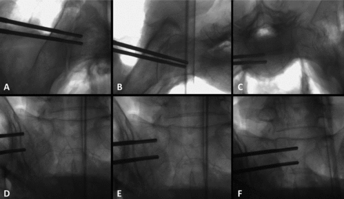
FIGURE 1 Radiographs obtained by use of a fluoroscan showing the both inserted Densiprobes at the measurement points (subcategories “superior” and “inferior”) on an inlet (A–C) or an outlet view (D–F) of the right hemipelvis at the level of the iliosacral joint (A, D), the lateral mass of the sacrum (B, E) and the center of the vertebral body of S1 (C, F).
The Densiprobe device has a minimal detection level set at 0.05 Nm that is given by internal friction and safety margin. To compensate for this, the minimal recorded value was noted at 0.05 Nm instead of 0 Nm. For reasons of practicability, we assigned all these potential measurements the value of 0.05 Nm as this has no clinical consequences as all values below 0.5 Nm are related to severe osteoporosis.
The obtained data was stored electronically and therefore the investigators were blinded at the time of the experiment. The torque measurements were performed on five human cadaveric specimens on both sides by an experienced spine surgeon Lorin Michael Benneker who was involved in the development of the device.
Statistical Analysis
To compare the potential differences between the breakaway torque measured at the different sites of the posterior pelvic ring, a repeated-measures analysis of variance (ANOVA) with matched samples was performed. Mauchly test for sphericity indicated a model violation, therefore the Greenhouse–Geisser correction was applied and pairwise comparisons with Bonferroni adjustment of p-values were performed between multiple groups. Wilcoxon signed-rank test was performed for pairwise comparison between two groups only. Data were presented as the median (range) and the mean ± SD. The level of significance was set at p < .05. Analyses and graphs were performed with SPSS software for statistical analysis (SPSS 14.0, SPSS Inc., Chicago, IL).
RESULTS
Radiographic evaluation of the cadaveric specimens did not reveal any pathologies that would impact on the measurements mentioned above, however, in one cadaver osseous defects impeded measurements at all inferior measurement points on one side and at the “iliosacral joint (inferior)” on the other side. The extent of degeneration at the iliosacral joint between specimens was comparable with mainly partial or complete ankylosis noted (Grade 3 in three cases, Grade 4 in two cases).
On four occasions, the breakaway torque was below the detection level of the device (three times at the caudal part of the lateral mass, once at the inferior part of the body of S1). The measured median (range) breakaway torque in pelvises of human cadavers was 0.63 Nm (0.31–2.52 Nm) at the iliosacral joint, 0.14 Nm (0.05–1.22 Nm) at the lateral mass of the sacrum, and 0.57 Nm (0.05–1.42 Nm) at the center of the vertebral body of S1. The breakaway torque obtained at the lateral mass was significantly lower compared to that obtained at the iliosacral joint (p < .001) or at the center of the vertebral body of S1 (p < .001). The median (range) breakaway torque measured at all superior measurement points was 0.52 Nm (0.10–2.52 Nm), and 0.48 Nm (0.05–1.18 Nm) at all inferior measurement points. The observed differences were statistically significant (p < .05).
The detailed results of the assessment of breakaway torque (in Nm) in relation to each measurement point are listed as peak values (median, range; mean ± SD) in Table and the results of the statistical analysis are illustrated in Figure . The breakaway torque at the “lateral mass (superior)” was lower than that obtained at the “iliosacral joint (superior)” (p < 0.05) or at the “S1 center (superior)” (p < 0.01). In addition, the breakaway torque at the “lateral mass (inferior)” was also lower when compared to “iliosacral joint (inferior)” (p < 0.05) and the “S1 center (superior)” (p < 0.05). No differences were observed in the breakaway torque between SI joint with partial or complete ankylosis.
FIGURE 2 Box and whisker plots show the values for the breakaway torque [in Nm] in pelves of human cadavers as measured at the iliosacral joint, the lateral mass of the sacrum or at the center of the vertebral body of S1. These measurement points were further divided in the subgroups “superior” and “inferior.” The horizontal line indicates the median value, the top and bottom borders of the box show the 75th and 25th percentiles, the whiskers show the 10th and 90th percentiles; differences with statistical significance are indicated with a p-value <.05 (*) or p-value <.01 (**).
![FIGURE 2 Box and whisker plots show the values for the breakaway torque [in Nm] in pelves of human cadavers as measured at the iliosacral joint, the lateral mass of the sacrum or at the center of the vertebral body of S1. These measurement points were further divided in the subgroups “superior” and “inferior.” The horizontal line indicates the median value, the top and bottom borders of the box show the 75th and 25th percentiles, the whiskers show the 10th and 90th percentiles; differences with statistical significance are indicated with a p-value <.05 (*) or p-value <.01 (**).](/cms/asset/b54b839e-e83f-4dc4-8546-a274077b884e/iivs_a_1016249_f0002_b.gif)
TABLE 1. Breakaway peak torque values (in Nm): Median (range) and mean ± SD in relation to the measurement points
DISCUSSION
An increasing incidence of osteoporotic pelvic injuries as a result of low-energy trauma has previously been noted and was found to occur particularly frequently in patients older than 60 years of age [Citation21]. Besides the risk for complications secondary to immobilization, immobilizing pain was considered to be the most significant symptom experienced by this patient group leading to its previous name “the fracture disease” [Citation22]. As a treatment option, percutaneous screw fixation was feasible, allowed for early mobilization with less pain and was recommended in selected cases [Citation1, Citation22, Citation23]. However, the loss of fixation at the posterior pelvic ring and the subsequent need for revision surgery resulting in recurrent surgical morbidity and/or residual instability at the fracture site leading to chronic pelvic pain may be the most significant failure in the management of geriatric pelvic trauma. Data concerning failures (e.g., loss of reduction, screw breakage/cut out, loosening) of posterior pelvic ring fixation using iliosacral screws in the elderly, have to our knowledge, not been reported. In contrast, poor implant anchorage in osteoporotic bone has been noted as a cause of failure of osteosynthesis as osteoporosis precludes the screw purchase necessary for fixation of fractures [Citation5, 6].
The presented report shows that in cases using iliosacral screws, the lowest bone breakaway torque is located within the lateral mass of the sacrum as opposed to the center of S1 or the iliosacral joint. The peak breakaway torque values of our presumably osteopenic specimens (mean age of 87 years) are within the expected range based on the experimental and clinical experience with the DensiProbeTM principle in spinal surgery. Areas with extremely poor breakaway torque (below detection level of the device) were seen three times at the inferior lateral mass (16%) and once at the inferior corpus of S1 (5%) (compared to 8% and 7% [Citation16, Citation24] in lumbar and thoracic vertebrae respectively). Although the validation of the relevant cut-off levels is still ongoing, a torque value of less than ca. 0.5 Nm seems to be associated with osteoporosis and a higher risk for screw loosening [Citation16, Citation24]. These results are in accordance with those obtained in an experimental setup using fresh frozen human cadaveric pelvises: the lowest extraction strength was required if short-threaded cancellous screws were placed in the lateral mass of the sacrum; in contrast, the required extraction strengths were about five times higher when short-threaded screws were placed in the sacral body instead [Citation10]. As a consequence, especially in the elderly, a very poor fixation was expected in the lateral mass of the sacrum [Citation10]. The implant purchase may be enhanced by increasing the bone-implant contact area using modificated screws and techniques [Citation7–11], or by the application of bone cement [Citation12–14]. The use of cement-augmentation of standard iliosacral screws has been reported earlier [Citation25], with promising preliminary outcomes noted when employing the use of iliosacral screws with perforations at the tip of the screws [Citation26]. Based on our presented results, placement of screw perforations can be recommended also at the center of the screw to allow for additional cement application, especially at the lateral mass of the sacrum where a significantly diminished bone quality is expected. However, the surgeon has to be aware of the potential complications related to cement leakage, especially at this site [Citation27]. Cement leakage was noted within the fracture gap in 27% of the 63 cases who underwent sacroplasty in the treatment of their sacral fracture. In one of these, radiculopathy of the 5th lumbar nerve root occurred as a consequence of leakage, however, there was no neurological deficit and the patient's symptoms were treated successfully by conservative means [Citation27]. To minimize the risk of cement leakage the “two stage injection technique” was reported earlier [Citation28]: Highly viscous cement is applied in a first stage to seal sites of the lowest resistance, such as the fracture gap, in order to avoid cement leakage during the second stage of application during which a cement of a lower viscosity used to fill the defect void. Instead of using bone cement as a void filler in elderly patients with osteoporotic bone, calcium phosphate (CaP)-chitosan formulations might be applied to the bony defect as a void filler as these formulations were previously identified to stimulate initial bone formation [Citation29]; in addition, in a senescence-accelerated osteoporotic mouse model (SAMP6) delayed fracture healing was noted to occur in advanced age SAMP6 mice [Citation30].
As a second finding of our measurements, a further option to prevent iliosacral screw failure may be to place an iliosacral screw as superior as possible within the safe tunnel as, in general, a higher bone breakaway torque was observed in the superior portion of the sacrum compared to the inferior portion.
The performed measurements also revealed that the breakaway torque values at the iliosacral joint were unexpectedly low. Although measurement at the two cortices was performed simultaneously, and all specimens showed relevant sclerotic changes, the values were not different from the data for the cancellous bone within the body of S1. A possible explanation might be the relatively high bone quality at the upper region of S1 as a reaction to the degenerative changes often present in this segment. In addition, in advanced degeneration of the iliosacral joint, the sclerosis might not be distributed uniformly and in case of complete ankylosis of the joint; a reduction of bone mass by stress shielding may be reasonable. Moreover, the principle of peak breakaway torque as a measure of bone quality has been used and validated for cancellous bone. Therefore, these first data for cortical bone should be interpreted with care.
The objective of the presented report was to provide data regarding the ideal site for screw-based fixation methods in posterior pelvic ring injuries using an objective measurement device for the resistance of trabecular bone against destruction [Citation16–18]. One limitation may be that no correlation to bone mineral density (BMD) is provided since BMD can only be measured using dual energy X-ray absorptiometry (DXA) or quantitative computer tomography (QCT). However, destructive assessment of bone using measurements of the peak breakaway torque of the cancellous bone has previously been shown to correlate with BMD and implant failure [Citation16–18]. A further limitation may be that the comparison of our results in Thiel embalmed cadavers to previous biomechanical studies using the densiprobe device in fresh frozen cadavers [Citation16, 17] or clinical data [Citation24] may be limited. Unger et al. reported the influence of preservation methods on the mechanical properties of bone and recommended Thiel embalmed specimens for pilot studies only [Citation31]. However, as all of our specimen were embalmed using the same technique, this shortcoming might be minimal for internal comparison but should be considered for clinical use.
The specimens used presented with advanced degeneration of the sacroiliac joint which may impact the results. However, degeneration of the iliosacral joint begins in the 20s and progresses with age [Citation32], so for the purpose of the study with focus on geriatric pelvic trauma, the overrepresentation of advanced degeneration of the sacroiliac joint may not impact the results and the clinical relevance. The simplified cadaveric test setup may be a further limitation, as evidence for a true clinical benefit under physiological weight-bearing conditions in elderly patients cannot be provided. Moreover, we did not perform a power analysis ahead of time to determine the sample size of experiments. The decision to perform ten experiments was instead based on previous reports performing measurements in ten cadavers [Citation16] or patients [Citation17], respectively, and influenced by the limited availability of human cadavers in our country.
In conclusion, the highest bone breakaway torque is located at the vertebral body of S1, the iliosacral joint and in the superior portion of the sacrum compared to the inferior portion. The lateral mass of the sacrum provides the lowest bone quality for implant anchorage. In geriatric patients presenting with osteoporotic pelvic fractures requiring surgical management using iliosacral screws—based on the presented data—the screws should be placed as superiorly as safely possible, should bridge (using fully-threaded screws) the iliosacral joint and may allow for cement application at the lateral mass of the sacrum through perforations.
ACKNOWLEDGMENT
We thank P. Rodham, Newcastle, U.K., for linguistic help in the preparation of this manuscript. We thank Dr. D. Dietrich, Institute of Mathematical Statistics and Actuarial Science, University of Bern, for performing the statistical analysis and his assistance in the interpretation of the results.
Declaration of interest: The authors report no conflicts of interest. The authors alone are responsible for the content and writing of the article. Ronald Schywn is employed by the AO foundation which is holder of the intellectual property of the densiprobe device (DensiProbe™ Spine, AO Research Institute Davos, Switzerland).
REFERENCES
- Culemann U, Scola A, Tosounidis G, et al. Concept for treatment of pelvic ring injuries in elderly patients. A challenge. Unfallchirurg. 2010;113:258–271.
- Meyhoff CS, Thomsen CH, Rasmussen LS, et al. High incidence of chronic pain following surgery for pelvic fracture. Clin J Pain. 2006;22:167–172.
- Routt ML, Jr., Kregor PJ, Simonian PT, et al. Early results of percutaneous iliosacral screws placed with the patient in the supine position. J Orthop Trauma. 1995;9:207–214.
- Schildhauer TA, Josten C, Muhr G. Triangular osteosynthesis of vertically unstable sacrum fractures: a new concept allowing early weight-bearing. J Orthop Trauma. 2006;20:S44-S51.
- Chapman JR, Harrington RM, Lee KM, et al. Factors affecting the pullout strength of cancellous bone screws. J Biomech Eng. 1996;118:391–398.
- Seebeck J, Goldhahn J, Morlock MM, et al. Mechanical behavior of screws in normal and osteoporotic bone. Osteoporos Int. 2005;16(Suppl 2):S107-S111.
- Asnis SE, Ernberg JJ, Bostrom MP, et al. Cancellous bone screw thread design and holding power. J Orthop Trauma. 1996;10:462–469.
- Daniels AH, Magee W, Badra M, et al. Preliminary biomechanical proof of concept for a hybrid locking plate/variable pitch screw construct for anterior fixation of type II odontoid fractures. Spine (Phila Pa 1976). 2012;37:E1159-E1164.
- DeCoster TA, Heetderks DB, Downey DJ, et al. Optimizing bone screw pullout force. J Orthop Trauma. 1990;4:169–174.
- Kraemer W, Hearn T, Tile M, et al. The effect of thread length and location on extraction strengths of iliosacral lag screws. Injury. 1994;25:5–9.
- Wheeler DL, McLoughlin SW. Biomechanical assessment of compression screws. Clin Orthop Relat Res. 1998;237–245.
- Becker S, Chavanne A, Spitaler R, et al. Assessment of different screw augmentation techniques and screw designs in osteoporotic spines. Eur Spine J. 2008;17:1462–1469.
- Chang MC, Liu CL, Chen TH. Polymethylmethacrylate augmentation of pedicle screw for osteoporotic spinal surgery: a novel technique. Spine (Phila Pa 1976). 2008;33:E317-E324.
- Frankel BM, D'Agostino S, Wang C. A biomechanical cadaveric analysis of polymethylmethacrylate-augmented pedicle screw fixation. J Neurosurg Spine. 2007;7:47–53.
- Thiel W. The preservation of the whole corpse with natural color. Ann Anat. 1992;174:185–195.
- Deckelmann S, Schwyn R, Van der Pol B, et al. DensiProbe Spine: A Novel Instrument for Intraoperative Measurement of Bone Density in Transpedicular Screw Fixation. Spine (Phila Pa 1976). 2010;35:607–12.
- Suhm N, Hengg C, Schwyn R, et al. Mechanical torque measurement predicts load to implant cut-out: a biomechanical study investigating DHS anchorage in femoral heads. Arch Orthop Trauma Surg. 2007;127:469–474.
- Suhm N, Haenni M, Schwyn R, et al. Quantification of bone strength by intraoperative torque measurement: a technical note. Arch Orthop Trauma Surg. 2008;128:613–620.
- Lee YH, Hong YS, Park W, et al. Value of multidetector computed tomography for the radiologic grading of sacroiliitis in ankylosing spondylitis. Rheumatol Int. 2013;33:1005–11.
- Hoppe S, Uhlmann M, Schwyn R, et al. Intraoperative mechanical measurement of bone quality with the DensiProbe. J Clin Densitom. 2015;18(1):109–116.
- Kannus P, Palvanen M, Niemi S, et al. Epidemiology of osteoporotic pelvic fractures in elderly people in Finland: sharp increase in 1970–1997 and alarming projections for the new millennium. Osteoporos Int. 2000;11:443–448.
- Rommens PM, Wagner D, Hofmann A. Surgical management of osteoporotic pelvic fractures: a new challenge. Eur J Trauma Emerg Surg. 2012;38:499–509.
- Gansslen A, Hufner T, Krettek C. Percutaneous iliosacral screw fixation of unstable pelvic injuries by conventional fluoroscopy. Oper Orthop Traumatol. 2006;18:225–244.
- Popp AW, Schwyn R, Schiuma D, et al. DensiProbe Spine: an intraoperative measurement of bone quality in spinal instrumentation. A clinical feasibility study. Spine J. 2013;13:1223–1229.
- Tjardes T, Paffrath T, Baethis H, et al. Computer assisted percutaneous placement of augmented iliosacral screws: a reasonable alternative to sacroplasty. Spine (Phila Pa 1976). 2008;33:1497–1500.
- Wahnert D, Raschke MJ, Fuchs T. Cement augmentation of the navigated iliosacral screw in the treatment of insufficiency fractures of the sacrum. A new method using modified implants. Int Orthop. 2013;37:1147–50.
- Bastian JD, Keel MJ, Heini PF, et al. Complications related to cement leakage in sacroplasty. Acta Orthop Belg. 2012;78:100–105.
- Wu ZX, Liu D, Wan SY, et al. Staged-injection procedure to prevent cement leakage during vertebroplasty: an in vitro study. J Surg Res. 2010;164:e253-e256.
- Geffre CP, Ochoa J, Margolis DS, et al. Evaluation of the osteogenic performance of calcium phosphate-chitosan bone fillers. J Invest Surg. 2010;23:134–41.
- Histing T, Kuntz S, Stenger D, et al. Delayed fracture healing in aged senescence-accelerated P6 mice. J Invest Surg. 2013;26:30–35.
- Unger S, Blauth M, Schmoelz W. Effects of three different preservation methods on the mechanical properties of human and bovine cortical bone. Bone. 2010;47: 1048–1053.
- Shibata Y, Shirai Y, Miyamoto M. The aging process in the sacroiliac joint: helical computed tomography analysis. J Orthop Sci. 2002;7:12–18.