
Abstract
This study intends to develop protocols for sampling and characterizing multi-walled carbon nanotube (MWCNT) aerosols in workplaces or during inhalation studies. Manufactured dry powder containing MWCNT’s, combined with soot and metal catalysts, form complex morphologies and diverse shapes. The aerosols, examined in this study, were produced using an acoustical generator. Representative samples were collected from an exposure chamber using filters and a cascade impactor for microscopic and gravimetric analyses. Results from filters showed that a density of 0.008–0.10 particles per µm2 filter surface provided adequate samples for particle counting and sizing. Microscopic counting indicated that MWCNT’s, resuspended at a concentration of 10 mg/m3, contained 2.7 × 10⁴ particles/cm3. Each particle structure contained an average of 18 nanotubes, resulting in a total of 4.9 × 10⁵ nanotubes/cm3. In addition, fibrous particles within the aerosol had a count median length of 3.04 µm and a width of 100.3 nm, while the isometric particles had a count median diameter of 0.90 µm. A combination of impactor and microscopic measurements established that the mass median aerodynamic diameter of the mixture was 1.5 µm. It was also determined that the mean effective density of well-defined isometric particles was between 0.71 and 0.88 g/cm3, and the mean shape factor of individual nanotubes was between 1.94 and 2.71. The information obtained from this study can be used for designing animal inhalation exposure studies and adopted as guidance for sampling and characterizing MWCNT aerosols in workplaces. The measurement scheme should be relevant for any carbon nanotube aerosol.
Acknowledgements
The authors would like to thank Amy Cumpston and Donny Leonard (NIOSH) for helping with the experiments and Mr. Owen Price and Dr. Bahman Asgharian for useful discussion on the MPPD model and the data interpretation.
Declaration of interest
The findings and conclusions in this report are those of the authors and do not necessarily represent the views of the National Institute for Occupational Safety and Health. The mention of any company names or products does not imply an endorsement by NIOSH, nor does it imply that alternative products are unavailable, or unable to be substituted after appropriate evaluation. The authors report no conflicts of interest. The authors alone are responsible for the content and writing of the paper.
Appendix A: Estimation of the number concentration of a MWCNT aerosol based on its mass concentration
The number concentration of a MWCNT aerosol can be calculated from its mass concentration if the particle size distribution follows a lognormal distribution. Then the average size and average mass of the particles can be estimated with this information (Hatch & Choate, 1929; Chen et al., 1990). The relationship is more complicated for fiber-like particles, because additional information is needed concerning their width and length. The following calculations use available information, describing the length and width of individual carbon nanotubes, to estimate their number concentration based on their mass concentration.
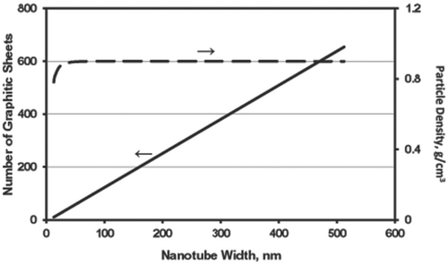
A description of an average single MWCNT was obtained from the lattice image of the graphene structure shown in the high resolution TEM micrograph [ in Porter et al. (2010)]. The width of the inner hollow core (WC) was 5.385 nm, the thickness of each graphene sheet (WT) was 0.166 nm, and the distance between two graphene layers (WD) was 0.222 nm. Even though these measurements were based on photographic images, they could be biased by the size limitation of their resolution. The measured values, however, seem to be in reasonable agreement with the information provided by the manufacturer. The data, that were made available, gave an average inner core dimension of 5.0 nm and the average distance from the center of one sheet to the center of the next sheet was 0.3385 nm (Kim et al., 2005). These measurements were used to determine the number of graphene sheets and the volume of a single MWCNT with a known length and width. As an example, MWCNT’s having widths of 21, 49, and 66 nm consisted of 21, 57, and 79 graphene sheets or layers, respectively. shows the relationship of the number of graphene layers versus the fiber density (ρf) of a single nanotube (with voids) observed in the study, as a function of the width of the nanotube. That relationship was calculated based on physical measurements of WC, WT, and WD from the electron micrographic images. The number of graphene sheets increases linearly with tube width (solid line), whereas the fiber density per nanotube (dashed line) increases asymptotically with tube width and quickly reaches a constant value of 0.9 g/cm3 for widths greater than 40 nm. The two curves in have the same shape for various combinations of WC, WT, and WD with the exception that the constant asymptotic value of particle density is a function of WC, WT, and WD.
Figure 11. The relationship of number of sheets and fiber density (with voids) of a multi-walled nanotube with respect to its tube width. The values of number of sheets and particle density were calculated based on the measurements of the nanotube from a high-resolution TEM images (Porter et al., 2010; ), in which the width of the inner hollow core (WC) = 5.385 nm, the thickness of a graphitic sheet (WT) = 0.166 nm, and the distance between two graphitic sheets (WD) = 0.222 nm. The material density of graphite was provided by the manufacturer as 2.1 g/cm3.
Results from Porter et al. (2010) indicate that individual MWCNT’s have a count mean width of 49 nm (SD = 13.4) and a count median length of 3.86 µm (GSD = 1.94). Assuming that a nanotube with these dimensions represents a fiber with an average mass, the average volume per nanotube (without voids) would be 6.0 × 10⁶ nm3 (Cheng, 1986). Note that this estimate is based on assuming a bivariate lognormal distribution for the width and length of fiber-like particles having a correlation value of 0.5.
The manufacturer indicated that the average MWCNT density (specific gravity) measured with a pycnometer was 2.1 g/cm3. Because the nitrogen molecules used in the device were able to penetrate through the voids between the graphene sheets, this value represents the material density (without voids) of the nanotube. Using this value of specific gravity, the average mass per nanotube (excluding voids) is 1.23 × 10⁻¹¹ mg.
Making the assumptions described above, a mass concentration of a 10 mg/m3 aerosol of total MWCNT’s would correspond to 8.0 × 10⁵ nanotubes/cm3. This value represents the total number of nanotubes in the aerosol, not the total number of particles. Since multiple nanotubes form agglomerate, which are counted as one particle (“CNT structure”), the number concentration of particles depends on the average number of nanotubes per particle. As an example, assume that each particle contains an average number of 10 nanotubes, then the aerosol concentration would contain about 8.0 × 10⁴ particles/cm3. Bear in mind that the accuracy of the results obtained from these calculations is based on the single MWCNT characteristics described by Porter et al. (2010). A more accurate value of particle concentration using the average value of 18 nanotubes per particle (described in the text) would result a concentration of 4.4 × 10⁴ particles/cm3.
Appendix B: Development of a sampling/operation protocol for microscopic observations of MWCNT particles
The NIOSH standard 7402 (NIOSH, 1994a) asbestos fiber measurement method using membrane filters and microscopic analysis was initially adopted as a guideline for quantitatively describing MWCNT aerosols. Initially, polycarbonate filters of 2.5 µm pore size (Whatman, Clinton, PA) were used for collecting MWCNT samples from the chamber at a flow rate of 1 L/min. Polycarbonate filters were used because of their smooth surface, which was advantageous for microscopic observation of particles. After sampling, the loaded filters were cut into four pieces and mounted onto aluminum stubs with double-stick carbon tape, and coated with gold/palladium using a SPI sputter coater (SPI-Module, Structure Probe Inc., West Chester, PA). The samples were then analyzed using a field emission scanning electron microscope (FE-SEM; Hitachi, S-4800, Tokyo, Japan). A FE-SEM, rather than a transmission electron microscope (TEM), was used to observe the samples. This FE-SEM is capable of providing information concerning the 3-dimensional morphology of the particle, as well as estimating the number of nanotubes incorporated in each particle. show the images of aerosol particles collected on filters of 2.5 µm pore size. The sample appears to contain particles of many different shape configurations including single smooth nanotubes with various aspect ratios (L/W, L = length and W = width or diameter), bundled nanotubes, nanotube nodules (fiber-like particles having nano-sized nodules attached), and isometric-shape or fiber-like agglomerates having nanotubes and/or compact particles attached. While the “individual nanotubes” are easy to identify as fiber-like particles that contain mainly elemental carbon, the agglomerates (“CNT structures”) could consist of nanotubes, nodules, and/or compact particles with heterogeneous compositions. shows the particles are primarily composed of elemental carbon using energy dispersive X-ray analysis (SEM-EDX; Princeton Gamma-Tech, Rocky Hill, NJ). illustrates that, at a magnification of ×40K, the nano-sized nodules (right in the figure) attached to the nanotubes are likely to be residual metal catalyst seeds from the growth process, and the micron-sized compact particles (left in the figure) with a smooth surface are believed to be soot resulting from the condensation of carbon-containing vapor.
Although the sample in contains a good representation of particles with diverse morphologies, the images of the particles distributed on the filter surface clearly illustrate that the sample was overloaded during collection. This condition makes it difficult for identifying individual particles and determining their size. This sample also indicates that the use of filters with a 2.5 µm pore size may allow ultrafine nanotube particles to deposit inside the pore () or even penetrate through the pores and cause underestimate when counting the nano-sized fraction of the MWCNT aerosol. Due to these shortcomings, modifications to the NIOSH standard 7402 are needed to provide adequate filter samples with high collection efficiency and optimal distribution for microscopic observations of MWCNT’s. The important issues related to filter sampling and microscopic operation are listed below:
Selection of filters: In order to improve the sampling techniques for particle quantification, filters with a 0.1 µm pore size (Whatman) were selected for this study. The filters were mounted in a closed-face filter holder with a 5-cm electrically conductive plastic extension cowl. This filter assembly was used for collecting MWCNT samples for particle counting, sizing, and morphological analysis with the FE-SEM (Hitachi). The small pore size polycarbonate filter has a collection efficiency of greater than 99% for ultrafine aerosol particles (Liu et al., 1983). As a tradeoff for the high pressure drop across the filter, however, the sampling flow rate was reduced to 0.4 L/min.
Microscopic observation: The FE-SEM images in indicate that MWCNT aerosol contains a mixture of individual nanotubes and particles with agglomerated structures which represent a broad size spectrum between 40 nm and 10 µm. This required an electron microscope operated under an extensive range of different magnifications (see ). The lower range was used to determine the micrometer-sized length of nanotubes and agglomerates under a lower magnification (e.g. ×2K and ×5K), while the upper range was used to determine the nanometer-sized width of individual nanotubes under a higher magnification (e.g. ×40K).
Optimal surface density: To minimize the sample overloading on filter during collection like the one shown in , an ideal range of surface density should be determined. Unlike the deposit range of 100–1300 fibers/mm2 as described in the NIOSH method for counting asbestos, the MWCNT aerosol contains particles of a smaller size and requires a denser deposit on filter for proper microscopic observations. At a flow rate of 0.4 L/min, different time intervals and sampling volumes were chosen to produce an optimal surface density on filter for microscopic observations. After several trials, a range of 0.008–0.10 particles/µm2 was selected based on the ease and ability of counting FE-SEM images of particles on the filter. The surface density was determined by microscopically counting the number of particles and particle structures across an effective sample area on the filter surface.
Sampling time interval: The ideal range of a sampling time was estimated using the number concentration of MWCNT particles in the sampling volume. Although the number concentration associated with the mass concentration targeted at 10 mg/m3 was not immediately available, it could be approximated by converting particle mass to particle number using previously published information (Porter et al., 2010) combined with assumptions that have been used previously for fibrous particles (Cheng, 1986). Details of the method of estimating particle number from mass measurements are described in Appendix A. Results of those calculations show that the estimate of the number concentration was 8.0 × 10⁴ particles/cm3 for a mass concentration of 10 mg/m3. Consequently, a series of sampling time intervals between 5 s and 2 min were selected for the initial trials. It should be noted that this estimation was based on an assumption that there was an average of 10 nanotubes per particle.