Abstract
Purpose: To report a case of a child with near-simultaneous onset of Vogt Koyanagi Harada disease (VKH) and insulin-dependent diabetes mellitus (IDDM). Design: Interventional case report.
Methods: An 11-year-old child with known psoriasis presented with headache and bilateral granulomatous panuveitis. Nine weeks later, he presented with diabetic ketoacidosis and IDDM. Diffuse choroidal depigmentation followed within months. HLA was positive for DRB1*0405. Despite aggressive local and systemic therapy, the ocular disease was complicated by bilateral cataracts, angle closure glaucoma, and choroidal neovascularization.
Results: The patient is currently pseudophakic in one eye and aphakic in the other, with best-corrected visual acuity of 6/24 and 6/5, respectively.
Conclusions: VKH may present in children with panuveitis in the setting of other autoimmune disorders. Treating such patients is complicated by the need to minimize systemic corticosteroid use. A combination of local therapy and systemic steroid-sparing agents should be the mainstay of treatment.
Vogt Koyanagi Harada disease (VKH) is a systemic autoimmune disease affecting the uvea, meninges, inner ear, and skin. Other systemic autoimmune phenomena are not commonly part of it, but insulin-dependent diabetes mellitus (IDDM), celiac disease, psoriasis, ulcerative colitis, and Graves disease have each been rarely described to occur in conjunction with VKH.Citation1–6 We report a rare case of a child with psoriasis and near-simultaneous onset of VKH and IDDM.
CASE REPORT
An 11-year-old child from a Philippine-English background presented to an ophthalmologist with headache and bilateral granulomatous panuveitis starting 3 weeks earlier. Fundus visibility was limited by posterior synechiae and vitritis. Visual acuity was 6/24 bilaterally. He was treated with topical steroids and homatropine for a month, with partial improvement. He had a prior history of polyuria and polydipsia, and blood tests performed by his pediatrician showed increased hemoglobin A1c (13%) and borderline-high fasting serum glucose. Oral prednisolone, 1 mg/kg, was added to control his uveitis and his vision improved to 6/6 in each eye within 2 weeks. He then presented to hospital with diabetic ketoacidosis and a diagnosis of IDDM was made. He had a prior history of psoriasis and thalassemia minor. An extensive infectious and rheumatological laboratory investigation failed to identify an etiology for his uveitis. Cerebrospinal fluid was not examined at that stage, as the patient had no signs of meningitis, and the diagnosis of VKH was not entertained. The fundi later showed multifocal, small, midperipheral choroidal scars. Oral methotrexate, 15 mg per week was added.
The patient was referred to the Royal Victorian Eye and Ear Hospital, Ocular Immunology Clinic 5 months after presentation, with visual acuity of 6/6, +3 anterior chamber cell, 360 posterior synechiae, and posterior subcapsular cataracts in both eyes. Bilateral multifocal choroidal scars were noted and the vitreous showed 1+ cell. He was taking oral prednisolone (25 mg daily) and methotrexate (15 mg weekly) and his diabetic control was poor due to inadequate compliance. He was treated with pulse intravenous methylprednisolone and bilateral periorbital triamcinolone injections. Oral prednisolone followed and was rapidly tapered down. Eight months following initial presentation, orbital floor steroid injections were repeated for increasing inflammation, methotrexate was discontinued due to lack of efficacy, and cyclosporine A was started.
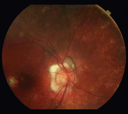
The diagnosis of VKH became apparent as diffuse choroidal depigmentation was noted, with “sunset glow” appearance and multiple peripheral punched-out choroidal lesions (). Bilateral periocular vitiligo was also noted, but there were no auditory symptoms. HLA typing was done to look for a possible connection between his IDDM and VKH,Citation3 and was indeed positive for DRB1*0405.
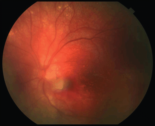
One year following uveitis onset he had an exacerbation with bilateral granulomatous anterior uveitis, recurrent iris bombé, and bilateral angle closure glaucoma, despite several laser iridotomies. He underwent right cataract aspiration with primary posterior capsulectomy and anterior vitrectomy. An acrylic, foldable intraocular lens was implanted in the capsular bag. Anterior synechiae were lysed and 4 mg of triamcinolone was injected into the anterior chamber. Due to uncontrolled glaucoma, trabeculectomy with mitomycin C was done in the right eye 6 months later. The uveitis increased bilaterally and oral prednisolone was started but stopped due to uncontrolled hyperglycemia. A right subfoveal choroidal neovascular membrane (CNVM) developed and responded to intravitreal ranibizumab. The left cataract worsened, with visual acuity of 6/60, and cataract surgery with intracameral triamcinolone injection was performed shortly after. Given the continuing postoperative formation of inflammatory deposits on the IOL in the right eye, a decision was made to leave the left eye aphakic. The patient’s postoperative left visual acuity with an aphakic contact lens was 6/5. Seven months later, a large macular hemorrhage and a pigment epithelial detachment were observed in the contralateral, left eye, due to a juxta-papillary CNVM (). Intravitreal tissue plasminogen activator (tpa), SF6, and bevacizumab were injected, with resolution of the macular hemorrhage and the visual symptoms within weeks.
The patient has been under our care now for 5 years. His corrected visual acuity is currently 6/24 in the right eye and 6/5 in the left eye. Intraocular pressure is controlled on topical brimonidine, timolol, bimataprost, and dorzolamide. He is taking systemic immunosuppressive therapy with cyclosporine A (125 mg daily) and methotrexate (15 mg weekly). Both eyes continue to show recurrent, low-grade anterior uveitis.
DISCUSSION
The exact pathogenesis of VKH disease is unknown.Citation7 An underlying T-cell-mediated autoimmune process directed against melanocytes has been postulated as the likely immunologic mechanism, with infectious agents as possible triggers in genetically susceptible individuals.Citation7 IDDM is also an autoimmune disorder characterized by lymphocyte-mediated destruction of insulin-producing β cells in the pancreatic islets of Langerhans. Genetic and environmental factors, including infections, are also described in the pathogenesis of IDDM. The coexistence of IDDM and VKH has been reported in the literature.Citation1-3,Citation5 Our patient was positive for HLA DRB1*0405, which is known to be associated with VKH and IDDM.Citation7,Citation8 HLA DRB1*0405 is strongly associated with diabetes mellitus in the Philippine population.Citation9 The small number of reports of simultaneous VKH and IDDM can be explained by other unknown HLA DRDQ haplotypes, which may significantly modify the risk for the disease.Citation9 The coexistence of VKH and psoriasis has also been independently reported.Citation4 Some specific HLA variants may confer susceptibility to epidemiologically associated autoimmune and autoinflammatory diseases, as illustrated, for instance, by the occurrence of vitiligo, autoimmune thyroid diseases, and psoriasis in several members of families with multiple autoimmune diseases associated with vitiligo.Citation10
The coexistence of IDDM and VKH precluded long-term systemic treatment with corticosteroids in our patient, who suffers from brittle IDDM and refractory uveitis. Although rare, diabetic ketoacidosis has been reported in children after steroid therapy.Citation11 Hyperglycemia can potentially be life threatening in such patients, as shown by Suzuki et al., reporting a case of hyperosmolar coma during prednisolone therapy for VKH in an IDDM patient.Citation1 We partially controlled our patient’s ocular disease with a combination of orbital and intraocular triamcinolone, systemic cyclosporine, and methotrexate. However, the prognosis for vision in such cases is unknown, given the patient’s young age and the prospect of severe glaucoma, CNVM recurrence, and future diabetic retinopathy.Citation2
The course of VKH is known to be more aggressive in children, and they tend to have more severe ocular complications.Citation2 Our patient had a CNVM, which responded well to intravitreal injections of ranibizumab and bevacizumab. We extrapolated from adult data when deciding to treat this child with anti-VEGF therapy, because only anecdotal evidence derived from case reports was available in children.Citation12–14,Citation16 As discussed in a recent editorial, more research to assess the safety and efficacy of anti-VEGF therapy in the pediatric population is required.Citation14 The use of intravitreal tPA and SF6 in children with macular hemorrhage has been reported to be safe in 4 eyes of 2 children with shaken baby syndrome.Citation17
VKH should be considered in children with panuveitis in the setting of other autoimmune disorders, in particular IDDM and psoriasis. The combination of a severe, potentially blinding uveitis with diabetes poses a difficult management challenge. Intensive medical (local and systemic) and surgical treatment may still result in favorable outcome.
Declaration of interest: The authors report no conflicts of interest. The authors alone are responsible for the content and writing of the paper.
REFERENCES
- Suzuki H, Isaka M, Suzuki S. Type 1 diabetes mellitus associated with Graves’ disease and Vogt-Koyanagi-Harada syndrome. Intern Med. 2008;47(13):1241–1244.
- Al Hemidan Al, Tabbara, KF, Althomali T. Vogt-Koyanagi-Harada associated with diabetes mellitus and celiac disease in a 3-year-old girl. Eur J Ophthalmol. 2006;16(1):173–177.
- Maruyama Y, Hayashi H, Takahashi K, et al. A case of insulin dependant diabetes mellitus following systemic treatment for Vogt-Koyanagi-Harada syndrome. Ophthalmic Surg Lasers. 2000;31(6):487–490.
- Kluger N, Mura F, Guillot B, Bessis D. Vogt-Koyanagi-Harada syndrome associated with psoriasis and autoimmune thyroid disease. Acta Derm Venereol. 2008;88(4):397–398.
- Futagami Y, Sugita S, Fujimaki T, et al. Bilateral anterior granulomatous keratouveitis with sunset glow fundus in a patient with autoimmune polyglandular syndrome. Ocul Immunol Inflamm. 2009;17(2):88–90.
- Al-Halafi A, Dhibi HA, Hamade IH, Bou Chacra CT, Tabbara KF. The association of systemic disorders with Vogt-Koyanagi-Harada and sympathetic ophthalmia. Graefes Arch Clin Exp Ophthalmol. 2011;249:1229–1233.
- Fang W, Yang P. Vogt-Koyanagi-Harada syndrome. Curr Eye Res. 2008;33:517–523.
- Gorodezky C, Alaez C, Murguía A, et al. HLA and autoimmune diseases: type 1 diabetes (T1D) as an example. Autoimmun Rev. 2006;5:187–194.
- Bugawan TL, Klitz W, Alejandrino M, et al. The association of specific HLA class I and II alleles with type 1 diabetes among Filipinos. Tissue Antigens. 2002;59:452–469.
- Jin Y, Mailloux CM, Gowan K, et al. NALP1 in vitiligo-associated multiple autoimmune disease. N Engl J Med. 2007; 356: 1216–1225.
- Cağdaş DN, Paç FA, Cakal E. Glucocorticoid-induced diabetic ketoacidosis in acute rheumatic fever. J Cardiovasc Pharmacol Ther. 2008;13:298–300.
- Cakir M, Cekic¸ O, Yilmaz OF. Combined intravitreal bevacizumab and triamcinolone injection in a child with Coats disease. J AAPOS. 2008;12:309–311.
- Cakir M, Cekic¸ O, Yilmaz OF. Intravitreal bevacizumab and triamcinolone treatment for choroidal neovascularization in Best disease. J AAPOS. 2009;13:94–96.
- Cakir M, Cekic¸ O, Yilmaz OF. Intravitreal bevacizumab for idiopathic choroidal neovascularization. J AAPOS. 2009;13:296–298.
- Avery RL. Extrapolating anti-vascular endothelial growth factor therapy into pediatric ophthalmology: promise and concern. J AAPOS. 2009;13:329–331.
- Lott MN, Schiffman JC, Davis JL. Bevacizumab in inflammatory eye disease. Am J Ophthalmol. 2009;148:711–717.
- Conway MD, Peyman GA, Recasens M. Intravitreal tPA and SF6 promote clearing of premacular subhyaloid hemorrhages in shaken and battered baby syndrome. Ophthalmic Surg Lasers. 1999;30:435–441.

