Abstract
Purpose: To describe a case of frosted branch angiitis in a patient with tuberculous meningitis.
Methods: Case report.
Results: A 27-year-old woman of tuberculous meningitis was referred to us complaining of blurred vision for 2 days. Prominent white sheathing of the retinal venules and, to a much lesser extent, arterioles, consistent with frosted branch angiitis were also observed in both eyes. And after treatment with systemic anti-tuberculosis medications and steroid, frosted branch angiitis showed resolution.
Conclusions: Frosted branch angiitis can be caused by Mycobacterium tuberculosis. Systemic anti-tubercular therapy and steroids were effective.
Keywords::
Frosted branch angiitis (FBA) was first reported in 1976 to depict the bilateral white perivascular retinal sheathing seen in a young, immunocompetent boy.Citation1 Classic findings in FBA included decreased visual acuity, preferential involvement of veins over arteries, lack of associated infectious or autoimmune disease, and a rapid response to systemic corticosteroids.Citation2 Later, cases resembling FBA in adult patients with human immunodeficiency virus (HIV), cytomegalovirus, and toxoplasmosis were reported. Mycobacterium tuberculosis infection is rare in these patients. Here we report a case of frosted branch angiitis with Mycobacterium tuberculosis.
A 27-year-old woman with headache and intermittent fever for half a month was referred to us in January 2011. Her C-reactive protein and erythrocyte sedimentation rate were raised. Cerebrospinal fluid (CSF) showed pleocytosis (212 cells, 180 lymphocytes), increased proteins (178 mg/dL) and low sugar levels (2.89 mmol/L) and normal LAM-IgG. Mantoux skin test was strongly positive. X-rays of the chest were normal. Brain magnetic resonance imaging showed meningeal lineal strengthening. She was diagnosed as having tuberculous meningitis in our hospital.
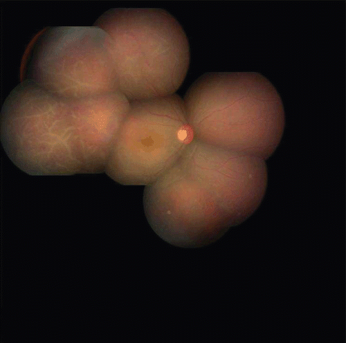
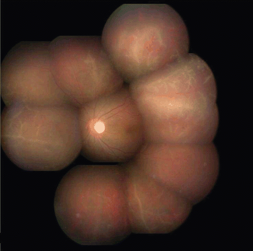
She complained of blurred vision for 2 days. Visual acuity was counting fingers at 20 cm right eye and 0.05 cm left eye. Slit-lamp examination showed mild keratic precipitates (+1) and aqueous flare and cells (+1) in the anterior chamber in both eyes. Both eyes showed vitreous haze. Prominent white sheathing of the midperipheral retinal venules and, to a much lesser extent, arterioles, consistent with frosted branch angiitis, was also observed in both eyes. Both eyes showed scattered areas of retinal hemorrhages and the right eye showed macular edema ( and ). Titers of antibody to Toxoplasma and HIV were within normal limits. Treponema pallidum hemagglutination test (TPHA) was negative.
FIGURE 1 Fundus photograph of right eye pre-therapy showing retinal vessels with perivascular sheathing with scattered areas of retinal hemorrhages along the macular edema before treatment.

FIGURE 2 Fundus photograph of left eye pre-therapy showing retinal vessels with perivascular sheathing with retinal hemorrhages before treatment.

Because of the tuberculous meningitis, she was treated with isoniazid 0.6 g/day, rifampicin 450 mg daily, ethambutol 0.75 g/day, and pyrazinamide 1.5 g/day. Oral prednisone 50 mg was also given. Topical dexamethasone and atropine sulfate eyedrops were given four times daily also.
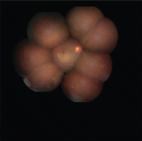
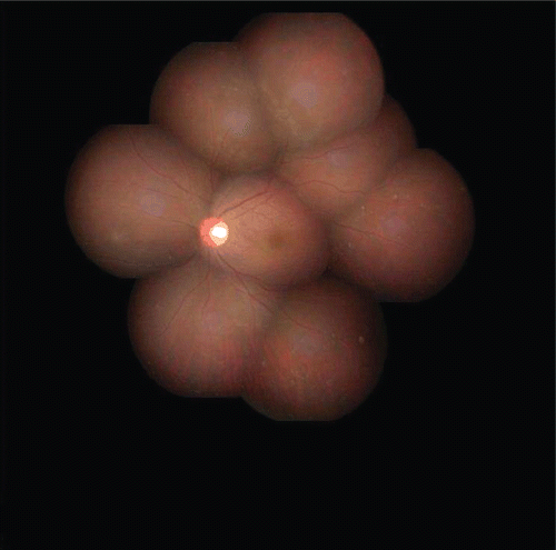
Twenty-five days after treatment, her headache and fever were reduced, C-reactive protein and erythrocyte sedimentation rate returned to normal, CSF showed normal pleocytosis (25 cells, 5 lymphocytes), proteins (61 mg/dL), and sugar levels (3.2 mmol/L). Her visual acuity was 0.2 in the right eye and 0.6 in the left eye. Keratic precipitate and aqueous flare disappeared in both eyes. Vitreous haze reduced. White sheathing of the vessels and hemorrhages alleviated. Macular hard exudates were observed in the right eye ( and ). Later she went to another city. She was interviewed by telephone half a year later, at which time her visual acuity was 0.2 in the right eye, and 0.8 in the left eye.
FIGURE 3 Fundus photograph of right eye post-therapy showing resolution of vasculitis and hemorrhage after 25 days of treatment.

FIGURE 4 Fundus photograph of left eye post-therapy showing resolution of vasculitis and hemorrhage after 25 days of treatment.

Ocular involvement in patients with systemic tuberculosis has traditionally been considered uncommon.Citation3 There have been several reports of retinal vasculitis in tuberculosis.Citation3,Citation4 Agarwal reported the first case of FBA along with tuberculosis.Citation5 We report a case of FBA with meningitis tuberculosis. The evidence favoring tuberculous etiology in this case was associated tuberculous meningitis and positive results of Mantoux testing. Its responsiveness to prompt treatment with anti-tuberculosis therapy also confirmed that pathogens are tuberculosis bacilli. In accordance with most patients with FBA, after treatment her visual acuity improved rapidly in the left eye. The right eye has no favorable visual acuity, and we inferred the reason is persistent macular edema. It is a pity that she never followed up again, and didn’t undergo fluorescein angiograms since she was allergic to fluorescein sodium injection, so we don’t know whether there is an ischemic nonperfused area in the retina.
We indicate that FBA could be a rare manifestation of intraocular tuberculosis. Prompt treatment with anti-tuberculosis therapy can lead to good vision recovery.
Declaration of interest: The authors report no conflicts of interest. The authors alone are responsible for the content and writing of the paper.
REFERENCES
- Ito Y, Nakano M, Kyu N, Takeuchi M. Frosted branch angiitis in a child. Jpn J Clin Ophthalmol. 1976;30: 797–803.
- Geier SA, Nasemann J, Klauss V, et al. Frosted branch angiitis in a patient with the acquired immuno deficiency syndrome. Am J Ophthalmol. 1992;113:203–204.
- Gupta V, Gupta A, Rao NA. Intraocular tuberculosis-an update: major review. Surv Ophthalmol. 2007; 52; 561–587.
- Gupta A, Gupta V, Arora S, et al. PCR positive retinal vasculitis: clinical characteristics and management. Retina. 2001;21:435–444.
- Agarwal M, Biswas J. Unilateral frosted branch angiitis in a patient with abdominal tuberculosis. Retinal Cases Brief Rep. Winter 2008:2:39–40.