TO THE EDITOR
Historically, there was not a more dreadful ophthalmologic disease than sympathetic ophthalmia (SO). A significant injury to one eye would have meant a lifetime of blindness. SO is a bilateral, granulomatous panuveitis that occurs after ocular trauma or surgical procedure to one eye, which ultimately threatens sight in the fellow eye. While SO is considerably rare, the significant morbidity of the disease makes it an important differential in cases of bilateral panuveitis.1 Immunosuppression, particularly with oral steroids, remains the staple for therapy. Treatment response is typically monitored by clinical findings, fluorescein angiography, ultrasonography, and optical coherence tomography. We present herein a case of SO and describe the use of wide-field imaging and enhanced depth imaging spectral domain optical coherence tomography (EDI SD-OCT) for diagnosis and monitoring of treatment response.
A 39-year old Hispanic male presented to our ER for evaluation of facial trauma, including significant injury to his left eye (OS). The patient had been involved in an altercation, and was punched in the left eye with car keys gripped within a fist. On initial examination, visual acuity was 20/25 in the right eye (OD) and no light perception (NLP) in the left. The left eye revealed diffuse chemosis, a nasal scleral laceration with exposed uvea, clear cornea, and total hyphema. There was no view of the posterior pole and an ocular ultrasound was not performed. The uninvolved right eye was unremarkable. The patient had immediate uncomplicated repair of scleral laceration and repositioning of uveal tissue OS.
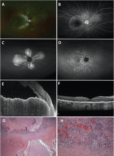
A month following initial trauma, the patient noted difficulty reading OD associated with headache that progressed to decreased distance vision later that day. Visual acuity OD was 20/60, while OS remained NLP. Intraocular pressures were normal in both eyes. Slit-lamp examination OD showed diffuse conjunctival hyperemia, 1+ anterior chamber cell and flare, and 2+ anterior vitreous cells. Dilated fundus examination revealed optic nerve edema, and a petaloid pattern of subretinal fluid accumulation centered on the optic disc that was better appreciated with wide-field fundus autofluorescence (, ). A subsequent exam revealed Dalén-Fuchs nodules in the right eye. Examination OS also showed diffuse conjunctival hyperemia with a significantly blood-stained cornea. Wide-field fluorescein angiography of the right eye exhibited multiple pinpoint areas of leakage in the posterior pole (). We obtained EDI SD-OCT imaging using the method described by Maruko and colleagues.2 The Heidelberg Spectralis instrument was placed close enough to the eye to obtain an inverted image and the zero delay was placed as posterior to the choroid as possible. The scans were then obtained in this fashion. Choroidal thickness was measured using the software (1.5.12.0) of the Heidelberg Spectralis OCT. EDI SD-OCT of the right eye demonstrated submacular subretinal fluid, irregularity of the retinal pigment epithelium (RPE), and thickening of the choroid to more than 500 μm (). Clinical findings and ancillary tests were consistent with SO.
FIGURE 1 A 39-year-old male presented with blurred vision and loss of accommodation in the right eye 1 month after suffering a ruptured globe from facial trauma in the left eye. (A, B) Right eye. Dilated fundus examination showed mildly hyperemic optic nerve associated with exudative retinal detachment in the posterior pole (A) as well as multiple pinpoint areas of leakage on wide-field fluorescein angiography (B). (C, D) Right eye. Wide-field fundus autofluorescence revealed a petaloid pattern of hyperautofluorescence centered on the optic nerve corresponding to areas of exudative retinal detachment (C) at initial presentation that would later evolve to speckled areas of hyper- and hypoautofluorescence resembling leopard spots (D) following resolution. (E, F) Right eye. Enhanced depth spectral domain optical coherence tomography demonstrated a massively thickened choroid measuring more than 500 μm, irregularity of the retinal pigment epithelium, submacular fluid accumulation, disruption of the IS-OS line, and hyperreflective spots within the area of subretinal fluid accumulation (E). Three months following treatment, there was restoration of the normal foveal contour with some residual focal areas of photoreceptor loss (F). (G) Left eye. There is diffuse choroidal thickening with sheets of lymphocytes focally interrupted by nests of epithelioid histiocytes (1.25×, hematoxylin & eosin). No Dalén-Fuchs nodules were identified. (H) Left eye. Monomorphic lymphocytic infiltration of the choroid with occasional nests of epithelioid histiocytes (20×, hematoxylin & eosin). Note that the lymphocytic infiltrate involves the choriocapillaris.

The patient was started on oral prednisone 60 mg daily and scopolamine drops twice daily in the right eye only. Over 4 months of treatment, there was significant decrease in anterior chamber and vitreous cells, along with reduction in subretinal fluid accumulation in the right eye. Fundus autofluorescence OD showed speckled hyper- and hypoautofluorescence in the previous areas of exudative detachment (). EDI SD-OCT showed a corresponding decrease in choroidal thickness to 237 μm OD (). The patient also elected to undergo enucleation OS for cosmetic reasons and pathology revealed granulomatous inflammation of uveal tissue and choroidal thickening consistent with SO (, ). Visual acuity during the last follow-up visit at 6 months was 20/40 OD without any evidence of inflammation. He continues at 60 mg of oral prednisone, as a previous attempt to slowly taper the steroids after a transition to methotrexate had resulted in a recurrence.
Prior to enhanced depth imaging technique in SD-OCT, visualization of the choroid was restricted to ultrasonography. EDI SD-OCT is uniquely able to provide high-resolution images of the choroid and allow thickness measurements. Apart from providing more detailed information regarding the choroid, EDI SD-OCT has been used to visualize optic nerve structures such as the lamina cribrosa.3 Vogt-Koyanagi-Harada disease (VKH) has a similar pathologic mechanism as SO without the appropriate clinical setting of ocular trauma or surgery and there have been reports describing the use of EDI SD-OCT in monitoring progress of treatment in VKH.2 Although there have been case reports describing SD-OCT findings in sympathetic ophthalmia and review of the images shows choroidal thickening at initial presentation, this was not performed with EDI SD-OCT as described by Spaide and coauthors.4-6 Further, the authors were not able to document improvement of choroidal thickening following treatment.Citation4,Citation5 Wide-field fundus autofluorescence also showed initial hyperautofluorescence corresponding to accumulation of subretinal fluid that turned to areas of speckled areas of hyper- and hypoautofluorescence similar to the leopard-spot appearance following resolution of VKH or SO well described in literature. Ours is the first report describing the use of wide-field fluorescein angiography, fundus autofluorescence, and EDI SD-OCT to the best of our knowledge.
In summary, we describe the use of multimodality imaging in a confirmed case of SO. Our findings suggest that EDI SD-OCT may be a useful modality in monitoring the efficacy of treatment.
Declaration of interest: The authors report no conflicts of interest. The authors alone are responsible for the content and writing of the paper. University of North Carolina Hospitals, Department of Ophthalmology, is a recipient of an unrestricted grant from Research to Prevent Blindness.
REFERENCES
- Reynard M, Riffenburgh RS, Maes EF. Effect of corticosteroid treatment and enucleation on the visual prognosis of sympathetic ophthalmia. Am J Ophthalmol. 1983;96 (3):290–294.
- Maruko I, Iida T, Sugano Y, et al. Subfoveal choroidal thickness after treatment of Vogt-Koyanagi-Harada disease. Retina. 2011; 31:510–517.
- Park HY, Jeon SH, Park CK. Enhanced depth imaging detects lamina cribrosa thickness differences in normal tension glaucoma and primary open-angle glaucoma. Ophthalmology. 2012 Jan;119 (1):10–20.
- Gupta V, Gupta A, Dogra MR, Singh I. Reversible retinal changes in the acute stage of sympathetic ophthalmia seen on spectral domain optical coherence tomography. Int Ophthalmol. 2011;31:105–110.
- Magalhaes FP, Lavinsky D, Rossi LV, et al. Sympathetic ophthalmia after penetrating keratoplasty: a case report evaluated by spectral-domain optical coherence tomography. Retina Cases & Brief Reports. 2012;6:11–15.
- Spaide RF, Koizumi H, Pozonni MC. Enhanced depth imaging spectral coherence tomography. Am J Ophthalmol. 2008;146:496–500.