Abstract
Objective: To report on ampiginous choroiditis of tubercular etiology with nPCR positivity for M. tb in aqueous sample and also to stress upon the usefulness of nPCR to consolidate the etiological diagnosis.
Material and methods: Case report of a patient presenting with active ampiginous choroiditis with no evidence of systemic TB but had hilar and para-tracheal lymphadenopathy on CT chest. QuantiFERON TB gold test was positive The patient responded inadequately to systemic anti-tubercular treatment and oral steroids. Nested PCR for M.tb was done on aqueous sample was positive for MPB64 gene of M.tb. Anti-TB treatment was continued inspite of initial poor response to which the patient responded and his choroiditis regressed.
Results: Ocular tuberculosis forms the essential differential diagnosis of any chronic or recurrent choroiditis, especially in high endemic areas. The presumed tubercular etiology can be substantiated using highly sensitive and specific tests such as nested PCR on intraocular fluid.
Ampiginous choroiditis has been described as a clinical entity involving features of both acute posterior multifocal placoid pigment epitheliopathy (APMPPE) and serpiginous choroiditis (SC).Citation1 Some consider it as a variant of SC with macular and peripheral involvement.Citation2 Jones et al.Citation3 were the one of the first groups to describe the condition and to use the term relentless placoid chorioretinitis. However, the first group to coin and use the term ampiginous choroiditis was Nussenblatt et al.Citation4Multiple etiologies, including autoimmunity, infection, vasculopathy, and degeneration, have been proposed for serpignous choroiditis per say, but only autoimmunity has been reported to date for the ampiginous variant of serpiginous choroiditis. To our knowledge no cases of ampiginous choroiditis have been reported with latent systemic infection (tuberculosis). We report a case of ampiginous choroiditis of tubercular etiology well supported with laboratory evidence of nested PCR positive for MPB64 genome of Mycobacterium tuberculosis in an aqueous sample obtained from an anterior chamber tap.
CASE REPORT
A 33-year-old Asian (Indian) male presented to our hospital with persistent redness, watering, and mild discomfort in his right eye for 15 days. These ocular complaints were associated with mild blurring of vision. He had had similar complaints in his left eye 3 years back, for which he was treated elsewhere with good response. No treatment details were available. There was no significant family history and his systemic examination was within normal limits.
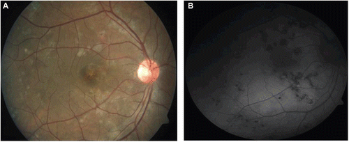
Ocular examination revealed a BCVA of 6/6, N6 in both eyes. Anterior segment evaluation with a slit-lamp biomicroscope was within normal limits in both eyes. Fundus examination in his right eye revealed multiple choroidal lesions within the arcade and spreading to the periphery along the vessels. The choroidal lesions within the arcade show central resolution and peripheral active border and exhibit features of typical serpiginous choroiditis lesion. On the contrary, the temporal midperiphery and periphery had lesions that were placoid, nearly a half disc diameter in size with features resembling APMPPE. The fovea was spared. The optic disc and blood vessels were normal. The left eye fundus showed multiple healed choroidal atrophic patches occupying the posterior pole and periphery with varying degree of pigmentation and normal overlying retinal vasculature. The distribution and morphology of the healed lesions was similar to that described for the right eye. There were mild RPE alterations at the fovea in left eye. Fundus autofluorescence and fluorescein angiography showed early hypo- and late hyperfluorescence with blurring of margins in some choroidal lesions, suggestive of active choroiditis in the right eye and healed choroiditis with persistent hypofluorescence of choroidal lesions in left eye. We labeled this patient as having ampiginous choroiditis, as it followed the diagnostic criterion described by us in our previous paper.Citation1 His blood hemogram was within normal limits, with an ESR of 10 mm. HRCT chest revealed right hilar and paratracheal lymphadenopathy. Mantoux test was 10 mm at 48 h. QuantiFERON TB gold test was positive.
The results of the investigations were all suggestive of latent systemic tubercular infection and the choroiditis to be of probable tubercular etiology. The patient was put on a 4-drug systemic anti-TB treatment for 2 months followed by 3 drugs for a continuation phase of 4–6 months. Oral prednisolone starting at 60 mg/day was also begun, with tapering of 10 mg/week. At 6 weeks follow-up, right eye fundus () revealed new active choroiditis lesions within the arcade, whereas the left eye fundus was same as before. Due to the inadequate response and appearance of new lesions, the suspected tubercular etiology was questioned. Nested PCR for M.tb was done on aqueous tap sample targeting MPB64 genome of M.tb using primer targeting (), which was positive. This gave conclusive evidence of the presence of Mycobacterial DNA in aqueous sample and helped us to consolidate our decision to continue the patient on anti-tubercular therapy. At 12 weeks follow-up the right eye fundus () showed healed choroiditis lesions with varying degree of pigmentation. Fundus autofluorescence imaging suggested the same. The patient was advised to complete the continuation phase of anti-tubercular medications with 3 drugs, and systemic steroids were stopped successfully without recurrence.
FIGURE 1 (a) First follow-up fundus image of right eye showing whitish yellow choroiditis, new lesions, and atrophic lesions along vessels. (b) Late-phase FFA image of right eye showing the fuzzy margins due to leakage from the active lesions.

FIGURE 2 Gel photograph of positive nested PCR on aqueous sample for M.tb done against a positive (PC) and negative (NC) control.
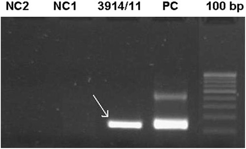
FIGURE 3 (a) Healed choroiditis lesions. (b) Fundus autofluorescence image shows no hyperautofluorescence within the lesions, suggesting no activity.
DISCUSSION
Ampiginous choroiditis is considered by many as a disease entity that exhibits features of both SC and APMPPE. The diagnostic criterion of ampiginous choroiditis described above in this case has been used by Biswas et al.Citation1 in their case series of ampiginous choroiditis. As described by them and others,Citation5 our patient had bilateral involvement. Also, only autoimmune etiology for ampiginous choroiditis has been described till now, although a case of ampiginous choroiditis following quadrivalent human papilloma virus vaccine has been reported only recently.Citation6 Except for this case we have not come across any case of ampiginous choroiditis described in literature of infective and none of tubercular etiology. In our case the suspected tubercular etiology was conclusively backed up by positive nested PCR for MPB64 gene of M.tb from aqueous sample. This helped us to continue with anti-tuberculosis treatment in spite of initial inadequate response. Identification of M.tb DNA in aqueous or vitreous sample is crucial in establishing a definitive etiological diagnosis in cases of infective choroiditis. nPCRs targeting the MPB64 gene of M.tb, which codes for an immunogenic protein specific for M.tb, are found only in the culture filtrates of M.tb strains and occasional strains of Mycobacterium bovis.Citation7 The antigen MPB64 is specific for M.tb. It has been found useful in studies on the pathogenesis, cell-mediated immunology and in the diagnosis of tuberculosis. The results indicate its higher sensitivity for M.tb DNA, so that an improved diagnosis of tuberculosis is possible by application of this nested PCR.Citation7–9
CONCLUSION
Ocular tuberculosis should be considered as a differential diagnosis in every case of posterior uveitis, especially in high endemic areas. Precise diagnosis is crucial for early eye-saving treatment. nPCR was sufficiently accurate to corroborate initial clinical suspicion, particularly in cases where the clinical picture is confusing.
Declaration of interest: The authors report no conflicts of interest. The authors alone are responsible for the content and writing of the paper.
REFERENCES
- Biswas J, Shafiq SJ, Sudharshan S, Badami K. Clinical profile and visual outcome of ampiginous choroiditis. Ocul Immunol Inflamm. 2010;18:46–51.
- Lim WK, Buggage RR, Nussenblatt RB. Serpiginous choroiditis. SOO. 2005;50: 231–244.
- Jones BE, Jampol LM, Yannuzzi LA. Relentless placoid chorioretinitis? Arch Ophthalmol. 200;118:931–938.
- Nussenblatt RB, Whitcup MW. Uveitis: Fundamentals and Clinical Practice. Philadelphia: Mosby;1996.
- Bisaws J, Kazi S. Relentless placoid chorioretinitis. AIOC 2006 proceedings.
- Khalifa YM, Monahan PM, Acharya NR. Ampiginous choroiditis following quadrivalent human papilloma virus vaccine. Br J Ophthalmol. 2010;94:137–139.
- Madhavan HN, Therese KL, Gunisha P, Jayanthi U, Biswas J. Polymerase chain reaction for detection of mycobacterium tuberculosis in epiretinal membrane in Eales’ disease. Invest Ophthalmol Vis Sci. 2004;41:822–825.
- Therese KL, Jayanthi U, Madhvan HN. Application of nested polymerase chain reaction (nPCR) using MPB64 gene primers to detect Mycobacterium tuberculosis DNA in clinical specimens from extrapulmonary tuberculosis patients. Indian J Med Res. 2005;122:165–170.
- Eisenach KD, Cave MD, Bates JH, Crowford JT. Polymerase chain reaction amplification of a repetitive DNA sequence specific for Mycobacterium tuberculosis. J Infect Dis. 1990;161(5):977–981.
- Marchetti G, Gori A, Catozzi L, et al. Evaluation of PCR in detection of Mycobacterium tuberculosis from formalin-fixed, paraffin embedded tissues: comparison of four amplification assays. J Clin Microbiol. 1998;36(6): 1512–1517.