Abstract
Purpose: To report a case of progressive outer retinal necrosis (PORN) presenting as a cherry red spot.
Methods: Case report.
Results: A 53-year-old woman with recently diagnosed HIV and varicella-zoster virus (VZV) aseptic meningitis developed rapid sequential vision loss in both eyes over 2 months. Her exam showed a “cherry red spot” in both maculae with peripheral atrophy and pigmentary changes, consistent with PORN. Due to her late presentation and the rapid progression of her condition, she quickly developed end-stage vision loss in both eyes.
Conclusions: PORN should be considered within the differential diagnosis of a “cherry red spot.” Immune-deficient patients with a history of herpetic infection who present with visual loss warrant prompt ophthalmological evaluation.
CASE REPORT
A 53-year-old woman presented with rapid sequential loss of vision in both eyes over 2 months. Three months prior to presentation, she had a recurrence of shingles on her left scalp and was subsequently diagnosed with acquired immune deficiency syndrome (AIDS). She had a CD4 count of 14/mm2 and HIV viral load of 943,142 copies/mL. A month later, she was hospitalized for aseptic meningitis. Serum testing showed presence of varicella-zoster virus (VZV) and Toxoplasma IgG, but not IgM. Tests for other infectious agents, including syphilis (Rapid Plasma Reagen (RPR) and Fluorescent Treponemal Antibody Absorption Test (FTA-ABS)), herpes simplex virus (HSV), hepatitis B virus (HBV), Coccidioides, Blastomyces, Histoplasma, Aspergillus, B. henselae, and B. quintana, were all negative. Cerebral spinal fluid evaluation confirmed the presence of VZV by polymerase chain reaction (PCR), but not Toxoplasma, HBV, HSV, or cytomegalovirus (CMV). During her hospitalization, she developed sudden loss of vision in the left eye with optic disc swelling, which was diagnosed by her local ophthalmologist as optic neuritis. When she presented to our institution after 2 months, she had no light perception in the left eye and was developing new vision loss to counting fingers in the right eye. Slit-lamp examination showed diffuse keratic precipitates, anterior chamber cells, and sectoral iris transillumination defects in the right eye. Fundoscopy revealed macular edema with a “cherry red spot” configuration in both eyes, as well as optic atrophy in the left eye (). Fluorescein angiography demonstrated choroidal hypoperfusion and a petaloid pattern of leakage consistent with macular edema in the right eye. The left macula showed diffuse late leakage (). Her CD4 count at this time was 67/mm2. After 1 week, the cherry red spot persisted in the right eye, but she developed significant pigmentary changes in the left macula, and peripheral retinal atrophy and pigmentary changes in both eyes (). Given the macular changes and peripheral scarring in the setting of recent VZV meningitis and cutaneous shingles, a diagnosis of progressive outer retinal necrosis (PORN) was given. Within days, the vision in the right eye deteriorated to no light perception also. Due to the end stage nature of her disease, the patient received only highly active antiretroviral therapy (HAART) and oral valacyclovir for the management of her AIDS and VZV meningitis, and was not placed on ganciclovir or foscarnet for the treatment of PORN.
FIGURE 1 Fundus photographs of the right (A) and left (B) maculae demonstrating “cherry red spot” in a patient with PORN on initial presentation.
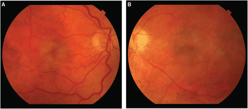
FIGURE 2 Fluorescein angiography of the right (A) and left (B) eyes during the late frames, demonstrating choroidal hypoperfusion and a petaloid pattern of leakage consistent with macular edema in the right eye and diffuse late leakage in the left eye.
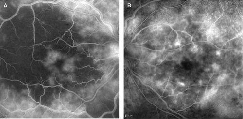
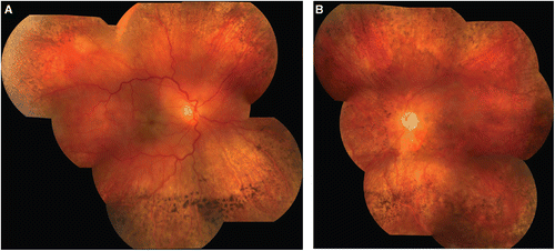
FIGURE 3 Composite fundus photographs of the right (A) and left (B) eyes 1 week after presentation, showing persistent “cherry red spot” and significant peripheral atrophy with sclerotic vessels and pigmentary changes in both eyes. There are also left optic atrophy and pigmentary changes in the left macula.
DISCUSSION
PORN is a rapidly progressive necrotizing retinopathy in immunocompromised patients most commonly caused by VZV, as well as other herpes viruses, including herpes simplex virus, cytomegalovirus, and Epstein-Barr virus. Diagnosis is made based on history, immune status, and clinical features, including the appearance of outer retinal opacification, minimal or absence of ocular inflammation, bilaterality, and multifocality.
Occasionally, subtle retinal whitening in the posterior pole may resemble a “cherry red spot.”Citation1 This clinical term is often used to describe the swelling and loss of transparency in the macula resulting from ischemia and retinal edema, as from a retinal artery occlusion, or from accumulation of glycolipids or sphingolipids, as in lysosomal storage disorders. The relative transparency of the foveola, where the retinal layers are thinnest, allows the normal choroidal vasculature to be seen more readily, hence resulting in the appearance of the red spot. Although uncommon, the retinal edema or whitening associated with inflammatory conditions may produce a similar appearance. Here, we present a case of PORN where the initial fundoscopic appearance was that of a “cherry red spot.”
The occurrence of VZV aseptic meningitis and optic neuritis has been reported to precede the onset of retinal necrosis,Citation2 suggesting that the infection may be transmitted from the central nervous system via the optic nerve. However, the temporal relationship between the neurologic and ophthalmic manifestations of herpes zoster is variable,Citation3,Citation4 and a causal relationship may thus be difficult to validate. Although intravenous and intravitreal foscarnet or ganciclovir are often used for the treatment of PORN,Citation5 prognosis and visual outcomes are poor, with retinal detachment occurring in up to 70% of cases.Citation6 Clinicians must promptly recognize the clinical features of herpetic retinopathies, particularly in patients who are immune-deficient or have a history of shingles or aseptic meningoencephalitis. Early and aggressive therapy is necessary to maximize the patient’s visual potential. The appearance of a “cherry red spot” may be an early clinical sign of PORN and warrants careful evaluation.
Declaration of interest: The authors report no conflicts of interest. The authors alone are responsible for the content and writing of the paper.
REFERENCES
- Blair MP, Goldstein DA, Shapiro MJ. Optical coherence tomography of progressive outer retinal necrosis. Retina. Nov-Dec 2007;27(9):1313–1314.
- Franco-Paredes C, Bellehemeur T, Merchant A, Sanghi P, DiazGranados C, Rimland D. Aseptic meningitis and optic neuritis preceding varicella-zoster progressive outer retinal necrosis in a patient with AIDS. AIDS. May 3 2002;16(7):1045–1049.
- Park KH, Bang JH, Park WB, et al. Retrobulbar optic neuritis and meningoencephalitis following progressive outer retinal necrosis due to CMV in a patient with AIDS. Infection. Oct 2008;36(5):475–479.
- Liu DT, Chan AY. Progressive outer retinal necrosis presenting with isolated optic neuropathy. Neurology. Nov 22 2005;65(10):1682–1683.
- Kim SJ, Equi R, Belair ML, Fine HF, Dunn JP. Long-term preservation of vision in progressive outer retinal necrosis treated with combination antiviral drugs and highly active antiretroviral therapy. Ocul Immunol Inflamm. Nov-Dec 2007;15(6):425–427.
- Engstrom RE Jr, Holland GN, Margolis TP, et al. The progressive outer retinal necrosis syndrome: a variant of necrotizing herpetic retinopathy in patients with AIDS. Ophthalmology. Sep 1994;101(9):1488–1502.