
Abstract
In recent years, mRNA-based vaccines have emerged to be a great alternative to DNA-based vaccines due to the safety of not inserting into host genome. However, mRNA molecules are single-stranded nucleic acids that are vulnerable under RNase existing in human skin and tissues. In this study, a self-assembled cationic nanomicelles based on polyethyleneimine-stearic acid (PSA) copolymer were developed to delivery HIV-1 gag encoding mRNA to dendritic cells and BALB/c mice. We evaluated the transfection efficiency and cell uptake efficiency of naked EGFP mRNA, PSA, PEI-2k and PEI-25k nanoparticles format on DC2.4 cell lines. Immune responses after sub-cutaneous administration of gag mRNA to BALB/c mice were notably induced by PSA as compared with naked gag mRNA. We found the PSA/mRNA nanomicelles were potent systems that can effectively deliver mRNA and induce antigen-specific immune response, stimulating various new vaccine strategies using mRNA.
Introduction
Over the past decades, gene vaccines held great promise for the treatment of numerous acquired and inherited diseases. Virus and plasmid DNA have been widely explored as potential vaccine strategies (Kariko et al., Citation1999; Qiu et al., Citation2015). However, worries have been raised regarding the clinical safety and none of them have been qualified for human use yet. Compared to the above strategies, there are great benefits in using messenger RNA (mRNA) for gene vaccine applications: such as high transfection efficiencies in non-dividing cells, easy production and high safety without integrating into host genome (Yamamoto et al., Citation2009; Tavernier et al., Citation2011).
mRNA, especially in vitro transcribed mRNA, has the potential as therapeutics and vaccines strategies due to its ability to active dendritic cells (DCs), which are the most efficient antigen-presenting cell (APC) in immune system and difficult to transfect (Palumbo et al., Citation2012), by interacting with the toll-like receptors (TLR): TLR3, TLR7, and TLR8 (Nishiya et al., Citation2005). Because of these advantages, mRNA has been designed for tumor vaccination (Steitz et al., Citation2006; Zhang et al., Citation2006; Amano et al., Citation2007; Mockey et al., Citation2007; Perche et al., Citation2011) and virus infection (Zarei et al., Citation2003; Yu et al., Citation2007; Petsch et al., Citation2012), even lead to some clinical progress (Weide et al., Citation2008, Citation2009; Rittig et al., Citation2011).
However, problems had emerged regarding the stability of mRNA: it was easily degraded by the presence of nuclease in tissues and skin (Probst et al., Citation2006). In addition to the stability matter, whether mRNA molecules could induce endosome escape also remains important (Pack et al., Citation2005). To solve these problems, several kinds of non-viral carriers had been evaluated for efficient mRNA administration. Most of them were based on cationic lipids. One of the studies developed an efficient and reproducible method for mRNA transfection by using DOTMA-lipofectin that leading to high luciferase activity (Malone et al., Citation1989), which was also achieved by complexed mRNA with an organic-inorganic polymer DOTAP-apatite (Zohra et al., Citation2007). Meanwhile, Su et al. (Citation2011) developed a biodegradable core-shell nanoparticle with a poly-(β-amino ester) core enveloped by a phospholipid bilayer shell. The uptake of mRNA-loaded nanoparticle by dendritic cells led to approximately 30% translation efficiency (Su et al., Citation2011). Besides lipids, cationic polymers have also been widely investigated for the delivery of mRNA. By adding melittin to PEI-2k could mediate high levels of mRNA transfection in vitro (Bettinger et al., Citation2001). PEG-poly [N9-[N-(2-aminoethyl)-2-aminoethyl]aspartamide] (PAsp[DET]) were able to cause endosome escape of mRNA because of pH-responsive membrane destabilization by PAsp[DET] (Uchida et al., Citation2013). However, more potent carriers for mRNA delivery are highly required to mediate the application of mRNA vaccine strategies.
Cationic micelles, which were based on hydrophobic modification of hydrophilic polymers, had emerged to be an efficient gene and peptide delivery system. The micelles can self-assemble in aqueous phase and offer unique advantages including protection of nucleic acids, enhanced cell interaction, higher gene transfection and less toxicity (Navarro et al., Citation2015). However, to our best knowledge, cationic micelles have never been reported as an efficient vehicle for mRNA vaccine delivery. We had previously reported that stearic acid and branched PEI-2k conjugates (PSA) could form self-assemble cationic micelles. The micelles were able to encapsulate tyrosine's-related protein to peptide and accumulate in the draining lymph nodes after subcutaneous injection (Zeng et al., Citation2014). The objective of the present work was to evaluate the capability of PSA cationic micelles for mRNA vaccines delivery.
To prove the potency of PSA based cationic micelles to be an mRNA vaccine vehicle, we first designed a stable in vitro transcribed mRNA with the anti-reverse cap analogue (ARCA), the poly (A) tail and the 5′ and 3′-globin untranslated region (UTR) of Xenopus. We examined the potential of PSA for stable condensation of mRNA in nanoparticle format and the ability to deliver mRNA into dendritic cell line DC2.4, by investigating the cellular uptake, the endosome escape and the transfection efficiency of PSA/mRNA nanoparticles. In immunological tests, the maturation of murine bone marrow-derived dendritic cells (BMDCs) induced by PSA was verified and the antigen-specific antibody, interferon (IFN)-γ and interleukin (IL)-4 levels were also measured. This study provides valuable information regarding the design and formulation of stable and effective mRNA delivery vehicles, as well as a potent mRNA vaccine delivery system that inducing strong immune response ().
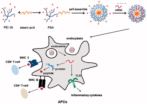
Figure 1. Schematic representation of how PSA micelles loaded with mRNA was delivered to APCs. PSA/mRNA nanoparticles were hypothesized to translate into protein, thus can lead to CD4+ and CD8+ T cell response. Gag mRNA alone can also induce immune response by interaction with the TLRs.
Materials and methods
Cell culture and animals
DC 2.4 cells (Chinese Academy of Sciences, Shanghai, China) were cultured in RPMI 1640 medium (Hyclone, Life Technologies, Grand Island, NY), supplemented with 10% fetal bovine serum, 100 units/ml penicillin and 100 g/mL streptomycin at 37 °C in a humidified atmosphere with 5% CO2.
BALB/c (female) and C57BL/6 (male) mice between six and eight weeks old were obtained from Dashuo Biotechnology Co. Ltd (Chengdu, China). All experiments were conducted according to the animal welfare legislation of China and the institutional animal care of Sichuan University.
Plasmids
pVAX1/5′UTR/gag/3′UTR/A30 and pVAX1/5′UTR/EGFP/3′UTR/A30 plasmids encoding the HIV-1 gag antigen and EGFP gene respectively were designed by our research group and synthesized by Sangon, China. Supercoiled plasmid DNA was isolated from E. coli DH5α super competent bacteria (Life Technologies, Grand Island, NY) by alkali lysis and purification with Omega Plasmid Mini Kit (Omega Bio-tek, Doraville, GA).
In vitro mRNA transcription
Plasmids were linearized with Xba I (Fermentas, Vilnius, Lithuania) and used as DNA template for the in vitro transcription reaction. Briefly, ACRA (3′-0-Me-m7G(5′)-ppp-(5′)G RNA Cap structure analog) mRNA was obtained after transcription by T7 High yield RNA synthesis kit (NEB, England, UK) allowing a capping with ACRA. mRNA was purified by DNase I digestion, followed by the phenol/chloroform and alcohol extraction. Then, mRNA was checked by 4% agarose gel electrophoresis at 80 V. The concentration of RNA was determined by absorbance at 260 nm using 1 UA=40 μg/mL. mRNA was stored at −80 °C in nuclease-free water.
PSA/mRNA nanoparticles
PSA was synthesized by amide reaction and was reported previously by our group (Zeng et al., Citation2014). Briefly, stearic acid (71.2 mg) was dissolved in 10 mL DMSO, then, EDCI (57.5 mg) and NHS (34.5 mg) were added. The mixture was stirred at room temperature for 2 h. To form the nanoparticles, PSA were dissolved in RNase-free water at the concentration of 1 mg/mL. The stock solutions were stored at 4 °C. PSA/mRNA nanoparticles were prepared at the indicated PEI/mRNA ratio (W/W) by adding mRNA to PSA, mixed within mild vortex, followed by an incubation time of 30 min. As the control for mRNA delivery, PEI-25k/mRNA and PEI-2k/mRNA complexes were prepared at the same ratio.
Gel retardation assay
A gel retardation assay was employed to confirm the mRNA binding ability to PSA. The PSA/mRNA complexes were prepared at PSA/mRNA (W/W) ratios of 0.05:1, 0.1:1, 1:1 and 2:1. Then samples were mixed with 6 × loading buffer (Tiangen, Beijing, China) and analyzed by gel electrophoresis on a 4% agarose gel containing 0.0005% Goldview nucleic acid stain (Wolsen, Xian, China). After electrophoresis at 80 V for 30 min, the bands of mRNA were visualized and photographed by molecular imager (ChemiDoc™ XRS system; Bio-Rad, Hercules, CA).
Size and zeta potential measurements
Size and zeta potential of the nanoparticles were determined by Malvern zetasizer Nano ZS90 instrument (Malvern Instruments Ltd, Worcestershire, UK). Prior to measurement, 500 μL PSA complexes and 500 μL mRNA solutions were mixed with mild vortex in RNase-free water to form the complexes. The total volume of 1 mL for each sample was used for determination.
Transmission electron microscope
Transmission electron microscope (TEM) was used to study the morphology of PSA alone and PSA/mRNA-nanoparticles. Samples were dispersed onto a copper grid and then stained with phosphotungstic acid (1%) for 20 s before observation using TEM instrument (H-600, Hitachi, Tokyo, Japan).
Cytotoxicity assay
The cytotoxicity of PEI derivate were evaluated by 3-(4, 5-dimethylthiazol-2-yl)-2, 5-diphenyltetrazolium bromide (MTT) assay according to the method of Mosmann et al (Citation1983). The PEI/mRNA complexes was prepared at the indicated mass ratios and then added to DC2.4 cells in 96-well plates. After incubation for 24 h, MTT (5 mg/mL, 20 μL) was added and reacted for 4 h at 37 °C. Following this, the supernatants were carefully removed and the formazan crystals were dissolved in DMSO (150 μL). After incubating at 37 °C for 30 min, the absorbance at 570 nm was determined. Cells not exposed to PEI samples were used as control with 100% viability.
Cell Uptake of PSA/Fl-mRNA in DC2.4 Cells
To detect the uptake efficiency of PEI nanoparticles on DC2.4, mRNA was labeled by Fluorescein Labeling Kit (Mirus, Madison, WI). Cell uptake study was performed by the method reported previously (Shi et al., Citation2012). Briefly, DC2.4 cells were maintained in RPMI 1640, supplemented with fetal bovine serum (10%), penicillin (100 units/mL), and streptomycin (100 µg/mL). Cells were seeded on 12 wells culture plates 1 d before treated with Fl-mRNA or PEI/Fl-mRNA in serum free medium for 2 h. As the positive control, Fl-mRNA was complexed with PEI-25k (Sigma, St Louis, MO). Then cells were washed three times with phosphate-buffered saline (PBS) containing heparin (20 U/mL). After that, cells were analyzed by flow cytometry (Beckman Coulter, Inc, Brea, CA).
Expression of EGFP mRNA (mEGFP) on DC2.4 cells
Transfection experiments were conducted on DC2.4 cell lines. Cells were seeded at 2 × 10⁵ cells /well on 12-well plates with complete growth medium and incubated at 37 °C, 5% CO2 until confluence reached ∼70%. Then cells were washed with PBS. After that, 1 mL serum-free medium containing PEI/mEGFP complexes and naked mRNA were added and incubated with the cells for 4 h at 37 °C, 5% CO2. Serum-free medium was replaced by fresh serum medium and cells were incubated for another 24 h. At last, cells were analyzed using flow cytometer.
Endosome escape assay
DC2.4 cells were seeded on class bottom cell culture dish (Nest, Wuxi, China) in culture plate 1 d before treated with PSA/Fl-mRNA (3 μg) polyplexes. After 2 h, 6 h times of incubation, cells were washed three times with PBS containing heparin (20 U/mL) and then once with serum-free medium, before exposed to Lyso-tracker (Beyotime, Nantong, China) for 1 h. After that, samples were washed with PBS and observed using a FV1000 confocal microscopy system (Olympus, Osaka, Japan).
In vitro BMDCs maturation
BMDCs were generated as previously described (Inaba et al., Citation1992). Briefly, 6–8 weeks old C57BL/6 male mice were purchased from Dashuo Biotechnology Co. Ltd (Chengdu, China). Bone marrow progenitor cells were acquired from the femurs. The cell suspension was filtered through a cell strainer and centrifuged at 1000 rpm for 10 min. The cell pellets were re-suspended in R-10 complete medium (RPMI medium supplemented with 10% FBS, 100 U/mL penicillin, 100 μm/mL streptomycin, 2 mM L-glutamine, 50 μM 2-mercaptoethanol and 10 mM Hepes) and cultivated in 100 mm petri dishes. The medium was carefully changed with fresh medium containing 20 ng/mL mGM-CSF every two days. On day six, the semi-adherent DCs were collected.
Before further experiments, the purity of BMDCs was detected by stained with FITC-anti-mouse CD11c antibody and 90–100% positive cells were acquired. To determine the effect of PEI/mRNA particles on DC maturation, the acquired DCs were seeded on 12-well plates. PSA/mRNA was added to 12-well plates with mRNA amount of 2 μg per well. After 24 h of incubation, cells were washed using PBS and stained with FITC-labeled CD80, PE-Cy5-labeled CD86 antibodies (eBioscience, San Diego, CA) before analyzed by the flow cytometer as above. For controlled group, cells were treated with equivalent amount of PEI-25k/mRNA or PEI-2k/mRNA. Cells treated with LPS (2 μg/mL) were the positive control.
Immunization protocol
BALB/c 6–8 weeks old female mice were used for all the experiments. Mice were anesthetized by diethyl ether and injected subcutaneously with PSA, PEI-25k and PEI-2k complexed with HIV-1 gag mRNA, PSA complexed with random mRNA (mRandom) and gag mRNA (mGag) alone. mRandom was the mRNA with random sequences, which was regarded as the negative control. At the first and third week, mice were boosted with the above complexes again.
Antibody ELISA
Levels of anti-HIV-1 gag specific antibodies (IgG, IgG1, IgG2a) were determined in serum samples by ELISA. 96 wells polystyrene plates (Corning Inc., Corning, NY) were coated overnight at 4 ºC with HIV-1 gag p24 recombinant IIIB protein (Biorbyt, Cambridgeshire, UK). After that, plates were blocked at 37 ºC for 1 h with PBS containing 1% BSA. After blocking, plates were washed three times with PBS containing 0.5% Tween-20. Then serum samples of PEI nanoparticles and mRNA alone immunized mice were diluted with PBS containing 1% bovine serum albumin (at a dilution ratio of 1:32) and added to the plates for 2 h of incubation. After washing three times again with PBS containing Tween-20, samples were incubated with HRP-conjugated anti-mouse antibodies against IgG (1:10000), IgG1 (1:5000) and IgG2a (1:5000) for 1 h at 37 ºC. After incubation, plates were washed and tetramethylbenzidine substrate (Biotime Inc, Beijing, China) were added to it for incubation at room temperature for 30 min. At last, 2 M H2SO4 were added to the plates to stop the reaction. To determine the optical absorbance, plates were measured at 450 nm using a Microplate Reader (Varioskan, Thermo Fisher Scientific, Waltham, MA).
Intracellular cytokine staining
Spleens were harvested from immunized mice on day 28 with softly homogenizing through a 70 μm cell strainer (BD Falcon, Franklin Lakes, NJ) in 10% RPMI 1640 medium to prepare splenocytes. Red blood cells were lysed using ACK buffer. Splenocytes were then incubated in the presence of 2 μL/mL HIV-1 gag peptide for 2 h at 37 °C and 5% CO2. After that, brefeldin A was added to a final concentration of 1 μL/mL and incubated for 5 h. The splenocytes were re-suspended in staining buffer (eBioscience, San Diego, CA) and incubated in the dark for 40 min at 4 °C with FITC-labeled anti-mouse antibody against CD8a or CD4 (1:100 dilution) in a final staining volume of 100 µL. They were washed, fixed, and permeabilized with fixation and permeabilization buffers (eBioscience). Then, cells were incubated in permeabilization buffer containing anti-mouse IFN-γ-PE or anti-mouse IL-4-PE (1:100 dilution), respectively. Cells were washed again, diluted in PBS with 1% BSA and analyzed for CD8+/IFN-γ+ and CD4+/IL-4+ by flow cytometry as above.
Statistical analysis
All experiments were performed in triplicate or quadruplicate. One-way ANOVA followed by Student's t-test was used to compare differences between two groups, and a p value < 0.05 was considered to be indicative of statistical significance.
Results
In vitro transcription of mRNA and PSA/mRNA nanoparticles formation
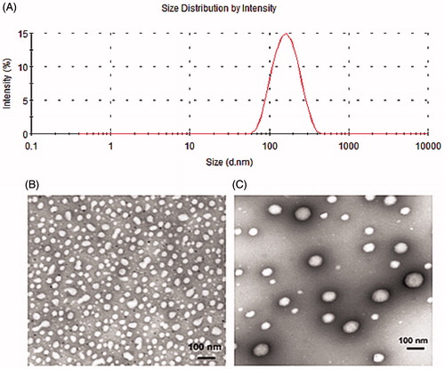
To accomplish the synthesis of functional mRNA transcripts, a plasmid DNA vector pVAX1 containing the HIV-1 gag gene was constructed as illustrated in . Linearized pVAX1-gag plasmid was the template for gag mRNA transcription. Synthesis of the transcripts was accomplished by in vitro transcription reaction using standard methods. As shown in , after in vitro transcription, mRNA was analyzed by agarose gel electrophoresis with the length of approximately 1700 nt, which was in accordance with the predicted length after adding the human β-globin untranslation regions and poly (A) sequences to plasmid skeleton. Gel retardation assay was performed to investigate the mRNA packaging by PSA (). With the W/W ratio of PSA/mRNA higher than one, the migration of mRNA was completely retarded, indicating a good encapsulation efficiency of PSA/mRNA. As shown in , mean size and polydispersity index of PSA/mRNA complexes were 117.77 ± 3.894 nm and 0.13 ± 0.017 respectively at the mass ratio of four, which suggested the nanoparticle formulation was homogeneous. TEM also illustrated that after packaging with mRNA, the size of PSA enhanced. The PSA/mRNA complexes were spherical in shape and approximately 100 nm in size ().
Figure 2. (A) Schematic representation of pVAX1-gag used in the study. The in vitro transcription template contained human β-globin-specific 5′ and 3′ UTRs and a 30 nucleotide-long poly A tail. The start (AUG) and stop (UAA) codons were added. (B) Detection of mRNAs. In vitro transcripts were tested in 4% nuclease-free agarose gel. The position and size of the mRNAs were indicated. (C) Gel retardation assay of PSA/mRNA polyplexes in 4% nuclease-free agarose gel. Polyplexes were prepared at different weight to weight ratio of PSA/mRNA. Naked mRNA was the control without any complexation.
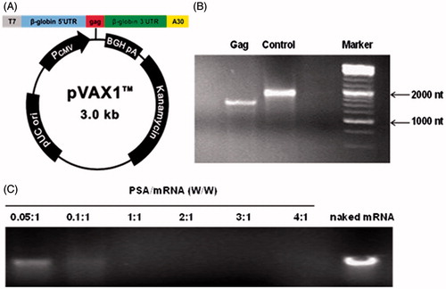
Figure 3. Morphology and size distribution of PSA and PSA/mRNA nanoparticles were evaluated at PSA:mRNA (W/W)ratio of 4. (A) Size and distribution of PSA/mRNA nanoparticles by intensity. TEM image of (B) naked PSA and (C) PSA/mRNA nanoparticles (1% phosphotungstic acid-negative staining).
PSA/mRNA delivery into DC2.4 cells
The cytotoxicity of PEI usually limits their applications both in vitro and in vivo, thus three kinds of PEI polymers were necessarily evaluated by MTT assay on DC2.4 cells. As shown in , when the ratio of PSA/mRNA was less than 4:1, the PSA formulations showed low toxicity, and the cell viabilities were more than 80%, showing no significant difference in comparison to PEI-2k polyplexes treated groups. Therefore the rest of our study was based on the PSA/mRNA ratio of 4:1. However, when the mass ratio of PSA: mRNA reached 5:1, PSA formulations induced more cell deaths. By contrast, the cell viability was much lower after being treated with PEI-25k, which hampered its application in cell experiments and animal tests.
Figure 4. Cytotoxicity and cell uptake of PSA/mRNA nanoparticles. (A) Cell viability of PEI polymers was determined on DC2.4 cells. Untreated cells were defined as 100% viability cells. Data are presented as mean ± SD (n = 3), **p < 0.01, ***p < 0.005. (B) Flow cytometry analysis for cell uptake efficiency of Fl-mRNA complexed with PEI-2k, PSA or PEI-25k at the indicated mass ratio after incubation with DC2.4 cells for 2 h. (Data are shown as mean ± SD (n = 3). ***p < 0.005) (C) Confocal laser scanning microscopy showed the endosome escape of PSA/Fl-mRNA nanoparticles. DC2.4 cells were incubated with Fl-mRNA complexed with PSA for the indicated times. Green fluorescence was Fl-mRNA and red fluorescence was endosome stained with Lyso-tracker, yellow fluorescence was the co-localization site of the Fl-mRNA and endosome.
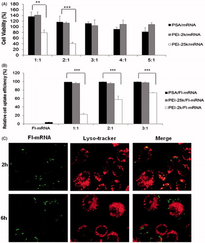
To detect the cell uptake efficiency of PEI nanoparticles on DC2.4, mRNA was labeled by Fluorescein Labeling Kit (Mirus, Madison, WI). The internalization of fluorescent PSA/mRNA was assayed by flow cytometry. PEI-25k, which was proven to be an efficient vehicle for gene transfer, was the positive control. As shown in , PSA could significantly improve the cell uptake efficiency of Fluorescein-mRNA (Fl-mRNA) on DC2.4 as compared to PEI-2k (p < 0.005), which was only slightly lower than that of PEI-25k. We only tested three ratios of the cell uptake efficiency of PSA/mRNA nanoparticles, which was because it could not increase anymore at higher mass ratio.
Nano sized particles are easily taken up through endocytosis, thus endosome escape is a key biological barrier in nanoparticle delivery system. As indicated in , after incubation with PSA/Fl-mENA complexes at the indicated time points, the escape ability of the nanoparticles on DC2.4 cells were identified by CLMS. In these images, Fl-mRNA was green and endosome stained with Lyso-tracker was red in color. As indicated in the figures, nanoparticles located in endosome within 2 h incubation. After 6 hours, the yellow fluorescence disappeared, indicating that more nanoparticles entered into cytoplasm. This phenomenon illustrated the PSA could help mRNA escape from endosome after being uptaken by cells, which was also a time-dependent process.
In vitro transfection efficiency of PEI/mRNA complexes on DC2.4 cells
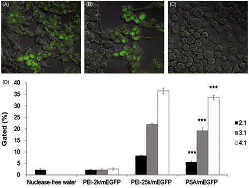
Confocal microscopy images () illustrated the expression of EGFP on DC2.4 cells after being treated with different PEI/mEGFP nanoparticles and naked mEGFP. PEI-25k exhibited the most effective delivery of EGFP mRNA in these experiments. PSA showed approximately the same level of EGFP expression as PEI-25k. However, no green fluorescence was observed in naked mRNA treated group. The ability of PEI polymers to mediate mEGFP transfection in vitro was also evaluated by flow cytometer (). Cells were incubated with mRNA nanoparticles for 24 h prior to the analysis of EGFP expression by flow cytometry. We have previously reported that naked mRNA failed to induce any transfection efficiency by flow cytometer (data not show). In these tests, the optimal mass ratio of PSA/mRNA was 4:1, showing the highest transfection efficiency. As the positive control, the transfection efficiency of PEI-25k/mEGFP maintained the highest among three kinds of polymers. With the mass ratio increased, cells transfected with PSA/mEGFP performed gradually increased transfection efficiency and were consistently significantly higher as compared to PEI-2k/mEGFP (p < 0.005). These results suggested that by conjugating STA to PEI-2k, the PSA polymer would sharply increase the efficiency for mRNA delivery, which was in accordance with the cellular uptake study. However, PEI-2k polyplexes failed to produce significant EGFP mRNA transfection (<5% EGFP positive cells) at each ratio.
Figure 5. DC2.4 cells after transfection with (A) PEI-25k/mEGFP W/W=4, (B) PSA/mEGFP W/W=4 and (C) naked mEGFP. (D) Transfection efficiency of naked mGag and the indicated PEI complexes. Transfection efficiency of cells treated with PSA/mEGFP was consistently significantly higher as compared to that of PEI-2k/mEGFP at the indicated mass ratio. (Data are shown as mean ± SD (n = 3). ***p < 0.005).
In vitro BMDCs maturation
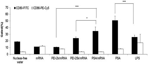
The maturation of DCs is an important parameter to consider the effect of vaccination and gene delivery, thus we investigated the maturation level of BMDCs at 24 h post-cultured with PEI complexes in vitro. shows the results of CD80 or CD86 positive BMDCs after incubation with different PEI/mRNA nanoparticles. PSA/mRNA complexes are able to dramatically enhance the CD80 level, which was even significantly higher than that of the positive control: lipopolysaccharide (LPS) (p < 0.005). As for the CD86 level, mRNA alone showed higher level than other groups except LPS, which may due to the ability of mRNA to activate TLRs.
Figure 6. Expression level of co-stimulatory surface markers (CD80 and CD86) in BMDCs treated with naked mRNA alone and complexes. Data are presented as mean ± SD (n = 3), *p < 0.05, ***p < 0.005.
Determination of anti-HIV1 gag specific antibody
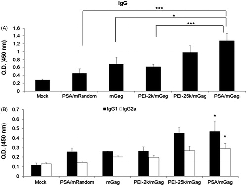
To evaluate the potency of the PSA/mRNA vaccine, we detected the HIV1-gag specific antibody. After immunization, serum of mice was obtained and levels of IgG, IgG1, and IgG2a were assessed. As shown in , serum from the group of PSA/mGag immunized mice indicated the highest IgG level, which were significantly higher as compared to PSA/mRandom (p < 0.005), naked mGag (p < 0.05) and PEI-2k/mGag (p < 0.005). These results illustrated the immune response was specific to the encoded antigen HIV1-gag. Furthermore, as shown in , both the IgG1 and IgG2a levels in PSA/mGag immunized mice was significantly higher than that of PSA/mRandom, mGag alone and PEI-2k/mGag group (p < 0.05). However, no significant difference was observed between PSA/mGag and PEI-25k group. These results demonstrate that the PSA/mGag complex could induce an enhanced humoral immune response in immunized mice.
Figure 7. Serum levels of IgG, IgG1 and IgG2a after immunization. Sera from PEI/mGag complexes and naked gag mRNA immunized mice were collected post-immunization. The serum levels of (A) IgG (B) IgG1 and IgG2a antibodies were assayed by ELISA. Both the IgG1 and IgG2a levels in PSA/mGag immunized mice were significantly higher than that of PSA/mRandom, gag mRNA alone and PEI-2k/mGag group (p < 0.05). Data were represented as mean ± SD values (n = 3), *p < 0.05, ***p < 0.005.
Monitoring HIV gag-specific T cells responses by intracellular cytokine staining (ICS)
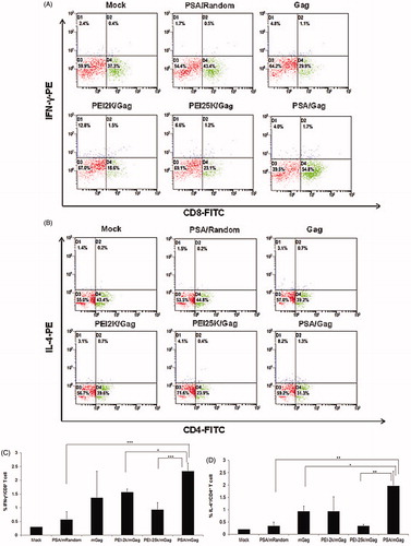
As cytokine levels in T cells plays a vital role in controlling virus infection (such as HIV), we detected the CD8+ and CD4+ T cell responses in different nanoparticles immunized mice by intracellular cytokine staining (). As shown in , after incubation with HIV-1 gag peptide for 5 h, the average percentage of IFN-γ expressing CD8+ T cells and IL-4 expressing CD4+ T cells in PSA/mGag complexes immunized group (2.33 ± 0.29% and 1.97 ± 0.59%, respectively) were considerably higher than those of PSA/mRandom complexes immunized group (0.57 ± 0.29% and 0.33 ± 0.15%, respectively), indicating that CD8+ and CD4+ T cell responses were specific to HIV-1 gag. Moreover, all of the PSA nanoparticle formulations were able to show greater performance in comparison with naked mGag format. The average percentage of IFN-γ positive cells in PSA/mGag immunized mice was significantly higher than those of PEI-2k/mGag (p < 0.05) and PEI-25k/mGag (p < 0.005). In addition, the average percentage of IL-4 positive cells in PSA/mGag immunized mice was significantly higher than those of mGag alone (p < 0.05) and PEI-25k/mGag (p < 0.01).
Figure 8. Intracellular cytokine staining of splenocytes after immunized with naked gag mRNA and different PEI/mGag complexes. (A) The CD8+/IFN-γ+ staining, (B) the CD4+/IL-4+ staining, the double-positive percentage of (C) CD8+/IFN-γ+ and (D) CD4+/IL-4+. Data were shown as mean ± SD (n = 3), *p < 0.05, **p < 0.01, ***p < 0.005.
HIV gag-specific T cells responses in splenocytes culture supernatants
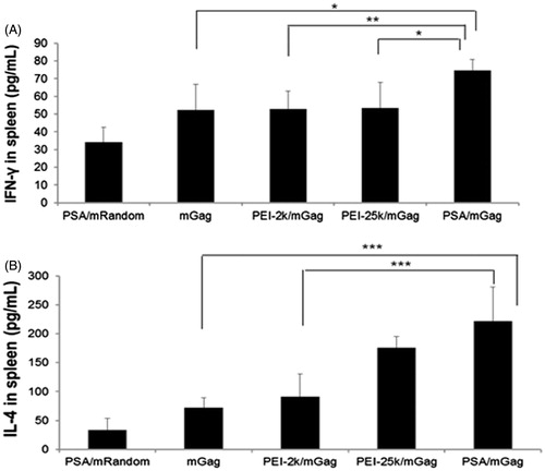
To further observe the T cell response, IL-4 and IFN-γ levels in splenocytes culture supernatants were determined. Cytokine levels in splenocytes culture supernatants were quantified by ELISA. As demonstrated in , the supernatants in PSA/mGag group showed the highest IFN-γ levels (74.74 pg/mL±6.03), which was dramatically higher than that in PSA/mRandom (34.13 pg/mL±8.55). The IL-4 levels in naked mGag and PEI-2k/mGag group were also significantly lower than that in PSA/mGag group (52.26 pg/mL±14.58 and 53.00 pg/mL±9.99 respectively). The same trend can be found in IL-4 level. The above data implied that PSA was able to promote the HIV-1 gag-specific CD8+ T cell and CD4+ T cell response, which was specific to HIV-1 gag.
Figure 9. Cytokine production of mice immunized with naked gag mRNA and different complexes. (A) IFN-γ levels in splenocytes supernatants. (B) IL-4 levels in splenocytes supernatants. Data were shown as mean ± SD (n = 4), *p < 0.05, **p < 0.01, ***p < 0.005.
Discussion
In recent years, mRNA-based vaccines had appeared to be a promising alternative to classical DNA-based immunization approaches, especially in the field of tumor derivate antigen-encoding mRNA vaccines (Bringmann et al., Citation2010; Kreiter et al., Citation2011; Perche et al., Citation2011). mRNA molecules were able to induce strong immune response as the binding ability to pattern recognition receptors, stimulating innate immunity (Fotin-Mleczek et al., Citation2011). However, it is not easy for mRNA vaccines to achieve sufficient protein expression in clinical tests due to the instability of single-stranded RNA molecule in the presence of RNase. To overcome this problem, a wide range of efforts had been made to protect mRNA from degradation, such as the progress in developing efficient carriers for mRNA delivery.
In this work, we assessed the ability of cationic micelles: PSA to successfully deliver mRNA to DC2.4 cells and BALB/c mice. Our finding indicated that PSA were able to condense mRNA with efficient binding affinity (). This was because the fatty acids could offer a relatively flexible three-dimensional conformation, thus enhancing physical encapsulation of RNA. (Alshamsan et al., Citation2009). The hydrophobic modification of PEI could also promote cell membrane interactions (Wong et al., Citation2007; Liu et al., Citation2010). This was confirmed by much higher cell uptake efficiency of PSA nanoparticles as compared to PEI-2k polyplexes (). The greater transfection efficiency on DC2.4 cells was able to prove the hypothesis as well.
Although PEI-25k was the most well-studied and potent gene transfection agent, the high toxicity hampered its' in vivo or clinical application (Fischer et al., Citation1999; Godbey et al., Citation1999). The mechanism for the toxicity of high molecular weight PEI has not been fully understood yet, but S. Moein Moghimi reported that two types of toxicity were induced by PEI: membrane damage and cell apoptotic (Moghimi et al., Citation2005). In this study, PEI-25k was also able to perform well in transfering mRNA and induce immunization response to some extent, however, the toxicity was still high (). As fatty acids were safe materials, cationic micelles PSA significantly increased its transfection efficiency and cell uptake efficiency while keeping the cytotoxicity relatively low. The result was consistent with the previous report that fatty acids−PEI conjugates complexed with siRNA had relatively less toxicity (Aliabadi et al., Citation2011). Whether mRNA could be expressed to protein format greatly associates with the endosome escape of PEI complexes. The most well-known explanation of this phenomenon was the “proton sponge effect” (Cho et al., Citation2003). Our results provided encouraging evidence to demonstrate the concept that it is possible to cause endosome escape in DC2.4 cells using PSA nanoparticles, which illustrated the ability of PSA to overcome the major obstacle for mRNA in in vitro delivery. Whether PSA could induce BMDCs maturation had been evaluated by testing the secretion of co-stimulators such as CD80 and CD86, which could be found only in mature DCs. In our study, immature BMDCs were activated with different PEI complexes and the expression of the above two co-stimulators was evaluated. We found both PSA alone and PSA/mRNA nanoparticles could both induce high level of CD80, indicating that PSA had the potential to be an efficient vaccines vehicle ().
mRNA vaccines could also induce antigen-specific antibody secretion. We tested antigen-specific antibody IgG, and the subtypes IgG1 and IgG2a. The results showed that both the above three types of antibody levels were the highest after immunization by PSA/mGag (). T helper cells mediated responses can be classified into two types: Th1 and Th2 immune responses, which were associated with IgG1 and IgG2a subtype level (Li et al., Citation2013). In our study, the immune response induced by mRNA vaccine candidates was confirmed to be a mixed Th1 and Th2 response.
The secretion of pro-inflammatory cytokines greatly associates with the ability of mRNA to activate toll-like receptors. These cytokines were able to trigger strong innate immune response as well as the maturation of antigen presenting cells (Phua et al., Citation2014). Our work discussed the percentage of IFN-γ expressing CD8+ T cells and IL-4 expressing CD4+ T cells in spleen with different PEI polyplexes (). The IFN-γ and IL-4 levels in spleen supernatants of the mice immunized with PEI polyplexes were investigated as well (). As illustrated from the result, both IFN-γ and IL-4 positive cells of the PSA/mGag immunized mice showed the highest level in spleen. IFN-γ/CD8+ T cells seem to activate Th1 response while IL-4 expressing CD4 + T cells seem to induce Th2 response (Doria-Rose & Haigwood, Citation2003). These results were consistent with a previous report that cationic micelles PSA could promote cytokines secretion as compared to peptide alone (Zeng et al., Citation2014). The PEI-25k/mGag group could also induce high secretion of pro-inflammatory cytokines in spleen supernatants. Naked mGag group, however, was less efficient to induce cytokines secretion. As is known, after recognized by pattern recognition receptors in APCs, mRNA nanoparticle can mediate immune activation. Also, TLRs mainly located in the endosome of antigen presenting cells and some fibroblast cells (Phua et al., Citation2014). Thus, only polymers with endosome location can be recognized by TLRs, resulting to the induction of cytokine secretion. Naked mRNA molecules were vulnerable and easily degraded by RNase, leading to poor cytokines levels. In addition, we demonstrated that PSA/mRandom failed to induce any HIV-1 gag specific IFN-γ or IL-4, which confirmed the specificity of HIV-1 gag.
Conclusion
In this study, PSA was utilized as an efficient mRNA vaccine vehicle. Our results indicated that steric acid binding to PEI-2k influenced mRNA uptake efficiency and improved transfection efficiency. More importantly, PSA was less toxicity than PEI-25k. At the same time, PSA/ mRNA nanoparticles could escape from endosome and lead to BMDC maturation. The PSA/mRNA complexes could significantly enhance anti-HIV1 gag immune responses as compared to mRNA alone, PEI-2k/mRNA and PEI-25k/mRNA groups, resulting in high antigen-specific antibody secretion and pro-inflammatory cytokines production, which make this strategy a possible anti-HIV1 vaccine candidate. The PSA nanomicelles was shown to be and efficient delivery vehicle for mRNA vaccine delivery. Future research may focus on the mechanism of vaccination and evaluate its clinical efficacy.
Declaration of interest
This work was supported by grants from the National Natural Science Foundation of China (No. 81173011 & 81422044).
Related Research Data
References
- Aliabadi HM, Landry B, Bahadur RK, et al. (2011). Impact of lipid substitution on assembly and delivery of siRNA by cationic polymers. Macromol Biosci 11:662–72
- Alshamsan A, Haddadi A, Incani V, et al. (2009). Formulation and delivery of siRNA by oleic acid and stearic acid modified polyethylenimine. Mol Pharm 6:121–33
- Amano T, Kajiwara K, Yoshikawa K, et al. (2007). Antitumor effects of vaccination with dendritic cells transfected with modified receptor for hyaluronan-mediated motility mRNA in a mouse glioma model. J Neurosurg 106:638–45
- Bettinger T, Carlisle RC, Read ML, et al. (2001). Peptide-mediated RNA delivery: a novel approach for enhanced transfection of primary and post-mitotic cells. Nucleic Acid Res 29;3882–91
- Bringmann A, Held SAE, Heine A, Brossart P. (2010). RNA vaccines in cancer treatment. J Biomed Biotechnol 2010:623687--98
- Cho YW, Kim JD, Park K. (2003). Polycation gene delivery systems: escape from endosomes to cytosol. J Pharm Pharmacol 55:721–34
- Doria-Rose NA, Haigwood NL. (2003). DNA vaccine strategies: candidates for immune modulation and immunization regimens. Methods 31:207–16
- Fischer D, Bieber T, Li Y, et al. (1999). A novel non-viral vector for DNA delivery based on low molecular weight, branched polyethylenimine: effect of molecular weight on transfection efficiency and cytotoxicity. Pharm Res 16:1273–9
- Fotin-Mleczek M, Duchardt KM, Lorenz C, et al. (2011). Messenger RNA-based vaccines with dual activity induce balanced TLR-7 dependent adaptive immune responses and provide antitumor activity. J Immunother 34:1–15
- Godbey WT, Wu KK, Mikos AG. (1999). Size matters: molecular weight affects the efficiency of poly(ethylenimine) as a gene delivery vehicle. J Biomed Mater Res 45:268–75
- Inaba K, Inaba M, Romani N, et al. (1992). Generation of large numbers of dendritic cells from mouse bone marrow cultures supplemented with granulocyte/macrophage colony-stimulating factor. J Exp Med 176:1693–702
- Kariko K, Kuo A, Barnathan E. (1999). Overexpression of urokinase receptor in mammalian cells following administration of the in vitro transcribed encoding mRNA. Gene Ther 6:1092–100
- Kreiter S, Diken M, Selmi A, et al. (2011). Tumor vaccination using messenger RNA: prospects of a future therapy. Curr Opin Immunol 23:399–406
- Li M, Jiang YH, Xu, CQ, et al. (2013). Enhanced immune response against HIV-1 induced by a heterologous DNA prime-adenovirus boost vaccination using mannosylated polyethyleneimine as DNA vaccine adjuvant. Int J Nanomed 8:1843–54
- Liu ZH, Zhang ZY, Zhou CR, Jiao, YP. (2010). Hydrophobic modifications of cationic polymers for gene delivery. Prog Polym Sci 35:1144–62
- Malone RW, Felgner PL, Verma IM. (1989). Cationic liposome-mediated RNA transfection. Proc Natl Acad Sci USA 86:6077–81
- Mockey M, Bourseau E, Chandrashekhar V, et al. (2007). mRNA-based cancer vaccine: prevention of B16 melanoma progression and metastasis by systemic injection of MART1 mRNA histidylated lipopolyplexes. Cancer Gene Ther 14:802–14
- Moghimi SM, Symonds P, Murray JC, et al. (2005). A two-stage poly(ethylenimine)-mediated cytotoxicity: Implications for gene transfer/therapy. Mol Ther 11:990–5
- Mosmann T. (1983). Rapid colorimetric assay for cellular growth and survival: application to proliferation and cytotoxicity assays. J Immunol Method 65:55–63
- Navarro G, Pan J, Torchilin VP. (2015). Micelle-like nanoparticles as carriers for DNA and siRNA. Mol Pharm 12:301--13
- Nishiya T, Kajita E, Miwa S, Defranco AL. (2005). TLR3 and TLR7 are targeted to the same intracellular compartments by distinct regulatory elements. J Biol Chem 280:7–17
- Pack DW, Hoffman AS, Pun S, Stayton PS. (2005). Design and development of polymers for gene delivery. Nat Rev Drug Discov 4:581–93
- Palumbo RN, Zhong X, Wang C. (2012). Polymer-mediated DNA vaccine delivery via bystander cells requires a proper balance between transfection efficiency and cytotoxicity. J Control Rel 157:86–93
- Perche F, Benvegnu T, Berchel M, et al. (2011). Enhancement of dendritic cells transfection in vivo and of vaccination against B16F10 melanoma with mannosylated histidylated lipopolyplexes loaded with tumor antigen messenger RNA. Nanomedicine 7:445–53
- Petsch, B, Schnee, M, Vogel, AB, et al. (2012). Protective efficacy of in vitro synthesized, specific mRNA vaccines against influenza A virus infection. Nat Biotechnol 30:1210--16
- Phua, KKL, Nair, SK, Leong, KW. (2014). Messenger RNA (mRNA) nanoparticle tumour vaccination. Nanoscale 6:7715–29
- Probst J, Brechtel S, Scheel B, et al. (2006). Characterization of the ribonuclease activity on the skin surface. Genet Vaccines Ther 4:4--12
- Qiu Y, Guo L, Zhang S, et al. (2015). DNA-based vaccination against hepatitis B virus using dissolving microneedle arrays adjuvanted by cationic liposomes and CpG ODN. Drug Deliv 27:1–8
- Rittig SM, Haentschel M, Weimer KJ, et al. (2011). Intradermal vaccinations with rna coding for taa generate CD8(+) and CD4(+) immune responses and induce clinical benefit in vaccinated patients. Mol Ther 19:990–9
- Shi SJ, Zhong ZR, Liu J, et al. (2012). Solid lipid nanoparticles loaded with anti-microrna Oligonucleotides (AMOs) for suppression of microRNA-21 functions in human lung cancer cells. Pharm Res 29:97–109
- Steitz J, Britten CM, Wolfel T, Tuting T. (2006). Effective induction of anti-melanoma immunity following genetic vaccination with synthetic mRNA coding for the fusion protein EGFP.TRP2. Cancer Immunol Immunother 55:246–253
- Su XF, Fricke J, Kavanagh DG, Irvine DJ. (2011). In vitro and in vivo mRNA delivery using lipid-enveloped ph-responsive polymer nanoparticles. Mol Pharm 8:774–87
- Tavernier G, Andries O, Demeester J, et al. (2011). mRNA as gene therapeutic: how to control protein expression. J Control Release 150:238–47
- Uchida S, Itaka K, Uchida H, et al. (2013). In vivo messenger RNA introduction into the central nervous system using polyplex nanomicelle. PLoS One 8:e56220
- Weide B, Carralot JP, Reese A, et al. (2008). Results of the first phase I/II clinical vaccination trial with direct injection of mRNA. J Immunother 31:180–8
- Weide B, Pascolo S, Scheel B, et al. (2009). Direct injection of protamine-protected mRNA: results of a phase 1/2 vaccination trial in metastatic melanoma patients. J Immunother 32:498–507
- Wong SY, Pelet JM, Putnam D. (2007). Polymer systems for gene delivery-past, present, and future. Prog Polym Sci 32:99–837
- Yamamoto A, Kormann M, Rosenecker J, Rudolph C. (2009). Current prospects for mRNA gene delivery. Eur J Pharm Biopharm 71:484–9
- Yu H, Babiuk LA, Littel-Van Den Hurk SV. (2007). Immunity and protection by adoptive transfer of dendritic cells transfected with hepatitis CNS3/4A mRNA. Vaccine 25:1701–11
- Zarei S, Arrighi JF, Ongaro G, et al. (2003). Efficient induction of CD8 T-associated immune protection by vaccination with mRNA transfected dendritic cells. J Invest Dermatol 121:745–50
- Zeng Q, Jiang H, Wang T, et al. (2014). Cationic micelle delivery of Trp2 peptide for efficient lymphatic draining and enhanced cytotoxic T-lymphocyte responses. J Control Release 200:1–12
- Zhang HM, Zhang LW, Ren J, et al. (2006). Induction of alpha-fetoprotein-specific CD4-and CD8-mediated T-cell response using RNA-transfected dendritic cells. Cell Immunol 239:144–50
- Zohra FT, Chowdhury EH, Tada S, et al. (2007). Effective delivery with enhanced translational activity synergistically accelerates mRNA-based transfection. Biochem Biophys Res Commun 358:373–8