Abstract:
To investigate the diagnosis and microsurgical treatment of cavernous sinus hemangioma, the clinical data, including pathology, epidemiology, medical imaging, operation procedure, and post-operational complication of 12 cavernous sinus hemangioma patients undergoing operations in Affiliated Hospital of Medical College of Qingdao University from 1999 to 2008, were analyzed. There were 2 males and 10 females. The patients were aged from 28 to 61 years. Headaches and deficits of the cranial nerves coursing through the cavernous sinus were the principal symptoms at presentation. The common clinical manifestations were visual loss, diplopia, facial numbness, and extraocular muscle palsy. The radiological features in all patients were similar with a characteristic pattern of extension and encasement of carotid artery. CT showed the lesion as hypodense to isodense with marked enhancement after contrast administration. T1-weighted MR imaging showed the lesions as hypointense with marked enhancement after contrast administration. T2-weighted MR imaging showed the lesions as hyperintense. The maximum size of the lesion was 9 to 57 mm (mean 45mm). Basal pterional craniotomies were used for eight patients. Orbitozygomatic osteotomies were used for two patients. Pterional approach was used for two patients. The lesions were removed through incising the lateral wall of the cavernous sinus. The tumor was totally removed in five cases, subtotally removed in four cases, and partially removed in two cases. The main post-operational complications included oculomotor nerve paralysis (four cases) and trigeminal nerve lesions (three cases). No postoperative death occurred. Operation is the best choice for cavernous sinous hemangioma. It was helpful to control bleeding through intradura and incising the lateral wall of the cavernous sinus.
INTRODUCTION
Cavernous hemangioma of the cavernous sinus is an uncommon lesion accounting for 0.2–2% of all cavernous sinus tumors [Citation1]. Although the tumor is benign, it is a neurosurgical challenge due to the high vascularity, location within the cavernous sinus, and relationship to the intracavernous internal carotid artery and cranial nerves. Histologically, cavernous hemangioma of the cavernous sinus is similar to intracerebral cavernous angioma, but is a distinct clinical entity and the management issues are vastly different from those located within the cerebral parenchyma. Cavernous hemangiomas limit to the dura of the cavernous sinus, and frequently reach giant size before diagnosis. There were 12 patients with cavernous sinus hemangioma hospitalized in our hospital during 1999–2008. We analyze those cases of cavernous hemangiomas and the relative articles.
MATERIALS AND METHODS
General Materials
There were 2 males and 10 females. The ages ranged from 28 to 61 years old. The duration of symptoms at the time of presentation ranged from 3 months to 2 years.
Clinical Manifestation
Eight patients presented with symptoms of headache, four patients presented with symptoms of facial numbness. Two patients had visual deficits on the side of the tumor. The visual deficits were progressive in all patients. Two patients had oculomotor paralysis, and two patients had abducent nerve lesion. Two patients had eyeball protrusion.
Radiological Examination
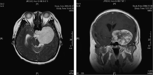
All patients were investigated with computed tomography (CT) and magnetic resonance (MR) imaging. CT showed the lesions as hypodense to isodense, and the lesions were demarcated and bell-shaped with marked enhancement after contrast administration (). T1-weighted MR imaging showed the lesions as hypointense with marked enhancement after contrast administration. T2-weighted MR imaging showed the lesions as highly hyperintense. The size of the tumor ranged from 9 to 57 mm in maximum dimension (mean 45 mm).
Figure 1. MRI imaging showed demarcated bell-shaped.
Treatment
The surgical treatment were performed in all 12 patients. We adopted various ways to expose the lesions under the dura, according to the size of the lesions. The lateral wall of cavernous sinus was incised and the lesion was removed. There were eight cases with zygomatic-pteroinal approach, two cases with pteroinal approach, and two cases with temporal-orbital-zygomatic approach in the surgical process. The sylvian fissures were dissected in six cases.
After head fixation in the Mayfield clamp, the skin incision was placed along or in the hair line starting in the preauricular region 1 cm below the zygomatic arc. Care was taken not to damage the frontal branch of cranial nerve VII by keeping the anterior dissection strictly on the fascia of the temporalis musculature. The skin flap developed was then turned anteriorly until the zygomatic process of the frontal bone and the frontal process of the zygoma were exposed. The fascia and the temporalis muscle were resected, and the zygomatic arc removed. The flap of the temporalis muscle and the zygomatic arc were then turned to the temporal fossa together. The frontal-temporal bone flap was removed after drilling holes on the cranium. The lateral sphenoid crest was drilled as largely as possible. In order to reach the base of middle fossa, it was necessary to remove a portion of temporal squamus and the spheniod wing.
After opening the dura, the sylvian fissure was opened in a lateral-to-medial manner and the carotid and chiasmatic cisterns were opened. This allowed gentle retraction of the frontal and temporal lobe. The parasellar region, edge of the tentorium, and the base of the middle fossa were exposed. The lateral wall of cavernous sinus was dissected under the microscope. Rapid debulking of the tumor using relatively powerful, graded, and controlled suction was carried out to remove the bulk of the tumor, to expose the sixth cranial nerve near the petrous apex, and to coagulate the feeders arising from the carotid artery early in the operation. Once the bulk of the tumor was removed and the branches from the major feeding channels were obliterated, the hemostasis was largely spontaneous and relatively simple. We used the bipolar coagulator and the absorbable hemostatic gauze to stop bleeding.
RESULTS
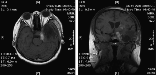
The lesions were totally removed in five patients (), sub-totally removed in four patients and partially removed in three patients. None of the patients died. In the immediate postoperative phase, two patients showed optic acuity recovery, three patients showed oculomotor paralysis, and six patients showed facial numbness. Extraocular movements completely recovered in two patients about three months following surgery. The function of the fifth nerve partially recovered. At follow-up after six months to two years, the lesions did not recur in five patients where they had been totally removed. Other patients were administered with radiotherapy, and the lesions controlled validly.
Figure 2. MRI showed the lesion was sub-total removed.
DISCUSSION
Cavernous angiomas are also known as cavernomas and cavernous hemangiomas. They are common lesions of the cerebral hemispheres, although they can occur anywhere in the central nervous system. Extra-axial cavernous angiomas are uncommon, and the cavernous sinus is one location in this group. It is most common in women in their fifth decade of life, being rarely multiple and hereditary [Citation2–6]. It is, in fact, a vascular malformation, which can behave like a real tumor when it grows up to the point where it compresses neighboring structures. Symptoms are exacerbated in pregnant women Cavernous hemangiomas are frequently seen in the fourth and fifth decades of life [Citation7,Citation8]. Ninety-four percent of reported cases occurred in women [Citation9]. In our series, 83% were females. Considering that females in their youth or middle age are more common victims of this tumor, the origin may be hormonal [Citation10]. Clinical presentation is usually in the form of symptoms related to the acute or subacute dysfunction of the nerves traversing the cavernous sinus and the optic nerve. Headache, which varied in intensity from moderate to severe, was present in all patients and was the most disabling clinical feature. Hemorrhage within the cerebral cavernous hemangiomas is a common feature, but is relatively uncommon in cavernous hemangiomas located in the cavernous sinus.
Radiographically, this entity appears as an iso/hyperdense mass on non-enhanced CT scans, enhancing intensely after infusion of iodinated media in most cases [Citation4]. Cavernous hemangiomas have a characteristic pattern of extension towards the sella, superior orbital fissure, and Meckel’s cave, which was observed in all our cases and in the majority of reported cases with radiography of the lesion [Citation8]. Calcification is an occasional finding, and it is most common in meningiomas [Citation6]. Erosion of the sphenoid bone can also be seen. DSA can be normal and show an avascular mass or a discrete to moderate tumoral blush, with feeding vessels originating from branches of the external carotid or cavernous internal carotid [Citation11]. The internal carotid artery is often encircled by the lesion in its cavernous portion, usually maintaining its normal caliber. Cavernous hemangioma is the only primary intra-cavernous sinus tumor. Irrespective of the size, the tumor has never been found to protrude out of the anatomical dural confines of the cavernous sinus. The extension towards the sella appears to be through the enlarged intercavernous sinus. Unlike cerebral cavernous angiomas, their cavernous sinus counterparts do not have a pathognomonic appearance on MRI. Findings usually are of well-delimited para-sellar lesions, hypo or isoin-tense in T1-weighted images and brightly hyperintense in T2-weighted images. A dumbbell-shaped mass can be seen, with a small supra-sellar component and most of the lesion within the cavernous sinus [Citation2,Citation5]. MRI allows superb evaluation of the relationships among the cavernous angioma and the surrounding structures [Citation2,Citation3]. Gradient-echo sequences may be useful to reveal the hemorrhagic component.
Surgical excision is the first choice to treat this benign lesion, and it could lead to potential curability [Citation12,Citation13]. Smaller lesions and those with mild symptoms can be clinically and radiologically observed. The main difficulty during surgery for cavernous hemangioma is the vascularity of the lesion [Citation14]. The risk of resection and uncontrollable bleeding is extensive. Surgical misadventures have resulted in high morbidity and mortality. Preoperative radiation showed the advantages of reducing tumor vascularity [Citation15]. Direct puncture and injection of sclerosing agent (alcohol) in the lesion has produced good results. Induced hypotension and hypothermia may be useful adjuncts for surgery. During the surgical procedure, we found that the lesions were red, soft, and containing thin vascular channels. The lesions should be relatively stripped from the dura. The large cavernous hemangiomas can be easily resected by rapid tumor decompression using powerful and controlled suction. In order to preserve the carotid artery and the cranial nerves, we did not sharply dissect in the cavernous sinus. It was found that hemostasis was achieved spontaneously after a large portion of the lesion was resected. The residual portion could also easily be removed from the corners. Radical resection of cavernous sinus hemangiomas is possible by the subdural route. Recurrence rates are very low after surgical resections.
Stereotactic radiotherapy should be considered in partially resected cavernomas and should be reserved for elderly or high-risk patients as a noninvasive treatment option to reduce the size of the lesion and decrease the risk of hemorrhage [Citation16–18]. Rigamonti [Citation19] reported that they abandoned total resection in two of three patients due to excessive bleeding. One patient responded favorably to an interphased course of radiotherapy (5000 rads in 5 weeks) before undergoing a successful total resection six months later and receiving an iridium-125 implant to prevent a local recurrence. The other patient showed no response to radiotherapy and underwent two additional surgeries to achieve gross total resection. This treatment was complemented with iridium-125 implants. The third patient underwent biopsy only and responded favorably to radiotherapy (5000 rads in 5 weeks) but elected to delay further surgery.
Declaration of interest: The authors report no conflicts of interest. The authors alone are responsible for the content and writing of the paper.
Related Research Data
REFERENCES
- Liu, Hong-wei, Ye, Hong-xing, Shen, Hong . (2005). Diagnosis and microsurgical treatment of cavernous sinus hemangioma. Journal of Chinese Neurosurgery 21(6):354–356.
- Sohn, C.H., Kim, S.P., Kim, I.M., Lee, J.H., Lee, H.K. (2003). Characteristic MR imaging findings of cavernous hemangiomas in the cavernous sinus. AJNR 24:1148–1151.
- Bristot, R., Santoro, A., Fantozzi, L., Delfini, R. (1997). Cavernoma of the cavernous sinus: case report. Surg Neurol 48:160–163.
- Momoshima, S., Shiga, H., Yuasa, Y., Higuchi, H., Kawase, T., Toya, S. (1991). MR findings in extracerebral cavernous angiomas of the middle cranial fossa: report of two cases and review of the literature. AJNR 12:756–760.
- Suzuki, Y., Shibuya, M., Baskaya, M.K., . (1996). Extracerebral cavernous angiomas of the cavernous sinus in the middle fossa. Surg Neurol 45:123–132.
- Gliemroth, J., Missler, U., Sephernia, A. (2000). Cavernous angioma as a rare neuroradiologic finding in the cavernous sinus. J Clin Neurosci 7:542–560.
- Goel, A., Nadkarni, T.D. (1995). Cavernous hemangioma in the cavernous sinus. Br J Neurosurg. 9: 77–80.
- Suzuki, Y., Shibuya, M., Baskaya, M.K. (1996). Extracerebral cavernous angiomas of the cavernous sinus in the middle fossa. Surg Neurol. 45: 123–132.
- Linskey, M.E., Sekhar, L.N. (1992). Cavernous sinus hemangioma: aseries, a review, and a hypothesis. Neurosurgery 30: 101–108.
- Ito, J., Takahashi, M., Saito, A. (1997). Cavernous hemangioma of the middle cranial fossa with an angiographically demonstrated feeding artery and tumor stain. Rinsho Hoshasen 22: 339–144.
- Shi, J., Hang, C., Pan, Y., Liu, C., Zhang, Z. (1999). Cavernous hemangiomas in the cavernous sinus. Neurosurgery 45: 1308–1312.
- Rosenblum, B., Rothman, A.S., Lanzieri, C. (1986). A cavernous sinus cavernous hemangioma. Case report. J Neurosurg. 65: 716–718.
- Sawamura, Y., Tribolet, N. (1990). Cavernous hemangioma in the cavernous sinus: case report. Neurosurgery 26: 126–128.
- Mori, K., Handa, H., Gi, H. (1980). Cavernous in the middle fossa. Surg Neurol. 14: 21–31.
- Rigamonti, D., Pappas, C.T.E., Spetzler, R.F. (1990). Extracerebral cavernous angiomas of the middle fossa. Neurosurgery 27: 306–310.
- Stea, R.A., Schicker, L., King, G.A., Winfield, J.A. (1994). Stereotactic linear radiosurgery for cavernous angiomas. Stereotact Funct Neurosurg. 63(1-4): 255–265.
- Kondziolka, D., Lunsford, L.D., Flickinger, J.C., Kestle, J.R. (1995). Reduction of hemorrhage risk after stereotactic radiosurgery for cavernous malformations. J Neurosurg 83:825–831.
- Shibata, S., Mori, K. (1987). Effect of radiation therapy on extracerebral cavernous hemangioma in the middle fossa. Report of three cases. J Neurosurg 67:919–922.
- Rigamonti, D., Pappas, T.E., Spetzler, R.F., Johnson, P.C. (1990). Extracerebral cavernous angiomas of the middle fossa. Neurosurgery 27:306–309.