Abstract
Abstract: This study aims to estimate the effects of using a portion of a nerve trunk to repair itself and the injured nerve simultaneously. Proximal 1/2 median nerve served as donor nerve to repair the distal 1/2 median and whole ulnar nerve. Four months postoperation, the number of myelinated axons and nerve conduction velocities of the distal half median and ulnar nerve were (2033 ± 135 and 24.6 ± 5.3 m/s) and (1138 ± 228 and 30.3 ± 7.2 m/s). It suggests that using a portion of a nearby nerve truck to reconstruct itself and the injured nerve simultaneously is a practical method for severe peripheral nerve injury.
INTRODUCTION
Severe nerve lesions characterized by the absence of a proximal nerve stump might be one of the most difficult clinical problems in the world. Traditionally, they can be remedied by many methods, including nerve transfer [Citation5–8], nerve grafts, artificial nerve conduit bridging [Citation9–12], and end-to-side neurorrhaphy [Citation13–20]. However, the reconstructive effects were very limited, especially for the distal small muscles, because of the long distance for the regenerated axons to go to the distal effectors. Failure to restore injured nerves can lead to loss of muscle function, impaired sensation, and/or painful neuropathies [Citation1–4], not only in the injured nerve but also in the donor site.
As we reported before, the peripheral nerve can regenerate more than one shaft in the regenerative distal stump. The use of one nearby nerve trunk can repair the injured nerve and the donor site simultaneously to shorten the regenerating distance and reduce the donor site morbidity. Although both donor and injured nerve can be restored to a certain degree, there is a great risk when cutting an intact nerve trunk nearby as a donor nerve.
Here, we focus on the possibility and reconstruction effects of using a portion of a donor nerve to reconstruct the injured site and the donor nerve simultaneously. Results here may give suggestions for the reconstruction of the peripheral nerve and for reducing the risk of donor site function loss.
MATERIALS AND METHODS
Animals
We used male New Zealand rabbits that weighed 2.5–3.0 Kg and that were maintained under specific pathogen-free laboratory conditions. The rabbits were separated into 4 groups at random (6 animals in each group). Every effort was made to minimize animal suffering and reduce the number of animals used, according to the Chinese guidelines for care and use of laboratory animals.
Surgical Procedures
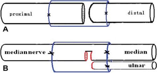
Surgical procedures were carried out under a binocular surgical microscope. Rabbits were anesthetized with sodium pentobarbital (30 mg/kg i.p.). After the anesthesia, rabbit limbs were treated in a sterile manner. An anterior approach was used to expose the median and ulnar nerves at the midarm level. In groups 2 and 3, the median/ulnar nerve was transected at the level of 2 cm above elbow, and the proximal nerve segment served as a father nerve to repair the distal nerve stump separately (Dor-Dor neurorrhaphy). In group 4, the proximal end of the ulnar nerve was ligated with 10-0 nylon sutures. Then, to provide a proximal median nerve with 1/2 numbers of axons, a 5-mm-long nerve segment with about 1/2 diameters was separated from the stem of the median nerve with its pedicel still connected with the proximal median nerve. The precise axon number was calculated 4 months later by histological methods. Then, the proximal half median nerve served as the donor nerve and was fixed with the distal 1/2 median and ulnar nerve. For this, we used biodegradable chitin conduits (patented by our laboratory and authorized by the state intellectual property office of China, No. ZL01 136314.2; it was an artificial nerve graft consisting of shell chitin showing satisfactory biocompatibility and degradation characteristics; the graft is now in a preclinical study). 10-0 nylon microsutures were used. The gap between the two nerve segments was kept at 1 mm. The biodegradable chitin conduits were 8 mm long, 0.1 mm thick, with an inner diameter of 4 mm. Subsequently, the muscle incision was sutured and the wound closed using 4–0 nylon sutures ().
Figure 1. Illustration of surgical precedures. A: Proximal median/ulnar nerve segment was served as father nerve to repair the distal nerve stump (Dor-Dor, Group 2, 3); B: Serving as a donor nerve, the proximal 1/2 median nerve was fixed to the distal stumps of 1/2 median and ulnar nerve simultaneously, using biodegegradable chitin conduits with a gap of 1 mm. (1/2Dor-1/2Dor+Rec, Group 4).
Tetanic Muscle Force
Tetanic muscle force was measured in the flexor carpi radialis, flexor digitorum superficialis muscle, and flexor carpi ulnaris. Four months post-operatively, the above-mentioned muscles were exposed, the tendons were cut as distally as possible, and ligated. The wrist, the elbow, and the shoulder joints were transfixed by Kirschner wires. The ligature was connected to a force transducer (PCLAB-UE, MicroStar Co. Ltd). The tetanic muscle force was measured at supramaximal stimulation (0.9 mA at 50 Hz) of the reinnervated median and ulnar nerves as previously described [Citation21]. The force was measured on the experimental and the contralateral side and expressed in percentage of the contralateral side.
Electrophysiological Study
Four months post-surgically, before sacrifice of the animals, the repaired median and ulnar nerves were exposed. The stimulating electrodes were placed proximal and distal to the repair site in each group. The recording electrode was placed in the distal reinnervated muscles, while the ground electrode went subcutaneously, between the stimulating and recording electrodes. Rectangular pulses (duration 0.1 ms, 0.9 mA, 10 Hz) were used to stimulate the repaired median and ulnar nerves. Compound muscle action potential was recorded and nerve conduction velocity (NCV, m/s) was obtained semiautomatically by dividing the distance between the two stimulating sites by the difference in the onset latency. The NCV of the contralateral normal median and ulnar nerves was recorded similarly.
Histological Study
The entire median and ulnar nerves were removed en bloc from each rabbit. Tissues were then harvested and fixed in 4% paraformaldehyde in 0.1 M phosphate buffer for 12 h at 4°C. After that, the nerves were rinsed twice in phosphate buffer, and 2 tissue blocks (approx. 5-mm long) were cut, one proximal to the surgical site and one distal to the surgical site. After this step, each sample was postfixed in 1% osmium tetroxide for 6 h, dehydrated through a graded series of ethanol, and embedded in paraffin. Specimen sections were then taken perpendicular to the long axis of the nerve fibers. Myelinated axons were quantified according to the unbiased counting rule. Finally, the total number of myelinated axons was estimated by multiplying the axonal density by the total cross-sectional area of the whole nerve.
Statistical Analysis
One-way analysis of variance was employed to compare the number of myelinated nerve fibers, NCV, and tetanic muscle force in all groups. A probability where p < 0.05 was considered significant for all statistical comparisons. All values are presented as the mean ± SD.
RESULTS
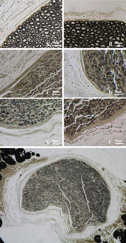
Four months post-operation, after exposure of operation site of the nerve, the biodegradable chitin conduits could not be clearly identified because of the absorption of the conduit and substitution by the fibrous tissues. The nerve stem was continued, which showed that the neonatal neurofibrils have regenerated to the distal part of the injured nerve. However, the regenerated nerves were a little thicker and much dimmer than the normal nerves. The results of quantitative stereological evaluations of the father nerve and the repaired nerve of different groups are summarized in . The single axon count for the sham control group was recorded as the same for the proximal and distal segment, for comparison. The morphologic preservation of all the tissues investigated was good and the myelinated fibers with various calibers were observed in all groups. In all experimental groups, regenerated axons could be observed in the distal part of both ulnar and median nerve. In group 4, there were regenerated fibers in half of the nerve trunk with the normal axons in the other intact nerve trunk. There was no significant morphologic difference of the regenerated axons among different experimental groups ().
Table 1. Comparison of myelinated axon numbers for all groups.
Figure 2. Histological sections through the reconstructed nerves 4 months after surgery. Proximal part of the median nerve (A) and ulnar nerve (B). Distal stump of transected median (C, Dor-Dor, Group 2) or ulnar (D, Dor-Dor Group 3) nerve neurorrhaphy; Distal regenerated median (E) and ulnar (F) stump of the group (1/2Dor-1/2Dor+Rec Group 4) in which half proximal median proximal median nerve was served as donor nerve to reconstruct the distal 1/2 median and ulnar nerve stump. (G) Distal stump of the reconstructed median nerve. Both regenerated fibers and original intact fibers could be observed.
Histomorphometrical evaluations revealed that the axon number of the distal segment of the median and ulnar nerve in the control group (group 1) are 2351 ± 274 and 1591 ± 257, respectively which represent the axon number of the normal median and ulnar nerves. These values are 2215 ± 338 and 1392 ± 281 in the groups of transected median and ulnar nerve neurorrhaphy (groups 2 and 3). The ratios of the regenerated axon number to the donor axon number (amplifying ratio as described before) in these two groups were 0.95 and 0.87 with an obvious ratio of distal acceptor axon number to the proximal donor axon number (RDP) of about 1. In group 4 “1/2Dor-1/2Dor+Rec neurorrhaphy,” the proximal half median nerve (donor nerve) number was 1133 ± 213, while the values of the regenerated fibers in the distal median and ulnar nerve were 900 ± 135 and 1138 ± 228, with the RDP value of 2.23 and amplifying ration of 1.79, respectively ().
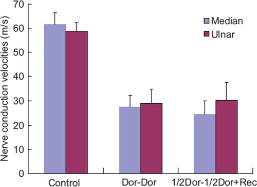
At 4 months post-operatively, electrophysiological assessment was conducted prior to the sacrifice of the animals. The motor nerve conduction velocity of the normal median and ulnar nerve was 61.6 ± 4.7 and 58.7 ± 3.6 m/s, respectively. In the groups of transected median and ulnar nerve neurorrhaphy (group 2 and 3), these values are 27.4 ± 4.9 and 29.1 ± 5.7 m/s separately, while the values are 24.6 ± 5.3 and 30.3 ± 7.2 m/s in the “1/2Dor-1/2Dor+Rec neurorrhaphy” group 4 ().
Figure 3. Nerve conduction velocities of rabbit regenerative median and ulnar nerve. Data were collected 4 months after reconstructive surgery. Electrophysiological recording was performed on the distal part of the median or ulnar nerve. There is no significant difference among the transected median/ulnar nerve neurorrhaphy groups (Dor-Dor, group 2, 3) and the group (1/2Dor-1/2Dor+Rec, group 4) in which proximal half median nerve served as donor nerve to reconstruct the distal median and ulnar nerve stump.
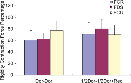
Electrical stimulation of the regenerated median or ulnar nerves resulted in muscle contraction of the forearm muscles innervated by the attached nerves (wrist/toe extension and wrist/toe flexion, respectively). Measurement of the tetanic muscle force in the flexor carpi radialis muscle, flexor digitorum superficialis, and the flexor carpi ulnaris, respectively, showed that the ipsilateral side had values of 61 ± 16% 63 ± 10% and 77 ± 17% of the contralateral side, after the distal ulnar and median nerves were repaired by their proximal stem. And the values were 71 ± 20% 80 ± 16% and 70 ± 16% in the “1/2Dor-1/2Dor+Rec neurorrhaphy” group 4 ().
Figure 4. The tetanic muscle force in the flexor carpi radialis muscle (FCR), flexor digitorum superficialis (FDS) and the flexor carpi ulnaris (FCU). It showed that the ipsilateral side had values of 61 ± 16% 63 ± 10% 77 ± 17% of the contralateral side, after the distal ulnar and median nerves were repaired by their own proximal stem (Dor-Dor, Group 2, 3). And the values were 71 ± 20% 80 ± 16% 70 ± 16% in the group where distal ulnar and 1/2 median nerves were reconstructed by the proximal half median nerve (1/2 Dor-1/2Dor+Rec, Group 4).
Discussion
In this research, we focused on the possibility of a partial donor nerve to reconstruct the injured site and the donor nerve simultaneously to reduce the loss of donor site function and the influence of the donor nerve axon number to the reconstruction effect of the injured nerve.
The biodegradable conduit used here was one patent of our lab authorized by the Chinese intellectual property office (No. ZL01136314.2). It was made from shell chitin with a partial de-acetyl procedure. Our previous work showed that that such a conduit could retain its structure for at least 6 weeks in vivo to support axon regeneration, and be absorbed completely within 3 months. It also showed good biologic compatibility with Schwann cells and neurons, and the degradation residue had no toxicity to the imbedding body. The suitable regeneration gap for the rat peripheral nerve was 1-2 mm, and it could not facilitate the nerve regeneration effects when the gap exceeded 5 mm [Citation23]. This product is now under a preclinical study. In this study, the gap between the proximal and distal segments was 1 mm, and the specimens were harvested 4 months after the operation. According to our previous results, with such a gap and the time period, the conduit could support axon regeneration from the donor side to the distal nerve stump with great advantage.
As has been described, it is possible to use one nerve to repair the injured nerve and donor site simultaneously (Dor to Dor+Rec neurorrhaphy). Here, we reduce the farther axon number to 1/2 of a donor nerve trunk. Half of the median nerve served as the father nerve to restore the distal half median and whole ulnar nerve at the same time (1/2Dor to1/2 Dor neurorrhaphy; group 2, 3). Thus, the ratio of proximal donor axon number to the distal acceptor nerve axon number is about 1:2.23 in group 4. This ratio was greater than the value in the previous experiment. However, both recovery of the donor site and injured nerve were again observed in the experiment. Such results showed the amazing compensative capacity of the peripheral. It was reasonable to suggest that within the ration limit for the acceptable reconstructive effect, the proximal donor axon number could be used as little as possible to get the same outcomes. That means separating a portion of a nearby intact nerve as a donor nerve to repair the injured nerve and donor site simultaneously can also get good reconstructive effects for the injured nerve and the donor site. It is obvious that there are many advantages in reducing the donor axon number. As described, donor site morbidity represents the great shortfall of nerve autografting and nerve transfer. Less using of the donor axons means less influence to the donor site function.
Based on the results of this experiment, surgeons could use a portion of a nearby donor nerve to repair the injured nerve and donor site simultaneously. It is very useful for severe nerve lesions (e.g. root avulsion of brachial plexus) characterized by the absence of a proximal nerve stump. We could choose the operation site as closely to the effectors as we can to reduce the reconstructive time. Therefore, both the donor site and the injured nerve effectors can be reinnervated quickly. The denervated effectors such as small muscles could be reinnervated before their degeneration to an unrecoverable level, and thus the reconstructive effects were enhanced. Although there seems to be an unavoidable injury to the nearby healthy nerve, if only a portion of a donor nerve trunk was used as the father nerve, the recovery of the donor site is within an acceptable level, and the injured fields are also restored, Partial Dor to Partial Dor+Rec neurorrhaphy may play a great role in such kind of nerve injuries.
Although the slices showed that the successful regeneration and electrical stimulation responded with contraction of the reinnervated muscles, it could only be a proof of structural reinnervation. The functional recovery needs not only the connection between the peripheral nerve and its effectors, but also the effective control from the central nerve system on the peripheral reinnervated effectors. It is obvious that when a donor nerve was used to repair the donor site and the injured nerve simultaneously, the original father nerve would control not only the effectors innervated by itself, but also control other effectors after reconstruction. Sometimes these effectors may have opposite functions like antagonistic muscles in the locomotor system. Therefore, brain reorganization related to major changes in the peripheral connections is a more important issue for the real function recovery of the reconstructed nerves. Are there any difficulties for a neuron to receive a message and send impulses through such a 1-axon-trunk several-collateral model? What are the brain plasticity mechanisms for adapting to environmental perturbations? More work needs to be done to reveal the nature of peripheral nerve reconstruction.
Declaration of interest: The authors report no conflicts of interest. The authors alone are responsible for the content and writing of the paper.
Related Research Data
REFERENCES
- Dahlin, L.B., Lundborg, G. (2001). Use of tubes in peripheral nerve repair. Neurosurg Clin N Am 12(2):341–52.
- Frostick, S.P., Yin, Q., Kemp, G.J. (1998). Schwann cells, neurotrophic factors, and peripheral nerve regeneration. Microsurgery 18(7):397–405.
- Lundborg, G., Dahlin, L., Danielsen, N., Zhao, Q. (1994). Trophism, tropism, and specificity in nerve regeneration. J Reconstr Microsurg 10(5):345–54.
- Seckel, B.R. (1990). Enhancement of peripheral nerve regeneration. Muscle Nerve 13(9):785–800.
- Chuang, D.C. (1995). Neurotization procedures for brachial plexus injuries. Hand Clin 11(4):633–45.
- Oberlin, C., Beal, D., Leechavengvongs, S., Salon, A., Dauge, M.C., Sarcy J.J. (1994). Nerve transfer to biceps muscle using a part of ulnar nerve for C5-C6 avulsion of the brachial plexus: Anatomical study and report of four cases. J Hand Surg Am 19(2):232–7.
- Sungpet, A., Suphachatwong, C., Kawinwonggowit, V., Patradul, A. (2000). Transfer of a single fascicle from the ulnar nerve to the biceps muscle after avulsions of upper roots of the brachial plexus. J Hand Surg Br 25(4):325–8.
- Tung, T.H., Mackinnon, S.E. (2001). Flexor digitorum superficialis nerve transfer to restore pronation: Two case reports and anatomic study. J Hand Surg Am 26(6):1065–72.
- Evans, G.R., Brandt, K., Widmer, M.S., Lu, L., Meszlenyib, R.K., Gupta, P.K., . (1999). In vivo evaluation of poly (L-lactic acid) porous conduits for peripheral nerve regeneration. Biomaterials 20(12):1109–15.
- Madihally, S.V., Matthew, H.W. (1999). Porous chitosan scaffolds for tissue engineering. Biomaterials 20(12): 1133–42.
- Matsumoto, K., Ohnishi, K., Kiyotani, T., Takashi, S., Ueda, H., Nakamura, T., . (2000). Peripheral nerve regeneration across an 80-mm gap bridged by a polyglycolic acid (PGA)-collagen tube filled with laminin-coated collagen fibers: A histological and electrophysiological evaluation of regenerated nerves. Brain Res 868(2):315–28.
- Vasconcelos, B.C., Gay-Escoda, C. (2000). Facial nerve repair with expanded polytetrafluoroethylene and collagen conduits: An experimental study in the rabbit. J Oral Maxillofac Surg 58(11):1257–62.
- Kostakoglu, N. (1999). Motor and sensory reinnervation in the hand after an end-to-side median to ulnar nerve coaptation in the forearm. Br J Plast Surg 52(5):404–7.
- Matsumoto, K., Ohnishi, K., Sekine, T. (2000). Use of a newly developed artificial nerve conduit to assist peripheral nerve regeneration across a long gap in dogs. Asaio J 46(4):415–20.
- McCallister, W.V., Tang, P., Trumble, T.E. (1999). Is end-to-side neurorrhaphy effective? A study of axonal sprouting stimulated from intact nerves. J Reconstr Microsurg 15(8):597–603; discussion 603–4.
- Okajima, S., Terzis, J.K. (2000). Ultrastructure of early axonal regeneration in an end-to-side neurorrhaphy model. J Reconstr Microsurg 16(4):313–23; discussion 323–6.
- Shah, M.H., Kasabian, A.K., Karp, N.S. (1997). Axonal regeneration through an autogenous nerve bypass: An experimental study in the rat. Ann Plast Surg 38(4):408–14; discussion 414–5.
- Viterbo, F., Trindade, J.C., Hoshino, K., Mazzoni, A. (1994). Two end-to-side neurorrhaphies and nerve graft with removal of the epineural sheath: Experimental study in rats. Br J Plast Surg 47(2):75–80.
- Yoleri, L., Songur, E., Yoleri, O. (2000). Reanimation of early facial paralysis with hypoglossal/facial end-to-side neurorrhaphy: A new approach. J Reconstr Microsurg 16(5):347–55; discussion 355–6.
- Zhao, J.Z., Chen, Z.W., Chen, T.Y. (1997). Nerve regeneration after terminolateral neurorrhaphy: Experimental study in rats. J Reconstr Microsurg 13(1):31–7.
- Scherman, P., Lundborg, G., Kanje, M., Dahlin, L.B. (2001). Neural regeneration along longitudinal polyglactin sutures across short and extended defects in the rat sciatic nerve. J Neurosurg 95(2):316–23.
- Jiang, B.G., Yin, X.F., Zhang, D.Y., Fu, Z.G., Zhang, H.B. (2007). Maximum number of collaterals developed by one axon during peripheral nerve regeneration and the influence of that number on reinnervation effects. Eur Neurol 58(1):12–20.
- Jiang, B.G., Zhang, P.X., Jiang, B.G. (2010). Advances in small gap sleeve bridging peripheral nerve injury. Artif Cells Blood Substit Immobil Biotechnol 38(1):1–4.