Abstract
Abstract: We investigated the neuroprotective effects of Rg2 in anoxic cultured hippocampal neurons of newborn rats. The cells were divided into a control group, nimodipine group (5 μmol/L), Rg2 (0.025 mmol/L), and Rg2 (0.05 mmol/L) group. The apoptosis rate of hippocampal neurons was measured by flow cytometry with staining of PI. The intracellular calcium ion [Ca2+]i was observed with fluorospectrophotometer determined by fluorescent probe Fluo-2/AM. The contents of MDA and NO and the activities of SOD in the supernatants of cells were determined by biochemical methods. The results demonstrated that Rg2 reduced the hypoxia-induced apoptosis, decreased the calcium overload in neurons, increased the activities of SOD, and decreased the contents of MDA and NO in the supernatants of cells. Our study suggests that Rg2 has a neuroprotective effect against hypoxia-induced neuronal damage in hippocampal neurons mediated by anti-apoptosis, blocking calcium over-influx into neuronal cells, eliminating the free radicals, and increasing the activities of anti-oxidative enzymes to inhibit the oxidative damages caused by anoxic.
INTRODUCTION
The body produced a large amount of oxygen free radicals after cerebral ischemia; meanwhile, the local concentration of NO increased. NO reacted with the surrounding oxygen free radicals, generated activated reactions such as nitric oxide, a strong oxidizer, which started lipid peroxidation. Free radicals medical research showed that the damage, apoptosis, and necrosis of neurons were closely related to lipid peroxidation. However, antioxidants could effectively block or prevent this process by scavenging free radicals, improving the activity of antioxidant enzymes [Citation1].
Apoptosis is the main form of neuronal cell death in cerebral ischemia [Citation2]. At present the exact mechanism of ischemic apoptosis is still unclear; some think that the increased intracellular [Ca2+]i is the start-up factors of cell apoptosis, and is the prerequisite to activate the degradation of DNA by endogenous nucleic acid enzymes [Citation3]. Research shows that calcium influx resulting in calcium overload, to trigger a series of biochemical changes and induce the related gene expression, may be contributing to the cell damage of the hippocampus [Citation4].
Ginseng is one of the most widely used herbal medicines and is reported to have a wide range of therapeutic and pharmacological applications. Ginsenosides, the major pharmacologically active ingredients of ginseng, appear to be responsible for most of the activities of ginseng. Ginsenosides have a four-ring, steroid-like structure with attached sugar moieties. About 30 different forms have been isolated and identified from the root of Panax ginseng. They are classified into protopanaxadiol or protopanaxatriol ginsenosides, according to the position of the sugar moieties at carbon-3 and -6. Ginsenosides regulate several types of ligand-gated ion channel activity. In cultured rat hippocampal neurons, ginsenoside Rb1 and Rg1 reduce glutamate-induced cell death. Ginsenosides and ginsenoside Rg3 also attenuate glutamate-induced neurodegenerations by inhibiting the overproduction of nitric oxide, formation of malondialdehyde, and influx of Ca2+ in rat cortical and hippocampal cultures. It has been well documented that ginsenoside Rg2 are important active principles of ginseng and have the effects of anti-apopotisis and blocking [Ca2+]i channel [Citation5].
In this study, we established an in vitro model of hypoxia damage in primary cultured hippocampal neurons to investigate the protective effects of Rg2 against hypoxia damage. Our results indicated that the neuroprotective effects of Rg2 against hypoxia-induced apoptosis in primary hippocampal neurons were associated with inhibition of [Ca2+]i influx.
MATERIALS AND METHODS
Materials
Newborn Wistar rats were provided by the Experimental Animal Center of ICAMA (Qingdao). Rg2 was provided by the Institute of Traditional Chinese Medicine and Materia Medica (Jilin). Nimodipine was provided by Bayer (Germany). Dulbeco's Modified Eagle Medium (DMEM) was from Gibco (USA). The other chemicals and regents of the highest grade available were from local commercial sources.
Cell Culture and Treatment
Rat hippocampal neurons were isolated as described previously [Citation6] with modifications. In brief, animals were euthanized by cervical dislocation under anesthesia, their brains were quickly removed, and the hippocampi were harvested on a cold stage. The procured hippocampal tissues were digested by 0.25% trypsin in Ca2+ and Mg2+ free Hank's balanced salt solution at 37 °C for 10 min, and the resulting cell suspension was passed through a filter and centrifuged for 5 min at 1000 rpm. The cells were re-suspended in DMEM supplemented with 10% FBS, and plated onto a poly-L-lysine-coated plate at a density of 1 or 2 × 10⁵ cells/cm2 to allow incubation in a humidified atmosphere of 95% air and 5% CO2 at 37 °C for 4 h. Then, the culture medium was replaced by Neural basal medium supplemented with 2% B27 0.5mM glutamine, 100U/ml penicillin, and 100U/ml streptomycin. The hippocampal neurons were incubated for 7–8 days to get the maturation hippocampal neurons. For the experiment, the hippocampal neurons were pretreated with nimodipine (5μmol/L), Rg2 (0.025mmol/L), and Rg2 (0.05mmol/L) for 4h, then were exposed to anoxic atmosphere (95% N2 + 5% CO2) [Citation7] for 4 h.
Assessment of Apoptosis
The cells were put into suspension, then were washed twice with ice-cold phosphate buffered saline (PBS), harvested, fixed with ice-cold PBS in 70% ethanol, and stored at -20 °C for 30 min. After fixation they were stained with propidium iodide, and we determined the content of DNA by Flow Cytometry (FCM).
DNA Fragment Detection
The ladder pattern of DNA fragment was detected by agarose gel electrophoresis. DNA from cultured cells was isolated following the instructions of the DNA ladder detection kit (BioDev-Tech., China). After staining with ethidium bromide (0.5 mg/ml), DNA bands were visualized by UV transillumination.
Intracellular Calcium Measurement
Before being loaded with fura-2, the cells were detached and put in suspension. Then loading was started by adding 15uL of fura-2/acetoxy-methylester (fura-2/AM) dissolved in DMSO to either the culture media or physiological medium (KRH). Final concentration of fura-2 in the incubation media was 5 μM, the incubation conditions were 37°C for 30min, then the concentration of Fura-2 was detected using fluorescence spectrophotometer with excitation wavelength of 500nm and emission wavelength of 340 and 380nm. Intracellular Ca2+ was calculated using the equation [Ca2+]i = Kd [(F– Fmin)/(Fmax– F)], where Kd = 400 nM [Citation8]. The maximal Fluo-2 fluorescence intensity (Fmax) was determined by adding 0.1% Triton X-100, and the minimal fluorescence (Fmin) was determined by quenching Fluo-2 fluorescence with the addition of 5 mM EGTA.F is the fluorescence measured without the addition of Triton-X-100 or EGTA.
Activity of SOD and the Content of MDA, NO
Measurements were performed as described before, following the commercially available kit (Nanjing Jiancheng Institute of Biological Engineering, China). The levels of MDA (malondialdehyde), a terminal product of lipid peroxidation, were measured as the indication of lipid peroxidation, using a commercially available kit (Nanjing Jiancheng Institute of Biological Engineering, China). The thiobarbituric acid reactive substrates were quantified using 1,1,3,3-tetraethoxypropane as the standard. The absorbance at 532nm was measured with colorimetry. Data were presented as the means ± SEM of each group. Each experiment was repeated three times.
Statistical Analysis
Data were presented as means ± SEM for three separate experiments (each in triplicate or duplicate). Comparisons were analyzed by one-way analysis of variance (ANOVA) and subsequent Bartlett's test. Differences were considered statistically significant at P < 0.05.
RESULTS
Rg2 Inhibited Hypoxia-induced Cell Damage of Hippocampal Neurons
Apoptosis rate were measured using propidium iodide (PI) FCM. The apoptosis rate of neurons in control group was significantly higher than the other groups; see .
Table 1. Induction of apoptosis.
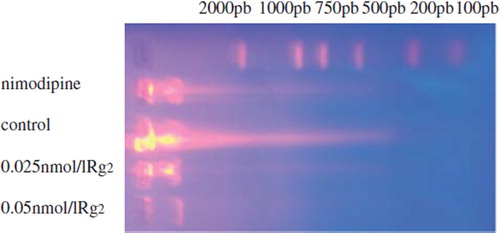
Rg2 Antagonized the DNA Fragment in the Hypoxia-induced Cell
We further detected DNA laddering in these cells. DNA extracted from the control group showed an apparent ladder pattern (), which indicated the formation of mono- and oligonucleosomes. No apparent laddering occurred in the other groups.
Figure 1. DNA fragmentin the hypoxia-induced cells.
Rg2 Inhibited Calcium Influx in the Hypoxia-induced Cells
The measurement of [Ca2+]i influx showed that hypoxia stimulation resulted in a prompt increase of [Ca2+]i influx in cultured hippocampal neurons, and treatment with Rg2 (0.025 or 0.05 mM) significantly lowered [Ca2+]i influx (F = 466.58, Q = 6.76∼48.82, P < 0.01), as shown in .
Table 2. The effects of Rg2 to [Ca2+]i(x ± s), n = 8).
Rg2 Increased the Activity of SOD and Reduced the Content of MDA and NO in Hypoxia-induced Cells
The measurement of the activity of SOD and the contention of MDA and NO showed that hypoxia stimulation resulted in a prompt increase of MDA and NO and a prompt decrease of SOD in cultured hippocampal neurons, and treatment with Rg2 (0.025or 0.05 mM) significantly antagonized those changes, as shown in .
Table 3. SOD and MDA, NO levels of groups (x ± s), n = 8).
DISCUSSION
This study showed the neuroprotective effects of Rg2 on hypoxia-induced hippocampal neurons and revealed the underlying mechanisms. We demonstrated that Rg2 could attenuate hypoxia induced neurotoxicity by anti-apoptosis and the effect of Rg2 was achieved by the regulation of calcium influx.
Cell apoptosis is the main form of neuronal cell death, whether in transient or permanent cerebral ischemia [Citation9,Citation10]. We found that treatment of hypoxia resulted in an increase of apoptosis rate in hippocampal cells, and could be reversed by pretreatment with Rg2. The results provided evidence for the anti-apoptosis role of Rg2 in hypoxia-treated hippocampal cells.
Under normal circumstances, the production of free radicals is within the physiological range, and could be removed by the antioxidant defense system to retain the balance of generation and elimination. On the conditions of acute hypoxia, the accumulation of free radical breaks the balance, and results in brain damage. The level of MDA can reflect the degree of injury; in the test, the content of MDA in control group was higher than the contents of MDA in the other groups, demonstrated that low-oxygen injured neurons, consistent with the literature [Citation11], but Rg2 could antagonize the injury. The activity of SOD can indirectly reflect the ability of scavenging oxygen free radicals. In experiments, SOD significantly decreased in the control group, indicating that hypoxia inhibited the activity of SOD, but Rg2 played a role in anti-oxidation injury. Recent research found that in a brain ischemic injury the over-expression of NOS can catalyze the production of NO, rapidly produce more peroxynitrite (ONOO22) with a superoxide anion radical (O22), and induce lipid peroxidation, which plays a key role in early injury [Citation12]. The results show that the content of NO in the Rg2 group was significantly lower than in the other groups, indicating that Rg2 could significantly protect against ischemic injury through reducing the level of NO.
The most valued theory of a hypoxia-injury-mechanism focuses on the neurotoxicity of excitatory amino acids and intracellular calcium overload. The presynaptic excitatory amino acids are largely released because of hypoxia, excessively activates the postsynaptic voltage-dependent NMDA receptors gating channel, which results in calcium over-influx into the cell. Calcium overload interferes with the mitochondrial oxidative phosphorylation process. The increase of cell plasma calcium ion concentration is considered the second messenger of apoptosis; it can activate some protease, endogenous nucleic acid enzyme, and phospholipase. The activation of these enzymes damages the nuclei structure, causing neuron damage. Therefore, intracellular calcium overload is considered the last pathway of hypoxia-injury in neurons [Citation13,Citation14]. Studies have shown that the possible mechanism of apoptosis was related with calcium overload; the increasing concentration of intracellular calcium was the initial factor of apoptosis [Citation15]. The results showed that the [Ca2+]i concentration of the control group was significantly higher than in the other three groups, indicating that low oxygen could destroy the balance of intracellular calcium, and result in significantly increasing of intracellular calcium. Rg2 could inhibit the calcium influx, reduce the concentration of intracellular calcium, and has neuroprotective effects against hypoxia-induced apoptosis in hippocampal neurons. These neuroprotective effects of ginsenosides on apoptosis might provide the theoretical and experimental basis for the clinical treatment of ischemic cerebrovascular disease.
Declaration of interest: The authors report no conflicts of interest. The authors alone are responsible for the content and writing of the paper.
REFERENCES
- Wang, P., Gu, Z.L., Zhou, W.X., . (2000). The protective effect of cardio-cerebral tong capsule on experimental cerebral ischemia. Proprietary Chinese Medicine. 22(9):636–639.
- Zeng, Y.S., Xu, Z.C. (2000). Co-existence of necrosis and apoptosis in rat hippocampus following transient forebrain ischemia. Neurosci Res. 37(2):113–125.
- Nitahara, J., Cheng, W., Liu, Y. (1998). Intracellular calium, DNase activity and myocyte apoptosis in aging Fischer 344 rats. Mol Cell Cardiol. 30(3):519–535.
- Cheng, C., Fass, D.M., Reynolds, I.J. (1999). Emergence of excitotoxicity in cultured forebrain neurons coincides with larger glutamate-stimulated [Ca2+]i increases and NMDA receptor mRNA levels. Brain Res. 849(1–2):97–108.
- Jiang, Y., Liu, W., Wang X.-M., . (1996). Calcium channel blockade and anti-free-radical actions of panaxatriol saponins in cultured myocardiocytes. Acta Phamacoloqica Sinica. 17(2):138–41.
- Brewer, G.J., Torricelli, J.R., Evege, E.K., Price, P.J. (1993). Optimized survival of hippocampal neurons in B27-supplemented Neurobasal, a new serum-free medium combination. Neurosci. Res. 35(5):567–576.
- Wang, F.Z., Ding, A.S., Liu, Z.W. (1995). Effect of ginsenosides against anoxic damage of hippocampal neurons in culture. Acta Pharmacol Sinica. 16(5):419–422.
- Okamoto, H., Inoue, K., Kamisaki, T., Takahashi, K., Sato, M. (1997). Regional differences in calcium sensitivity in the guinea-pig intestine. Pharm Pharmacol. 49(10): 981–984.
- Murakami, K., Nivzhe, H., Wang, T., . (1997). Advances in apoptosis research. Brain Res. 751:160–165.
- Nitatori, T., Sato, N., Waguri, S., . (1995). Delayed neuronal death in the CA1 pyramidal cell layer of the gerbil hippocampus following transient ischemia is apoptosis. Neuro Sci. 15:1001–1011.
- Wang, X.Y., Luo, J., Xing, H., . (1999). The toxicity of oxygen free radical and its mechanism in hypoxia-injury of neurons. Acta Biophysica Sinica. 15(2):375–380.
- Hara, H., Ayata, C. (1997). NG-nitroarginine binding after transient focal ischemia and NMDA-induced excitotoxicity in type I and type III metric oxide synthase null mice. J Cereb Blood Flow Metab. 17(5):515–526.
- Benveniste, H., Jorgensen, M.B., Diemer, N.H. (1988). Calcium accumulation by glutamate receptor activation is involved in hippocampal cell damage after ischemia. Acta Neural Scand. 78(6):529–536.
- Choi, D.W. (1988). Calcium-mediated neurotoxicity: relationship to specific channel types and role in ischemic damage. Trends Neural. 11(10):465–469.
- Nicotera, P., Zhivotovsky, B., Orrenius, S. (1994). Nuclear calcium transport and the role of calcium in apoptosis. Cell Calcium. 16(4):279–288.