Abstract
Objective: To evaluate the clinical use of a head-mounted display (HMD) for visualization in all neuroendoscopic procedures.
Materials and Methods: We retrospectively evaluated all endoscopic and endoscope-controlled procedures in which the HMD was used in our department between July 1999 and June 2002.
Results: A total of 269 endoscopic procedures were performed. In 147 cases intraventricular endoscopic procedures were carried out, mostly third ventriculocisternostomies, for which a fiberscope was used exclusively. Thirty intracranial cysts were fenestrated or removed (colloid cysts) with the help of various endoscopes. A total of 87 endoscopic transsphenoidal surgeries were performed with a lensscope. In only one case was it necessary to abandon use of the HMD due to inferior visualization; in all other cases visualization by the HMD was thought to be sufficient. Fatigue of the surgeon due to wearing the helmet did not occur. All surgeons had the impression that visual strain was decreased in comparison to looking at a monitor from a distance. The working position was considered to be more comfortable when wearing the HMD, and eye-hand coordination was improved. No technical problems occurred with the system.
Conclusions: The HMD is a new visualization tool in neurosurgery that may improve the ergonomics of neuroendoscopic and endoscope-controlled procedures.
Introduction
Over the past decade, neuroendoscopy and endoscope-assisted microsurgical procedures have gained in popularity, and indications for the use of an endoscope in neurosurgery are increasing Citation[1]. Endoscopic third ventriculocisternostomy has already become a standard procedure for treatment of obstructive hydrocephalus, and many intracranial cysts are now fenestrated endoscopically Citation[2–4]. Endoscopic pituitary surgery is also an evolving technique that promises good results Citation[5–7].
In macro- and microsurgery the head and eyes of the surgeon are directed towards the surgical field and the surgeon's hands. In endoscopy, however, the surgeon's head and eyes are directed towards a monitor, which in many cases requires turning the head away from the surgical field Citation[8], resulting in a divergence between the monitor viewing angle and the orientation of the surgeon's body and hands towards the patient. This has a direct impact on the eye-hand coordination of the surgeon, rendering manipulations less secure and less controlled, and is also the cause of some discomfort for the surgeon.
The use of a head-mounted display may eliminate these disadvantages by delivering the optical information directly to the surgeon's eyes independent of the head and body position and independent of the optical source (be it a microscope, endoscope or other video signals) Citation[8]. In this paper we present our clinical experience with the use of such a system for neuroendoscopy.
Material and methods
We retrospectively evaluated the use of a head-mounted display (HMD) in all neuroendoscopic and endoscope-controlled surgical procedures performed from July 1999 to June 2002, following the introduction of the HMD in our department.
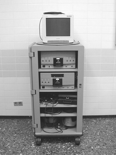
The HMD system used was the Stereosite™ Visualization & Information System (Vista Medical Technologies, Inc., Carlsbad, CA). This is a stereo microscope camera system combined with an HMD and digital information platform (). The Stereosite System displays stereoscopic (3D) images and any other video signal, such as a 2D endoscope signal, surgical videos, image guidance data or digitalized ultrasound signals. Surgical images, conventionally displayed on multiple monitors, may be presented in the HMD in picture-in-picture (PIP) format. By using a series of voice commands, the surgeon may select any combination of views in the PIP.
Figure 1. The Stereosite™ Visualization & Information System.
The Stereosite System's digital processor can support up to four HMDs, thus allowing the principal surgeon, as well as other members of the surgical team, to view the surgical site concurrently.
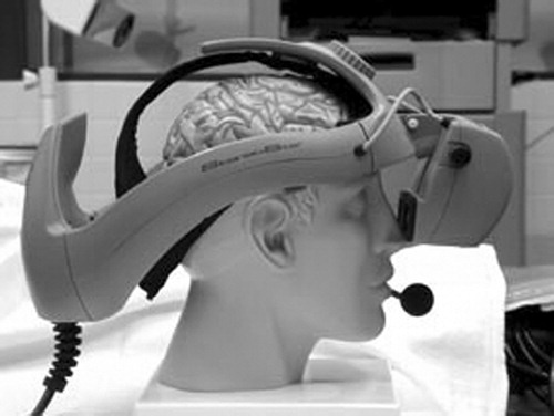
The HMD is a lightweight (950 g) personal surgical monitor that replaces conventional TV monitors and can be worn for hours without fatiguing the surgeon (). It has an optical resolution of 640×480 pixels with a 922K full-color VGA system. The HMD can also accommodate a wide variety of head sizes.
Figure 2. The head-mounted display with microphone.
In our endoscopic procedures only 2D video signals were used. However, in cases of videosurgery in which a switch from endoscopic to microscopic visualization may be anticipated in the event of complications, a 3D surgical camera system is mounted on the microscope and the microscope is sterile draped.
The HMD is placed on the head by the surgeon himself under sterile conditions. To achieve this, the surgeon puts on a second pair of sterile surgical gloves, places the HMD on his head, tightens the helmet, and positions both LCD screens of the helmet. The circulating nurse then pins the cable of the HMD to the back of the surgeon's coat to prevent pulling due to the cable's weight. The surgeon then removes the now-unsterile second pair of gloves.
Endoscopy was performed in the majority of neuroendoscopic procedures with the Channel™ neuroendoscope (Medtronic, Inc., Minneapolis, MN). This is an ultralight rigid fiberscope (30,000 pixel fibers) with an outer diameter of 4.22 mm and a working channel of 2.13 mm. The slightly inferior optical resolution of the fiberscope, as compared to that of lensscopes, is outweighed by its superior handling and maneuverability, since the camera is out of the operative field. In more complex neuroendoscopic procedures where optimal optical resolution and multiple working channels were required, lensscopes (MINOP system, Aesculap, Tuttlingen, Germany) were used. For endoscopic pituitary surgery, a lensscope with an auto-irrigation system (Richard Wolf GmbH, Knittlingen, Germany) was selected.
Results
In the period from 1 July 1999 to 1 June 2002, a total of 269 neuroendoscopic and endoscopy-controlled neurosurgical procedures were performed by four different neurosurgeons at the neurosurgical department of the University Medical Center St. Radboud. In 43% of cases the patients were children (aged 0–18 years).
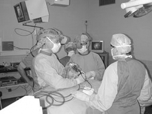
The majority of procedures were endoscopic third ventriculocisternostomies (ETVs) (133 procedures), all of which were performed with the Channel neuroendoscope (). The use of a fiberscope in conjunction with the HMD gives less optical resolution than with a lensscope and monitor. However, this is outweighed by the advantage of having lightweight equipment that is easy to handle and an ergonomic working position, which makes the guidance of the endoscope and surgical manipulations easier and faster. The usually simple and clear-cut anatomy of the ventricles means that optimum resolution is not necessarily required for this relatively straightforward procedure. The average operating time for an uncomplicated ETV was 20 min. In four cases of ETV, biopsy of third ventricular and/or aqueductal tumors was also performed, and in three cases a septum pellucidum fenestration was performed before the ETV was made. In addition, 6 endoscopic intraventricular tumor biopsies without ETV and 5 septostomies without ETV were performed. Endoscopic inspection of the ventricles was conducted in three cases.
Figure 3. The use of the HMD during endoscopic third ventriculocisternostomy.
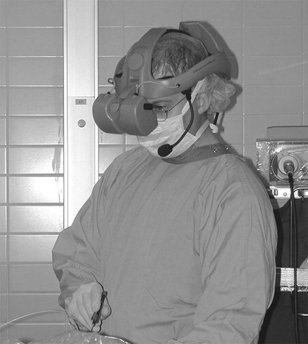
The second largest group of cases comprised 87 endonasal, transsphenoidal pituitary surgeries. In the majority of these cases the indication for surgery was pituitary adenoma (n=81). In addition, two Rathke's cleft cysts, one skull-base metastatic carcinoma, two craniopharyngiomas and a chordoma were operated in this way. Usually, a bi-nostril approach was used, in which the endoscope and an aspirator are introduced through the left nostril (for a right-handed surgeon) and surgical instruments (such as a high-speed drill and curettes) are inserted through the right nostril. The surgeon is positioned on the right of the patient (), and his orientation towards the operative field is oblique. Before introduction of the HMD, 22 cases of endonasal, transsphenoidal pituitary adenomectomies had been performed with the use of a monitor for visualization. The combination of the oblique position and the visual orientation relative to the monitor is not only uncomfortable and therefore fatiguing, but also hampers adequate hand-eye coordination. The use of the HMD significantly improved the surgeon's comfort and coordination, and contributed to the refinement of this specific endoscopic procedure.
Figure 4. The use of the HMD during endoscopic transnasal pituitary surgery.
A total of 30 cystic intracranial lesions were treated endoscopically or under endoscope control with the use of the HMD. There were 7 colloid cysts of the foramen of Monro, 8 arachnoid cysts of the cerebello-pontine angle, 9 intraventricular cysts, and 6 frontotemporal arachnoid cysts. All 7 colloid cysts were removed completely, and all other cysts were successfully fenestrated.
In four cases the endoscope with HMD was used to inspect cysts and ventricles prior to a microsurgical procedure being performed. In one patient a cystic recurrent low-grade glioma of the mesencephalon was biopsied endoscopically through the fourth ventricle and marsupialized. In this last case, the PIP function of the HMD was used to guide the endoscope through the craniotomy and between the cerebellar tonsils into the fourth ventricle while preserving the endoscopic view. From that point on, guidance was by endoscopic view only.
In only one of the above-mentioned surgical procedures did the use of the HMD have to be abandoned and a TV monitor used instead for visualization because of poor discrimination of the anatomy. After switching to the monitor it was discovered that this problem was caused by a loss of resolution in the fiberscope, which was compensated for by the better resolution of the monitor. In all other cases the optical resolution of the HMD was sufficient to allow successful completion of the surgery. Nevertheless, the optical resolution of the HMD was still considered to be sub-optimal by all participating surgeons. This was, however, fully outweighed by the improved ergonomics of the endoscopic procedure performed with the HMD.
No technical problems occurred with the HMD during any of the procedures and no equipment failures or complications attributable to the use of this display system were encountered. Although the surgeons had anticipated fatiguing of the head and neck in longer procedures due to the weight of the HMD, this did not occur: The HMD could be worn for up to 3 hours without any such problems. Indeed, surgeons usually tended to forget they were wearing an HMD. Neither was any visual strain or ocular fatigue observed. On the contrary, visual strain seemed to be decreased with the HMD; focusing on a monitor 2–3 meters away, with a small field of view and disturbances in the line of sight, was perceived as more fatiguing.
Neuroendoscopy is usually a single-surgeon procedure in which the surgeon takes care of visualization and instrumentation. An assistant is not required in the majority of cases. In the few cases where a third hand was needed to guide an instrument, usually during the beginning of our learning curve with endonasal transsphenoidal procedures, the assistant was confronted with a severe drawback of this generation of HMD: All the other HMD wearers have exactly the same view as the surgeon, even though the assistant's position relative to the patient and surgical field differs from that of the surgeon. This may make adequate eye-hand coordination almost impossible for the assistant, and may even be dangerous in some situations. In contrast to the surgical microscope, the assistant cannot turn and adapt his view through the HMD to match his position.
Discussion
Surgery is a profession that requires specific skills, dexterity and good eye-hand coordination. In microsurgery and endoscopic surgery the eye-hand coordination is uncoupled by the use of an external optical device. The surgeon compensates for this by relying more on vestibular and deep sensory feedback, a skill that is acquired by years of training. If the optical device displaying the operative field, such as a monitor, requires turning the head away from the operative field, there is a further uncoupling of the vestibular input, rendering eye-hand coordination even more difficult. One solution could be to position the monitor in such a way that the surgeon has an unobstructed view with a neutral head position at all times, so that the surgeon's head remains oriented towards his hands. However, the set-up of an OR, with its many instruments and devices, anesthesia equipment and circulating personnel, may not allow ideal positioning of a monitor. Also, complex procedures require shifting the visual field to the right or left, or even superiorly or inferiorly, during a procedure. This disruption in the hand-eye axis relationship is detrimental to efficient and safe operative progress. Furthermore, an unplanned shift in the direction of the operative field often requires the circulating nurse to move the video monitor from one sector of the OR to another to reorient the geometric relationship. In any case, the monitor only allows a relatively small field of view, with a lot of distraction for the surgeon surrounding the monitor.
The use of an HMD for visualization purposes was pioneered by Levy, Chen and Moffitt Citation[9]. The HMD provides an undisturbed view of the operative field at all times, regardless of the position of the surgeon. The surgeon may change his position relative to the operative field while maintaining optimal visual control, as well as remaining focused towards his hands, thus improving his situational awareness. Efficiency and versatility are thereby increased. The much wider field of view of the HMD enables the surgeon to be immersed in the surgery with less distraction. The field of view of the HMD is 35° wide, whereas a typically positioned monitor allows a field of view of only 8–12°. Circulating people in the OR cannot interrupt the line of sight of the surgeon, and the HMD also allows adequate peripheral vision, allowing the surgeon to maintain his/her orientation within the OR and simultaneously view the operative field and surrounding environment Citation[10].
The HMD also allows a neutral positioning of the head regardless of the position of the surgeon relative to the operative field. The surgeon is at all times oriented towards his hands, thus improving eye-hand coordination. Visual strain and ocular fatigue is decreased, as observed in our present study and also by other investigators Citation[10].
In a laparoscopy simulation model, the HMD did not enhance the task performance of the surgeons as compared to a regular 2D monitor, and thus was deemed to provide no advantage Citation[11]. However, in our opinion, the ergonomic advantages of the HMD are not yet appreciated. The neutral position of the head allows a straight and more relaxed posture, so that fatigue due to turning the eyes, head or body towards a monitor is decreased.
So far, the HMD has most commonly been used for entertainment applications, but it may also be employed for remote control and operations, maintenance, engineering and scientific simulations. The visual effects of the HMD on the user have been studied extensively and concerns regarding the ill effects of HMD use have been postulated Citation[12]. These effects may include simulator sickness resulting from vestibular-visual conflicts, accommodative difficulty, and binocular function difficulties with increased eye-strain, as well as postural instability Citation[12],Citation[13]. However, these applications usually involve a form of virtual reality for which the human visual system is not adapted. In surgery, the HMD shows the real world to which the visual system is already accustomed, and the effects of the HMD should therefore be no different from those of a normal desktop computer display.
The multimodality function of the HMD with PIP video images allows ancillary information to be made available to the surgeon by simple voice-control, without changing the working position or turning the head towards an additional monitor, thereby enabling a more ergonomic and safer procedure Citation[1]. In particular, guidance of the endoscope by neuronavigation benefits from this PIP mode of the HMD, since the view through the endoscope and the position of the endoscope in the 3D MRI or CT image are displayed simultaneously.
Levy et al. have gained considerable experience in the use of the HMD in neuroendoscopy and microsurgery Citation[14]. In microsurgery, however, they envisioned the primary role of the HMD as providing observers with a stereoscopic view, while the principal surgeon operated through the microscope Citation[8]. In our opinion, minimally invasive neurosurgery has virtually no role for a surgical assistant, so the HMD is more of a stereoscopic luxury for the observers.
Ergonomic considerations have entered and influenced many professions. Working hours are limited rigorously for truck drivers and pilots; limitations regarding weight lifting, bending and working position increasingly influence the workload in factories; more and more attention is paid to the sitting position of office workers, and regular pauses for working on computers is advised or even regulated. An increasing awareness of occupation-related illness or disability and RSI (repetitive strain injury), as well as of mistakes and loss of concentration resulting from fatigue, has already influenced the daily work of many professions. Surgery is not yet one of these professions, but it seems unlikely that a (neuro-) surgeon is not subject to fatigue and bodily strains while practicing surgery.
Although no reliable data exist regarding the influence of fatigue and strain on the quality of the surgeon's work and the rate of complications, self-criticism and reflection on the quality of one's surgical performance will reveal that this influence is very real. Therefore, tools that help decrease strain and fatigue may improve the quality of surgery and reduce the number of intraoperative complications. Surgeons' chairs that provide elbow and/or hand support, or head rings that enable hand support, are examples of such tools. An HMD may also be one of these tools that increases the ergonomics of the surgical procedure and thus may improve its safety Citation[8–10].
Despite the several advantages of the HMD, the associated disadvantages still limit its use in neurosurgery to neuroendoscopy. The optical resolution is still sub-optimal and less than that of the lensscope or microscope, although newer versions with a resolution of 800×600 pixels are appearing on the market. The weight of approximately 1 kg on the head should be decreased further to permit longer procedures to be performed on a routine basis. Another disadvantage is that all wearers of the HMD always see the same video image as the surgeon, regardless of their position relative to the operative field. The Stereosite System does not permit image rotation to compensate for differences in orientation and angles of access. This may hamper the ability of a surgical assistant to adequately assist in surgery, since optical and proprioceptive input are uncoupled. However, neuroendoscopy usually does not require an extra hand, so we have not yet been confronted with this problem. In microsurgery, however, it may be of some relevance to the surgical technique, and it may therefore be expected that newer versions of the Stereosite System will allow image correction. In neuroendoscopy, with its short-duration procedures and clearly delineated surgical anatomy, we found that the advantages of improved ergonomics outweigh these disadvantages.
Conclusion
The HMD is a new visualization tool in neurosurgery that may improve the ergonomics of neuroendoscopic and endoscope-controlled procedures. It is anticipated that further improvement of the optical resolution of the HMD will allow a role for the HMD in microneurosurgery as well.
Disclosure
The authors have no financial or other interest in the instruments and products described in this paper.
References
- Perneczky A, Fries G. Endoscope-assisted brain surgery: Part I: Evolution, basic concept, and current technique. Neurosurgery 1998; 42: 219–25
- Hopf N J, Perneczky A. Endoscopic neurosurgery and endoscope-assisted microneurosurgery for the treatment of intracranial cysts. Neurosurgery 1998; 43: 1330–6
- Hopf N J, Grunert P, Fries G, Resch K D, Perneczky A. Endoscopic third ventriculostomy: outcome analysis of 100 consecutive procedures. Neurosurgery 1999; 44: 795–804
- Vandertop W P, Verdaasdonk R M, van Swol C F.P. Laser-assisted neuroendoscopy using a neodymium-yttrium aluminum garnet or diode contact laser with pretreated fiber tips. J Neurosurg 1998; 88: 82–92
- Cappabianca P, Alfieri A, de Devitiis E. Endoscopic endonasal transsphenoidal approach to the sella: towards functional endoscopic pituitary surgery (FEPS). Minim Invas Neurosurg 1998; 41: 66–73
- Jho H-D, Carrau R L. Endoscopic endonasal transsphenoidal surgery: experience with 50 patients. J Neurosurg 1997; 87: 44–51
- Pillay P K, Leo C W, Sethi D S. Computer-aided/image-guided and video-endoscopic resection pituitary tumors. Stereotact Funct Neurosurg 2000; 74: 203–9
- Levy M L, Chen J C.T, Moffitt K, Corber Z, McComb J G. Stereoscopic head-mounted display incorporated into microsurgical procedures: technical note. Neurosurgery 1998; 43: 392–6
- Chen J C.T, Moffitt K, Levy M L. Head-mounted display system for microneurosurgery. Stereotact Funct Neurosurg 1997; 68: 25–32
- Geis W P. Head-mounted video monitor for global visual access in mini-invasive surgery. Surg Endosc 1996; 10: 768–70
- Herron D M, Lantis J C, II, Maykel J, Basu C, Schwaitzberg S D. The 3-D monitor and head-mounted display. A quantitative evaluation of advanced laparoscopic viewing technologies. Surg Endosc 1999; 13: 751–5
- Peli E. The visual effects of head-mounted display (HMD) are not distinguishable from those of desk-top computer display. Visual Research 1998; 38: 2053–66
- Nichols S. Physical ergonomics of virtual environment use. Applied Ergonomics 1999; 30: 79–90
- Levy M L, Day J D, Albuquerque F, Schumaker G, Giannotta S L, McComb J G. Heads up intraoperative endoscopic imaging: evaluation of techniques and limitations. Neurosurgery 1997; 40: 526–31