Abstract
We developed a new orthognathic surgical simulation system able to predict both occlusal correction and mandibular repositioning in three dimensions. This system uniquely integrates the real motion of the dental cast model with the virtual motion of the reconstructed cranio-facial model. The skeletal change of the mandibular osteotomy is simulated on the PC monitor while the occlusal change is confirmed by checking the cast model on the simulator. The simulation process is easily repeated and the operator can make several attempts to determine the final mandibular position. The occlusal relationship at the simulated mandibular posture is registered and the occlusal wafer splint, which ensures intermaxillary fixation, is fabricated on the simulator. This surgical simulation system appears to satisfy clinical demands well and is an important facilitator of communication between orthodontists and surgeons. Here, we outline the system and apply it to a demonstration case of orthognathic surgery.
Introduction
In orthognathic surgery, jaw osteotomy is used to correct jaw deformities and establish a normal occlusal relationship. To predict the outcome of jaw osteotomy, current orthodontic practice relies on simulations using lateral cephalograms, with the tracing overlay method being the simplest way to simulate mandibular osteotomy Citation[1]. This method attempts to visualize the skeletal and occlusal correction by superimposing a lateral cephalometric tracing of the jaw contour over the original image. Although convenient in clinical settings, it is not sufficiently sophisticated for three-dimensional (3D) prediction, especially in patients with facial asymmetry.
Clinically apparent facial asymmetry is reported in one third of patients with dentofacial deformity Citation[2], and it is common for patients with facial asymmetry and malocclusion to have mandibular lateral displacement and a subsequent 3D deformity Citation[3–5]. Characteristic dental asymmetries are also found in patients with mandibular lateral displacement Citation[6], Citation[7] and seem to be 3D dental compensations for skeletal discrepancies. To address the problem of asymmetry, therefore, the prediction of the jaw osteotomy should ideally be done in three dimensions.
A period of orthodontic treatment is necessary both before and after surgery to bring the teeth to their final position Citation[8]. Jaw osteotomy has two goals: morphological improvement of the jaw deformity and correction of the inter-occlusal relationship. After finishing the pre-surgical orthodontics, jaw osteotomy is then generally used to reposition the mandible, maxilla, or both so that the occlusal relationship between the upper and lower teeth is acceptably normal and stable. That is, the jaw is repositioned with reference to the occlusal correction. However, our clinical experience has led us to ask whether the two goals of skeletal and occlusal correction are compatible. In some asymmetry cases treated by jaw osteotomy, for example, despite correction of the occlusal asymmetry, such as denture midline discrepancy and right-left difference in the molar relationship, the mandibular asymmetry is not sufficiently improved. In other words, does occlusal correction correspond to skeletal correction? In our opinion, occlusal correction does not conflict with skeletal improvement. If pre-surgical orthodontics is carried out optimally to eliminate all dental compensations Citation[9], the occlusal correction will bring the jaw into the correct planned position. Any discrepancy indicates that 3D dental compensations were not adequately eliminated in the pre-surgical orthodontics, usually as a result of obscure treatment objectives and difficulties in controlling the teeth under daily occlusal contact. At the outset of the pre-surgical orthodontics, it seems to be important to diagnose the occlusal deviations and skeletal deformities in three dimensions.
Other models developed for diagnosis and prediction of orthognathic surgery have their own disadvantages. Articulator-mounted dental cast models Citation[1], for example, do not show overall jaw positioning, underscoring the need for simulations able to predict both jaw positioning and occlusal change. More recently, 3D solid models reconstructed from computed tomography (CT) have been used Citation[10–12], but while they precisely simulate the surgical procedure and easily demonstrate the eventual jaw position after correction of the occlusal relationship, their high cost and time requirements render them unsuitable for clinical use.
We have developed a new 3D simulation system, the mandibular motion tracking system (ManMoS), which predicts both occlusal correction and mandibular repositioning in orthognathic surgery Citation[13–15]. This system uses a computer-generated wire-frame model of the cranio-facial complex and representative points on the dental arches to integrate skeletal, dental and motion data. The computer-generated image can be arbitrarily moved and repositioned on the monitor, corresponding to the spatial orientation of the lower dental cast model mounted on a surgical simulator in real time. Here, we introduce the ManMoS surgical simulation system and discuss the significance of its prediction of jaw repositioning in three dimensions following mandibular osteotomy.
Materials and methods
ManMoS was originally developed using the programming language Visual Basic 6.0 (Microsoft Co.). The purpose of ManMoS in simulating mandibular osteotomy is as follows:
To represent the cranio-facial morphology and dental arches in virtual space on the PC monitor.
To visualize both the skeletal and occlusal change by arbitrarily translating and rotating the mandibular segment in the frontal, horizontal and sagittal planes.
To reflect the actual positioning of the lower cast model mounted on the simulator in the virtual positioning of the mandible represented on the monitor in real time.
Skeletal data of the cranio-facial complex.
Cast data of the representative points on both the upper and lower dental arches.
Motion tracking data of the lower dental cast.
Coordinates of the reference points to integrate the skeletal, cast and motion data.
Placement of reference points
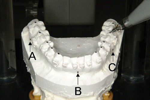
Prior to data acquisition, the reference points are established on the lower dental arch. As shown in , three metal spheres of diameter 1.0 mm are glued on the attached gingiva with an adhesive agent, approximately locating to the bilateral molar regions and incisal region. Lateral and postero-anterior (PA) cephalograms are then taken, followed by impressions of the dental arches. Following these recording procedures, the three reference spheres are represented on both the cephalometric images and the lower cast model, and their 3D coordinates are referenced to integrate the dental and skeletal morphology in a virtual space.
Cephalometric 3D modeling system of the cranio-facial complex
A 3D modeling system of the cranio-facial complex using conventional two-dimensional (2D) cephalograms has been developed Citation[16]. PA and lateral cephalograms are recorded using a cephalometric X-ray machine with two orthogonal X-ray sources to generate biplane projections, with the head of the patient fixed in the upright position by ear-rods during recording. The cephalometric films are scanned at a resolution of 100 dpi using an image scanner (ES-2200, Epson Co.) and the images are stored on a PC. To obtain 3D coordinates of the cranio-facial anatomical landmarks in a virtual space, the PA and lateral images are placed within a coordinate system, reproducing the geometrical arrangement relative to the X-ray beams and the patient's head under actual recording conditions (). A 3D wire mesh model with the standard morphology of the cranio-facial complex is placed between the ear-rods in the virtual space and projected onto both the lateral and PA images. The projected wire frame is then deformed to fit the skeletal contour on the lateral and PA cephalograms. Finally, an individualized 3D wire-frame model is reconstructed.
Figure 2. Coordinate system in 3D cephalometric modeling. The reference spheres (A, B and C) are recognized on the lateral and PA cephalometric images and their 3D coordinates are computed. [Color version available online.]
![Figure 2. Coordinate system in 3D cephalometric modeling. The reference spheres (A, B and C) are recognized on the lateral and PA cephalometric images and their 3D coordinates are computed. [Color version available online.]](/cms/asset/28106c24-2115-48a8-96bc-fb267d315c24/icsu_a_225300_f0002_b.gif)
Following these fitting procedures, the coordinates of the three reference spheres are measured. The coordinates of the bilateral porion and orbita are also measured to establish a global coordinate system for surgical simulation. The measurement error of the modeling system appears to be sufficiently small for clinical use Citation[16].
Three reference spheres, designated A, B and C to indicate the reference points placed at the right molar, incisal and left molar regions, respectively, are recognized on the lateral and PA cephalometric images and their 3D coordinates are computed.
Three-dimensional motion tracking
The ManMoS system uses the FASTRAK 3D motion tracking and digitizing equipment (Polhemus, Colchester, VT) (), consisting of a system electronics unit (SEU), a transmitter producing a magnetic field, and a receiver to detect its own position. During recording, the SEU sends the dataset of three coordinates (X, Y and Z) and three Euler angles (azimuth, elevation and roll) with a time resolution of 8.3 msec (120 Hz). Two types of receiver were prepared for this simulation. A stylus-type receiver is used to digitize the landmarks on the dental cast models, while to track cast motion during surgical simulation a box-type receiver is fixed on the lower dental cast, which is mounted on the surgical simulator. The accuracy and precision of the tracking device appear to be sufficient for clinical use Citation[15].
Computer-aided diagnostic system of dental casts
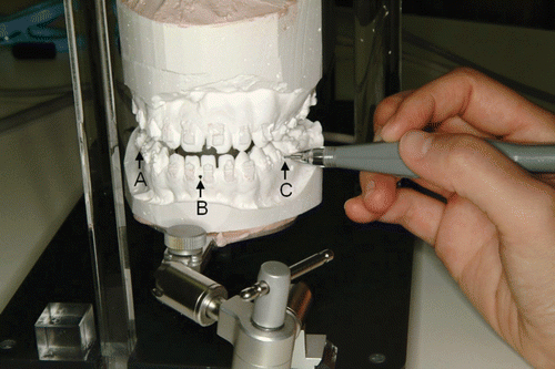
Facebow-transfer (a record of how the upper dental arch relates to the temporomandibular joint (TMJ)) is performed and the dental casts are mounted on an articulator, representing the location of the dental arches in relation to the cranium. Centric stops of the upper dental arch and their corresponding points on the lower arch are digitized by means of the stylus-type receiver (). A computer-aided diagnostic system of dental casts Citation[6], Citation[17] is used to analyze the dental arches in three dimensions (). The three reference spheres impressed on the lower dental arch are also digitized ().
Integration of skeletal and cast data
In order to process the data, we use three rectangular (Cartesian) coordinate systems; a global and two locals (). The original point (O) of the global system (G) is the midpoint between the ear-rods, and the X-axis directs from the origin to the left ear-rod. The Y-axis is chosen so that the XY-plane includes the left orbita and thus coincides with the Frankfort Horizontal (FH) plane. The local systems (L-1 and L-2) are set up by the transmitter and the box-type receiver. Their original points (O1 and O2) are located in the center of each device. As previously described, FASTRAK could measure the 3D coordinates of O2 and Euler's angles from L-2 to L-1 using the box-type receiver, and the 3D coordinates of arbitrary points in L-1 using the pen-type receiver.
Now, let us write the coordinates of the three reference points as column vectors a, b and c. Suppose a rotation matrix from L-1 to G is given by [R1], we getwhere the indices 0, 1 and 2 represent measures in G, L-1 and L-2, respectively. Let us make a matrix with the elements of a, b and c lined up in three columns, and write this as [a, b, c]. Then, the equations above are simply written as
As a, b and c are independent of one another, the matrix [a, b, c]1 has an inverse. Thus,
The coordinates of the centric stops (Cst)1, which are measured in L-1 using the pen-type receiver, are integrated into the skeletal data through the following transformation:
Inversely, any points in G, such as nodes of the mandibular frame (Nm), could be transferred to L-1 through
shows the obtained model of the cranio-facial skeleton with the centric stops of the dental arches. Frontal, horizontal and sagittal views are presented.
Pilot surgical prediction
In ManMoS, surgical prediction is performed at the initial case investigation and just prior to jaw surgery. At the initial recording, the 3D model is used to diagnose the skeletal deformity and its relationship to the dental deviation in three dimensions. The mandibular body with the lower centric stops can be moved in translation and rotation by handling the cursor arbitrarily on each of the frontal, horizontal and sagittal views (). The prediction of jaw repositioning is determined by confirming the mandibular position in relation to the midface and the occlusal relationship. The pilot surgical prediction is helpful in planning the orthodontic treatment during the pre-surgical period.
Real-time surgical simulation
The mandibular osteotomy is simulated in diagnosis before surgery. In ManMoS, repositioning of the mandible with the centric stops of the lower dental arch can be predicted through real-time simulation. shows the universal joint simulator as originally fabricated. Most of the body is composed of a plastic material which does not affect the magnetic field during FASTRAK recording, and the shape was modeled after an articulator (). The upper dental cast is mounted on the superior part and the lower dental cast is fixed on the inferior part through the universal joint. The box-type receiver is attached to the lower cast such that the motion of the lower cast can be detected as the motion of the receiver relative to the transmitter placed at the rear (). The procedure for real-time simulation is as follows:
Figure 9. Universal joint simulator. (a) Side view: 1 = universal joint, 2 = transmitter. (b) Superior view of the base section. A box-type receiver is placed on the lower cast model. [Color version available online.]
![Figure 9. Universal joint simulator. (a) Side view: 1 = universal joint, 2 = transmitter. (b) Superior view of the base section. A box-type receiver is placed on the lower cast model. [Color version available online.]](/cms/asset/af7fd3a3-222e-4dd2-b5c3-a25eeec4b1b0/icsu_a_225300_f0009_b.gif)
When the lower cast occludes to the upper cast at the centric occlusion, the universal joint is locked. Prior to the real-time simulation, the three reference points impressed on the lower cast are digitized using the stylus receiver ().
shows the real-time simulation. During the simulation, the serial dataset of the box receiver is sent to the PC. The motion data of the box receiver on the lower cast is transformed into mandibular motion data and finally demonstrated in the cephalometric global coordinates ().
The procedures to display motion of the mandible are as follows. By using the box-type receiver, we can obtain the coordinates of O2 and a rotation matrix from L-2 to L-1. Let us write these quantities as O2(t) and [R2(t)], respectively, where the suffix (t) means these are functions of time. Before jaw movement (at time t = 0), the mandibular frames in L-1 are expressed by Equation 3. The position of the mandible at time t is given by:Transferring the above equation to G through Equation 2, we finally obtain
and the motion of the mandibular frame is displayed in G.
The precision of ManMoS was assessed as follows. On a mandibular model, the center coordinates of the condyle were measured on both the right and left side. These condylar points were located approximately 90.0 mm away from the box-type receiver. When the lower cast model occluded to the upper one at the centric occlusion with a silicon biting material, motion data was acquired over 10 seconds (120 Hz). The x-, y- and z-coordinates of the condylar points in G were computed through the abovementioned transformation formulae. Static noise was calculated as the standard deviation of the 1,200 data samples (). Next, the lower cast was positioned at the centric occlusion 40 times with the orientation of the box receiver being recorded every time. The coordinates of the condylar points in G were then calculated and their standard deviation was formulated to evaluate the reproducibility ().
Table I. Static noise at ICP.
Table II. Reproducibility.
By releasing the universal joint, the lower cast can be arbitrarily positioned. When the operator moves the lower cast by confirming the occlusal relationship, the mandibular model with the lower centric stops is simultaneously moved on the PC monitor in correspondence with the cast motion. The desired mandibular position is determined by trial-and-error repetition in real time, with consideration being given to both the skeletal and the occlusal correction. For the skeletal correction, the chin should be positioned properly and symmetrically in relation to the cranium. The position of the ramus is important in ManMoS. In particular, medio-lateral displacement relating to the mandibular fossa and supero-inferior displacement should be checked. With regard to occlusion, the change in the occlusal relationship accompanying the mandibular positioning can be evaluated from the relationship between the upper and lower centric stops on the monitor, but the evaluation of representative points alone is apparently insufficient for deciding on the final mandibular position. Rather, the occlusal relationship of the real cast model provides more information for determining the final mandibular position, and consequently facilitates planning of the post-surgical orthodontic treatment. Thus, the ManMoS system provides for the simulation of skeletal prediction on the PC monitor on the one hand, and confirmation of the occlusal prediction using the cast model on the simulator on the other.
After deciding on the final mandibular posture, the occlusal relationship is registered using bite registration material () and an occlusal wafer splint to ensure that the intermaxillary fixation is correctly fabricated ().
Results
Precision
It has been reported that the accuracy and precision of the tracking device itself was sufficient for clinical use Citation[15]. When the box receiver with the lower cast model was motionless at centric occlusion, the stability of the condylar points in the global coordinate system (G) were assessed as the static noise. shows that the standard deviations of Cond_X, Cond_Y and Cond_Z in 1,200 data samples were extremely small. To examine the reproducibility of this system, the condylar points in G were also examined when the lower cast moved back to occlude at the centric occlusion 40 times. As shown in , the standard deviations of Cond_X, Cond_Y and Cond_Z in the 40 recordings were less than 0.1 mm. From these results, the precision of ManMoS seems to be sufficient for clinical use.
Clinical application
ManMoS has been applied in more than 50 cases of jaw deformity. An orthognathic surgical case showing mandibular prognathism was exemplified for presentation. and show the findings of the pre-surgical recordings. As shown in the intra-oral photographs (), anterior crossbite was found and the lower denture midline was displaced to the upper midline 2 mm to the left. No remarkable asymmetry was found in the facial photograph or PA cephalograms (). Sagittal splitting ramus osteotomy (SSRO) Citation[18] was planned and real-time simulation performed using ManMoS.
Figure 13. Soft tissue and skeletal findings in pre-surgical recording. [Color version available online.]
![Figure 13. Soft tissue and skeletal findings in pre-surgical recording. [Color version available online.]](/cms/asset/fe28f73a-64a3-41cb-b887-cfd1ff03be06/icsu_a_225300_f0013_b.gif)
Figure 14. Occlusal findings. (a) Pre-surgical recording. (b) Final mandibular positioning in ManMoS prediction. (c) Fabrication of the occlusal splint (vertical elastic was applied to extrude the upper molar on the right side during the intermaxillary fixation period). (d) Two weeks after surgery (at the end of the intermaxillary fixation). (e) Eight months after surgery (at the end of the post-surgical orthodontics). [Color version available online.]
![Figure 14. Occlusal findings. (a) Pre-surgical recording. (b) Final mandibular positioning in ManMoS prediction. (c) Fabrication of the occlusal splint (vertical elastic was applied to extrude the upper molar on the right side during the intermaxillary fixation period). (d) Two weeks after surgery (at the end of the intermaxillary fixation). (e) Eight months after surgery (at the end of the post-surgical orthodontics). [Color version available online.]](/cms/asset/35cdbc2a-b609-40ff-a0be-1d3e6a02b0c0/icsu_a_225300_f0014_b.gif)
shows the wire-frame model demonstrated on the monitor in the real-time simulation, with the lower cast set back to occlude in a normal relationship to the upper cast. Although the mandible seems to be placed properly in the sagittal view, the frontal and horizontal views show that the ramus was placed asymmetrically. When compared to the original mandibular model, the right ramus was displaced medially and the left ramus laterally. With regard to SSRO, medio-lateral displacement of the ramus demonstrated in ManMoS indicates displacement of the posterior portion of the distal tooth-bearing segment. In this situation, an osteotomy gap between the distal segment and condylar proximal segment would occur, in this case causing a flaring out of the condylar proximal segment on the left side.
Figure 15. ManMoS simulation with priority given to the occlusal correction. [Color version available online.]
![Figure 15. ManMoS simulation with priority given to the occlusal correction. [Color version available online.]](/cms/asset/7a9cdcd2-25ed-4253-84a5-48674596db71/icsu_a_225300_f0015_b.gif)
In ManMoS prediction, priority is placed on the skeletal rather than the occlusal correction. shows the final mandibular position determined by ManMoS prediction, with priority accordingly given to the skeletal correction. In the frontal view, superimposition of the predicted mandible on the original mandible indicated that the bilateral rami were directed to the mandibular fossae symmetrically without medio-lateral deviation. In the sagittal view, in contrast, occlusal clearance between the upper and lower centric stops is excessive on the right side. This finding was confirmed in the occlusal relationship of the cast model (), and it was reconfirmed in the frontal view that occlusal clearance on the right molar related to the elevation of the upper frontal occlusal plane toward the right. Following the SSRO, the occlusal splint was applied to guide the mandible into the simulated position and rigid internal fixation was accomplished bicortically with bone screws. In the right molar region, the inner surface of the splint covering on the occlusal table was trimmed out to allow extrusion of the upper molars, and up-down elastic was applied during the two-week intermaxillary fixation period ().
Figure 16. ManMoS simulation with priority given to the skeletal correction. [Color version available online.]
![Figure 16. ManMoS simulation with priority given to the skeletal correction. [Color version available online.]](/cms/asset/028f8d02-453b-43c6-8d02-db6864d502a0/icsu_a_225300_f0016_b.gif)
shows intra-oral photographs taken two weeks after surgery. The upper right segment from the canine to the first molar was successfully extruded. and , taken after eight months of post-surgical orthodontics, show that soft tissue and skeletal morphology was suitably improved and that an acceptable normal occlusal relationship was established. superimposes the lateral cephalograms taken at one day and eight months after surgery. With regard to skeletal change during post-surgical orthodontics, it was confirmed that there was no change in mandibular shape, and only a small change in mandibular body length from the condyle to the gnathion. Counter-clockwise rotation of the mandible was found during the post-surgical orthodontic period, and was approximately 3.0 degrees.
Figure 17. Soft tissue and skeletal findings after 8 months of post-surgical orthodontic treatment. (a) Soft tissue findings. (b) Cephalometric findings. (c) Superimposition of the lateral cephalometric tracings (solid line = 8 months after surgery; dotted line = 1 day after surgery). [Color version available online.]
![Figure 17. Soft tissue and skeletal findings after 8 months of post-surgical orthodontic treatment. (a) Soft tissue findings. (b) Cephalometric findings. (c) Superimposition of the lateral cephalometric tracings (solid line = 8 months after surgery; dotted line = 1 day after surgery). [Color version available online.]](/cms/asset/298f2346-329a-429d-bb7f-c7fea797ecb5/icsu_a_225300_f0017_b.gif)
Discussion
The purpose of orthognathic surgery is to improve not only the morphology of jaw deformities and facial aesthetics, but also the oral function (mastication, phonation, etc.) by correction of the inter-occlusal relationship. In patients with jaw deformities, therefore, accuracy in the diagnosis of skeletal and occlusal problems assumes particular importance.
We have developed a new surgical simulation system, ManMoS, which accurately and easily predicts in three dimensions the changes in occlusion and jaw positioning that occur after orthognathic surgery. The system operates by integrating the morphological data of the mandible and kinematic data of the lower dental cast. While other integration techniques that represent condylar motion in the mandibular fossae using an optoelectronic tracking system have been reported Citation[19], Citation[20], ManMoS uses the electromagnetic equipment to provide 3D motion tracking and digitization, with good precision and accuracy Citation[15].
Pilot surgical prediction
At the commencement of treatment, occlusal deviations and skeletal problems should be diagnosed in three dimensions and the treatment objectives in pre-surgical orthodontics should be clarified. Despite the significant advances in diagnostic equipment now available to orthodontists, it is difficult to say that 3D diagnosis of jaw deformity and occlusal deviation is generally undertaken. Although 3D reconstruction of the cranio-facial morphology using CT is useful for diagnosing jaw deformity Citation[10–12], this does not show the occlusal deviation in three dimensions very well.
In contrast, ManMoS provides a wire-frame model of the jaws and centric stops of the dental arches reconstructed three-dimensionally in a virtual space which can be used for both morphological assessment and pilot surgical prediction. As shown in , the position and inter-occlusal relationship of the centric stops are diagnosed in relation to the skeletal wire-frame model. This presentation clearly shows whether the occlusal deviations derive from the skeletal deformity or dental compensations. At the outset of the pre-surgical orthodontics, teeth alignment should be planned with reference to the jaw correction in the later osteotomy. Pilot surgical simulation allows the spatial orientation of the mandible with the lower dental arch to be arbitrarily changed on the PC monitor, thus allowing the results of the planned jaw osteotomy to be predicted (). Jaw repositioning is approximately simulated, revealing the change in the inter-occlusal relationship. This simulation helps to clarify the teeth movement required in pre-surgical orthodontics.
Compromise treatment goals in pre-surgical orthodontics
Theoretically, if pre-surgical orthodontics is carried out perfectly, post-surgical orthodontics will not be necessary. The extra effort involved may not be time-effective in a real clinical situation, however, and it has been stated that it is neither necessary nor desirable to set things up so perfectly pre-surgically that no post-surgical orthodontics will be required Citation[8]. Occlusal contact between the upper and lower teeth is a daily occurrence; even if treatment objectives are clearly indicated in the pilot surgical prediction, any attempt to move the teeth without regard to habitual occlusal contact will be problematic. It is fundamental to our approach that pre-surgical orthodontic treatment is oriented to the planned treatment objectives and is usually terminated within one year. Planned teeth alignment is therefore not fully achieved before jaw osteotomy and will be completed in post-surgical orthodontics.
Jaw repositioning with priority given to skeletal correction
If the planned teeth movement is insufficiently terminated and the dental compensations are not eliminated adequately before surgery, mandibular repositioning directed toward the occlusal correction will not ensure the skeletal correction. In the case presented here, the cant of the frontal occlusal plane was not corrected in pre-surgical orthodontics. The simulation showed that the mandible was repositioned asymmetrically when the lower dental arch was placed in a normal occlusal relationship with the upper arch (). In this situation, the concern is that the jaw deformity will not be sufficiently corrected or that an accidental displacement of mandibular bony segments will occur. In ManMoS, the mandibular repositioning is simulated with priority being given to the skeletal correction rather than the occlusal correction.
Real-time simulation
The unique feature of ManMoS is its integration of the real motion of the cast model with the virtual motion of the reconstructed cranio-facial model. The skeletal change is simulated on the PC monitor, while the occlusal change is confirmed by checking the cast model on the simulator. Mandibular repositioning with priority given to the skeletal correction may produce an imbalance in the occlusal relationship between the upper and lower dental arches. Although the occlusal change is also evaluated on the PC monitor by checking the centric stops, the information provided by the representative points is limited. Checking the occlusal relationship using the real cast models provides additional information for determining the final mandibular position, and can thus be used to plan the post-surgical orthodontic treatment. Another advantage of using actual cast models is that the surgical occlusal splint used to transfer the occlusal relationship provided by the simulated mandibular posture is easily fabricated on the model.
Rigid internal fixation
It is common in ManMoS simulation directed at skeletal correction that little occlusal contact occurs and the occlusion is unstable at the final mandibular posture. One concern is that such unstable occlusion will produce oral discomfort and the relapse of the bony segments. To ensure the skeletal morphology is corrected by the osteotomy, the surgical occlusal splint is applied and the rigid internal fixation (RIF) of the bony segments with bicortical titanium screws is essentially proposed in ManMoS.
Ramus positioning in SSRO
Following the development of SSRO and RIF, surgical orthodontics has assumed a more central position in the practice of clinical orthodontics Citation[21], Citation[22]. With regard to SSRO, medio-lateral displacement of the posterior portion of the distal segment impinges on the condylar proximal segment, and results in a large osteotomy gap between them Citation[23]. From an aesthetic point of view, this interference results in the flaring out of the condylar proximal segment and the consequent bulging of the facial contour at the gonion region. Asymmetry of the gonion has been described as a subjective determinant of facial asymmetry Citation[24]. Concerning the TMJ, fixation techniques should be carried out with great care Citation[25]. Inattentive RIF (not passive fixation) to reduce the osteotomy gap will displace the condyle in the glenoid fossa, and result in the development of temporomandibular joint disorders or the relapse of the bony segments after the inter-maxillary fixation is released. These post-SSRO problems seem to occur when repositioning of the distal tooth-bearing segment is done with preference being given to the occlusal correction, and are associated with a lower predictability of skeletal change. Especially in the distal segment, the positioning of the ramus portion to the corresponding mandibular fossae seems to be an important determinant of post-SSRO problems Citation[11]. ManMoS allows for the original and predicted positions of the ramus to be compared in both the frontal and horizontal planes. Accurate prediction of ramus positioning clearly prevents many of the problems associated with SSRO.
Conclusions
Surgical simulation produces virtual rather than real surgical conditions. The appearance of reality is especially useful for trial-and-error testing of operations. A simulation system should therefore allow the simulation to be repeated easily, so that the operator can make a number of attempts before deciding the final mandibular position.
ManMoS seems to meet these clinical demands well. It also plays an important role as a communication tool between orthodontists and surgeons, allowing orthodontists to clearly present to surgeons the final mandibular position in three dimensions. In turn, the surgeon is able to evaluate problems that are likely to follow the planned osteotomy and prepare a way to prevent accidents. The resulting feedback from the surgeon may induce the orthodontist to try the simulation again. Thus, ManMoS provides a common language for orthodontists and surgeons, and facilitates communication between them. Such collaboration will certainly contribute to the development of surgical orthodontics.
References
- Proffit WR, Sarver DM. Treatment planning: Optimizing benefit to the patient. Contemporary treatment of dentofacial deformity. 1st, WR Proffit. Mosby, St. Louis 2003; 172–244
- Severt TR, Proffit WR. The prevalence of facial asymmetry in the dentofacial deformities population at the University of North Carolina. Int J Adult Orthodon Orthognath Surg 1997; 12(3)171–176
- Fushima K, Akimoto S, Takamoto K, Kamei T, Sato S, Suzuki Y. Morphological feature and incidence of TMJ disorders in mandibular lateral displacement cases. J Jpn Orthod Soc 1989; 48: 322–328
- Hall HD. Facial asymmetry. Surgical correction of dentofacial deformities: New concepts. 1st, WH Bell. W.B. Saunders, Philadelphia 1985; III: 153–168
- Tsurumi F, Takagi H, Fushima K. A multivariate analysis for classification of craniofacial morphology in facial asymmetry. Bull Kanagawa Dent Col 2000; 28: 15–27
- Fushima K, Odaira Y, Saito N, Tsurumi F, Sato S. Dental asymmetry in facial asymmetry. Bull Kanagawa Dent Col 1998; 26: 15–21
- Proffit WR, Turvey TA. Dentofacial asymmetry. Contemporary treatment of dentofacial deformity. 1st, WR Proffit. Mosby, St. Louis 2003; 574–644
- Proffit WR, White RP. Combining surgery and orthodontics: Who does what, when?. Contemporary treatment of dentofacial deformity. 1st, WR Proffit. Mosby, St. Louis 2003; 245–267
- Bell WH, Jacobs JD. Mandibular excess with vertical maxillary excess or deficiency. Surgical correction of dentofacial deformities: New concepts. 1st, WH Bell. W.B. Saunders, Philadelphia 1985; III: 107–152
- Fuhrmann RA, Frohberg U, Diedrich PR. Treatment prediction with three-dimensional computer tomographic skull models. Am J Orthod Dentofac Orthop 1994; 106(2)156–160
- Fushima K, Kobayashi M, Kubota E, Sato S. Simulation of mandibular osteotomy utilizing 3D CT solid model and 3D dental cast analyzing system. Japan J Oral Diagnosis/Oral Medicine 2000; 13: 275–283
- Hibi H, Sawaki Y, Ueda M. Three-dimensional model simulation in orthognathic surgery. Int J Adult Orthodon Orthognath Surg 1997; 12(3)226–232
- Fushima K, Konishi H, Minaguchi K, Kobayashi M, Sato S. Mandibular motion scanning system. Part II – Real time visualization of mandibular motion. Presentation at the annual meeting of the American Academy of Orofacial Pain. Washington, DC March 2000
- Kobayashi M, Fushima K, Konishi H, Kubota E. Mandibular motion scanning system. Part III – Application to interocclusal appliance. Presentation at the annual meeting of the American Academy of Orofacial Pain. Washington, DC March 2000
- Minaguchi K, Fushima K, Kobayashi M, Sato S. Mandibular motion scanning system. Part I – Precision of the system. Presentation at the annual meeting of the American Academy of Orofacial Pain. Washington, DC March 2000
- Konishi H, Fushima K, Matsunari A, Sato S. Development of three dimensional modeling system utilizing postero-anterior and lateral cephalograms. Proceedings of the European Orthodontic Society 1999; 21(5)598
- Takahashi S, Fushima K, Hasegawa E, Sato S. Computer-aided diagnostic setup system to visualize treatment outcome of dental arches. Bull Kanagawa Dent Col 2000; 28(1)29–34
- Blakey GH, White RP, Jr. Mandibular surgery. Contemporary treatment of dentofacial deformity. 1st, WR Proffit. Mosby, St. Louis 2003; 312–344
- Fushima K, Krebs M, Palla S. Three-dimensional reconstruction and animation of the temporomandibular joint. J Japan Orthod Soc 1996; 55: 528–538
- Fushima K, Gallo LM, Krebs M, Palla S. Analysis of the TMJ intraarticular space variation: A non-invasive insight during mastication. Med Eng Phys 2003; 25(3)181–190
- Dal Pont G. Retromolar osteotomy for the correction of prognathism. J Oral Surg 1961; 19: 42–47
- Trauner R, Obwegeser H. The surgical correction of mandibular prognathism and retrognathia with consideration of genioplasty. Oral Surg 1957; 10: 787–792
- Sinclair PM, Thomas PM, Tucker MR. Common complications in orthogonathic surgery: Etiology and management. Modern practice in orthognathic and reconstructive surgery. 1st, WH Bell. W.B. Saunders, Philadelphia 1992; 48–83
- Nishiyama M, Fushima K, Sato S. A study of soft tissue facial asymmetry in the cases with skeletal class III. Japan J Jaw Deform 2005; 15: 8–20
- Arnett GW, Tamborello JA, Rathbone JA. Temporomandibular joint ramifications of orthognathic surgery. Modern practice in orthognathic and reconstructive surgery. 1st, WH Bell. W.B. Saunders, Philadelphia 1992; 522–593
![Figure 1. Reference points on the lower dental arch. A, B and C are reference points glued to the attached gingiva with an adhesive agent at the right molar, incisal and left molar regions, respectively. [Color version available online.]](/cms/asset/11af5453-0e81-49fe-b439-5b7367e2e059/icsu_a_225300_f0001_b.gif)
![Figure 3. FASTRAK 3D motion tracking and digitizing equipment. (1) System electronics unit (SEU). (2) Transmitter. (3) Box-type receiver. (4) Stylus-type receiver. [Color version available online.]](/cms/asset/adce68c8-328c-4fbc-9f18-ad18dcf1b3eb/icsu_a_225300_f0003_b.gif)

![Figure 5. Three-dimensional analysis of centric stops. [Color version available online.]](/cms/asset/33be28a4-0e12-46dc-959e-42f9377ba674/icsu_a_225300_f0005_b.gif)
![Figure 6. Three-dimensional coordinate system in ManMoS. [Color version available online.]](/cms/asset/b35156b3-1054-4e4e-9442-ea90e198d23a/icsu_a_225300_f0006_b.gif)
![Figure 7. Three-dimensional wire-frame model available for ManMoS. [Color version available online.]](/cms/asset/2f392f26-ce06-48ad-ab27-aaa7f8f28a71/icsu_a_225300_f0007_b.gif)
![Figure 8. Pilot surgical simulation. [Color version available online.]](/cms/asset/967a146a-e91d-4dd5-a500-21dda8bfc313/icsu_a_225300_f0008_b.gif)

![Figure 11. Real-time simulation. [Color version available online.]](/cms/asset/22e82bc8-b86c-4e16-847e-37d6605e3ad5/icsu_a_225300_f0011_b.gif)
![Figure 12. Fabrication of the occlusal splint. (a) Bite registration. (b) Occlusal wafer splint. [Color version available online.]](/cms/asset/b5cf1ad1-e4c8-43ad-b7ed-67bb1b1b85f8/icsu_a_225300_f0012_b.gif)