Abstract
Objective: This study compared the repeatability and reproducibility of acetabular component positioning using imageless and fluoroscopic-referenced navigation methods.
Methods: A single cadaveric pelvis had a modular acetabular component securely fixed. Cup position was evaluated using imageless and fluoroscopic registration techniques. These were compared to measurements of a coordinate measuring machine (CMM) and a validated CT scan protocol.
Results: The CMM-determined anatomical acetabular inclination measurement was 46.02° (SD = 1.07), while the CMM-determined anatomical anteversion (pubic symphysis) was 15.79° (SD = 0.41). Computed tomography revealed inclination of 42.2° (SD = 0.65); anteversion with pubic tubercle referencing of 12.1° (SD = 0.14); and anteversion with pubic symphysis referencing of 14.3° (SD = 0.89). Evaluation of repeatability (one surgeon; n = 8) with the imageless system (pubic tubercle) revealed inclination of 41.8° (SD = 0.46) and anteversion of 11.2° (SD = 0.8). For the fluoroscopic system (pubic symphysis), inclination was 42.8° (SD = 1.6) and anteversion was 17.6° (SD = 3.1). Evaluation of reproducibility (three surgeons; n = 24) with the imageless system revealed inclination of 41.8° (SD = 0.82) and anteversion of 15.2° (SD = 1.06). For the fluoroscopic system, inclination was 48.5° (SD = 0.9) and anteversion was 17.8° (SD = 2.5). Imageless referencing of cup inclination and anteversion were found to be process capable using the Six Sigma Cp and Cpk capability indices. Fluoroscopic referencing was process capable for cup inclination but not for cup anteversion (Cp − 1.1; Cpk − 1.0). An F-test revealed significantly greater variance with fluoroscopic referenced anteversion (p < 0.002).
Conclusions: Imageless referencing was process capable for computer navigation of cup placement in the ex-vivo setting. Fluoroscopic referencing for pelvic landmarks is problematic as locating points from radiographic images is difficult, especially for cup anteversion.
Introduction
Optimal acetabular component orientation in total hip arthroplasty (THA) is a complex three-dimensional (3D) problem, with failure leading to increased wear and instability Citation[1–6]. Although the exact frequency of acetabular component malpositioning and the quantitative linkage to hip reoperation is uncertain, it is clear that at least some reoperations could be avoided through more reliable acetabular component positioning at the time of surgery. Extremes of component malpositioning are associated with an increased risk of dislocation and loosening. In the investigation by Lewinnek et al., the acetabular cup “safe zone” was radiographically identified as being within 15° of anteversion and 40° of opening angle in the performance of routine THA. The risk of dislocation increased from 1.5% to 6.1% if the cup was placed outside this safe zone Citation[7]. The tolerance associated with optimal cup positioning was thought to be similar for both anteversion and opening angle at ±10°. Computed tomography (CT) studies of postoperative cup insertions have shown that a large percentage of cases have unacceptable positioning when placement depends on freehand or conventional mechanical instrumentation Citation[8], Citation[9]. In a recent European investigation of THA cups positioned using manual instrumentation and evaluated using CT, it was found that only 27/105 (26%) fell within Lewinnek's safe zone Citation[10].
Computed assisted orthpaedic surgery has been recently defined as the ability to use sophisticated computer algorithms to allow the surgeon to determine the 3D placement of total hip acetabular implants in situ. Computer assisted navigation for acetabular cup placement requires a registration that defines the anterior pelvic plane (APP). McKibbin first defined the APP as a plane connecting the ventral surfaces of the anterior superior iliac spines and the pubic tubercles of the pubic rami Citation[11]. Basically, a cadaver pelvis was placed “table down” with these points contacting the table. Inclination and anteversion of the acetabulum were then measured in relation to this plane.
From the beginning, CT was the most accurate and reliable imaging modality for defining these 3D relationships and has a proven precision of approximately 1 mm or 1° Citation[12–15]. Other methods have been sought due to the amount of resources and time required to use CT for navigation. Two promising alternatives are fluoroscopic and imageless registration where simple anatomical referencing can be performed at the time of the operation Citation[16–21]; however, laboratory validation of these clinical applications is lacking. This study compared the precision, repeatability and reproducibility of these methods against known metrological and CT standards.
Methods
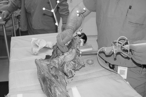
An embalmed cadaveric pelvis that had been stripped of superficial soft tissues was used for this anatomical validation. The testing was performed using a setup in an operating room (OR) to duplicate the operative procedure, including the use of a radiolucent operating table and typical OR positioning of the system camera and the C-arm for image acquisition (). For the fluoroscopic referencing, the cadaver was secured in the lateral decubitus position for image acquisition and cup insertion. The imageless referencing protocol required that the trackers be attached with the pelvis supine, and referencing was performed in that position (). For the acetabular cup insertion, the pelvis was then “flipped” into the lateral decubitus position. An acetabular component (Modular Trilogy Metal Cup, Zimmer, Inc.) was securely fixed into the cadaveric pelvis with two screws. An instrumented insertion device was attached to the cup using the threaded hole in the cup apex. A prior study had validated the accuracy of this step, indicating that there was virtually no error or toggle associated with device reattachment. Initially, a target position of insertion was 45° of cup inclination and 17.5° of anteversion. The insertion device could then be removed and reattached after the referencing protocol had been repeated.
Figure 1. Simulation of the operating room setting with placement of the cadaveric pelvis in the lateral decubitus position for fluoroscopic image acquisition.
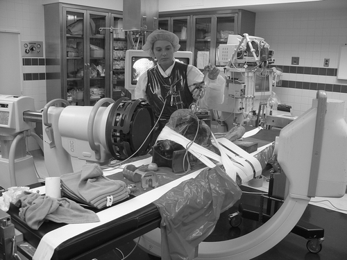
Figure 2. Cadaveric pelvis placed supine for imageless referencing of the pubic tubercle. Note the cup inserter placed for measuring cup position.
The system evaluated was the StealthStation Tria (Medtronic, Louisville, CO) with software protocols that allowed for fluoroscopic or imageless referencing of acetabular component insertion. The important navigational references for this system are the anterior pelvic plane (APP) and the anatomical center of the affected acetabulum. For each trial, the APP was determined which represented the coronal plane of the anterior pelvis in the standing position or the longitudinal axis of the patient. The APP's three reference points included the two anterior superior iliac spines and the most anterior surface of the pubic symphysis for fluoroscopy or the pubic tubercle for imageless referencing. For all trials of computer navigation, the APP was re-established using the chosen reference method.
To assess the accuracy of the system, the position of the cup in the cadaveric pelvis was measured by a NIST-traceable Brown and Sharpe coordinate measuring machine (CMM) (accuracy = 0.038 mm). This was done using a direct point-matching protocol, as all soft tissues over the anterior pelvis were removed and the reference points, including the ventral surface of the pubic symphysis, were directly visible. The specific coordinate system used was the acetabular anatomical pelvic plane reference, which measured the acetabular inclination angle at the center of the cup compared to the coronal plane. Similarly, acetabular anteversion was the angle at the center of the cup compared to the transverse axis. Five trials were used to calculate the acetabular position.
A CT study of the pelvis was undertaken using a published referencing protocol Citation[22], Citation[23]. This required that the pelvis be scanned from distal to proximal with a scan slice interval of 1 mm and a scan thickness of 1 mm. Specific details included the use of a square pixel, a field of vision of 400 mm, and a matrix of 512 × 512 pixels. The DICOM file of the pelvis was then analyzed using a referencing protocol established for acetabular component navigation. As the referencing protocols were slightly different for cup anteversion, an average of five trials was computed using either the pubic tubercle reference point or the most ventral margin of the pubic symphysis.
Assessment of repeatability of the referencing systems was done by an experienced surgeon. The measuring protocol used for fluoroscopic and imageless referencing determined the “anatomical” position of the acetabular component. Specifically, the acetabular inclination was defined as the angle of the polar axis of the cup projected onto the coronal plane; and the anteversion angle was defined as the angle of the polar axis of the cup projected onto the axial plane. For fluoroscopic referencing, the entire referencing protocol (Medtronic, Inc., Louisville, CO) was repeated including the initial fluoroscopic image acquisition. This protocol required the use of a grid on the fluoroscopic C-arm that calibrates the images to compensate for errors arising from the earth's gravitational forces. Images were acquired to represent each of the reference points in two perpendicular radiographic planes including the contralateral side anterior superior iliac spine (ASIS), the pubic symphysis, and the acetabular cup center. Comparable to the surgical setting, the affected side ASIS was determined by a direct point-matching method. The acetabular center was referenced on the computer screen from the concentric center of the acetabular component. The imageless referencing protocol (Orthosoft, Inc., Montreal, Canada) required direct touch pointing of the anterior iliac spines and one pubic tubercle (). This was done with the structures exposed and devoid of overlying soft tissue. The pelvis was then placed into the lateral decubitus position for cup measurement. Once the system had been referenced, the navigated acetabular cup positioner was calibrated to the computer using a reference block and geometric data for the specific cup-size chosen. The cup positioner was then fixed to the acetabular component in the cadaveric pelvis and measurements were made for eight consecutive trials.
Reproducibility of the cup insertion method was assessed by three surgeons who performed eight trials of referencing with either the fluoroscopic or imageless protocols. Fluoroscopic referencing was done using one set of images that had been acquired into the computer system using the C-arm. Multiple trials of referencing were then conducted from this single set of images. Imageless referencing was performed in a fashion similar to the reproducibility study. The navigated cup positioner was then fixed into the acetabular component that had been inserted into the cadaveric pelvis, and inclination and anteversion were measured. Each surgeon performed eight trials with each system.
Statistics
MS Excel 2003, SP1 and Minitab, version 14.13, were used for statistical analysis. Absolute dimensions are reported with variability measures of one standard deviation and the absolute range. Variance of the two samples was compared using the F test. Six Sigma process capability analysis was done using the process capability index (Cp):This is mathematically formulated as a ratio that basically compares a control limit about the mean that is considered to be clinically acceptable divided by the standard deviation (θ) multiplied by six. For our example, the boundary upper and lower limits were 10° around the ground truth standard for cup inclination and anteversion. The denominator becomes the 99% confidence intervals about the mean, or a bell-shaped curve with three standard deviations at either end. Industrial applications require capability indices (Cp) of greater than 1.3. Cp does not adequately reflect capabilities of the process when the distributional mean is offset. This is particularly problematic when calibration error, creep, or other systematic biases are present. To address the limitations of Cp, Six Sigma programs use the offset capability index or Cpk:
Although it is not clear what specific level of quality is appropriate for a given medical process, very high quality manufacturing is associated with processes that are capable of producing at levels where the Cpk exceeds 2.0.
Results
Benchmark precision
Following insertion, the acetabular cup position was assessed by comparing it with measurements from the NIST-traceable coordinate measuring machine. The mean CMM inclination measurement of the acetabular cup position was 46.02° (SD = 1.075; range: 43.32 to 46.84°). The mean CMM anteversion measurement of the cup position referenced to the pubic symphysis was 15.79° (SD = 0.411; range: 15.07 to 16.38°) (). The validated CT protocol revealed acetabular inclination of 42.2° (SD = 0.65) and anatomical anteversion of 12.1° (SD = 0.14) with pubic tubercle referencing, and anatomical anteversion of 14.3° (SD = 0.89) with pubic symphysis referencing ().
Table I. Measurement precision and variability. (Cp benchmark > 1.3; Cpk benchmark > 2.0)
Repeatability
Fluoroscopic referencing of the acetabular component position revealed a mean inclination of 42.8° (SD = 1.5; range: 39.5 to 44.5°). The mean anteversion was 17.5° (SD = 3.0; range: 14.5 to 22.5°). When using the imageless system, inclination was reported at 41.8° (SD = 0.46; range: 41 to 42°) and anteversion was 11.2° (SD = 0.8; range: 10.1 to 12.4°). Fluoroscopic referencing was process capable for inclination (Cp = 2.2; Cpk = 4.8) but not for anteversion (Cp = 1.1; Cpk = 1.0), while imageless referencing was process capable for both inclination (Cp = 8.3; Cpk = 69.7) and anteversion (Cp = 4.1; Cpk = 17.3). The F ratio was 13.7, demonstrating significantly higher variance for cup anteversion referenced by fluoroscopy (p < 0.002).
Reproducibility
For three surgeons assessing reproducibility using the fluoroscopic referencing technique, the mean overall group inclination was 48.5° (SD = 0.9; range: 46 to 50°). The mean overall group anteversion was 17.8° (SD = 2.5; range: 13.5 to 23.5°). When using the imageless system, the displayed inclination was 41.8° (SD = 0.82; range = 40.1 to 43.2°), while anatomical anteversion was presented as 15.2° (SD = 1.06; range: 13.6 to 16.8°). For fluoroscopic referencing, process capability was demonstrated for inclination (Cp = 3.7; Cpk = 7.5) but not for anteversion (Cp = 1.3; Cpk = 1.5). Imageless referencing was process capable for both inclination (Cp = 4.2; Cpk = 17.0) and anteversion (Cp = 3.2; Cpk = 7.7). The F ratio was 5.4, demonstrating significantly greater variance for fluoroscopic anteversion referencing (p < 0.0001).
Discussion
In computer assisted surgery, the basic precision of the computer system in providing numerically correct output data and the ability of the referencing protocol to reliably create the virtual operating field for the surgeon's interpretation are fundamental requirements. Variability in the process of measurement can potentially be a function of multiple random or systematic errors. For example, factors such as obesity or anatomic variation may interfere with the user's ability to reliably perform the referencing tasks. The potential success of improvements in positioning through the use of computer assisted surgery will therefore be dependent upon multiple factors.
McKibbin first established the anterior pelvic plane (APP) concept by creating a “table down” position of the pelvis, basically resting the pelvis on a flat surface Citation[11]. This presumes that the most anterior or ventral positions of the anterior superior iliac spines and the pubic tubercles are chosen for the APP. The computer navigation referencing protocols in this study used either fluoroscopically acquired images or a direct imageless touch point referencing to establish the APP and to determine the affected cup position. For fluoroscopy, the APP was the plane defined by the two anterior superior iliac spines and the most anterior point of the pubic symphysis. The pubic symphysis was chosen as the pubic tubercles are not easily identified fluoroscopically. As the fluoroscopic referencing points are picked from an X-ray image, it is possible that the pubic symphysis points are more difficult to determine reliably, and that the output cup position will not be comparable with the pubic tubercle method. In fact, CT measurements revealed an increase of 2° to 3° in anteversion following pubic symphysis referencing. This may be explained by the fact that the pubic symphysis may be several millimeters more posterior in the APP as defined by the pubic tubercles (). The fluoroscopic system determined cup position using the anatomical position definition as described by Murray et al. Citation[24]. The imageless system used the two anterior superior iliac spines and one of the pubic tubercles by direct touch pointing. The imageless system used the radiologic calculation from Murray et al., and data was converted to the anatomical measurement using previously described formulae. The anatomical scheme is typically applied by CT protocols for measuring cup position Citation[8], Citation[9], Citation[12] ().
Figure 3. Position of the more anterior pubic tubercle (A) compared to the pubic symphysis (B).
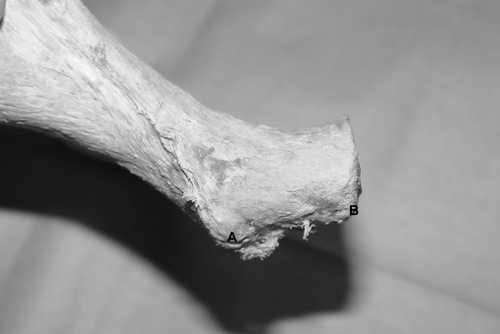
Figure 4. Anatomical reference planes of the pelvis used to calculate anteversion and abduction. [Color version available online.]
![Figure 4. Anatomical reference planes of the pelvis used to calculate anteversion and abduction. [Color version available online.]](/cms/asset/3df45f07-e2ed-49b7-ba0a-ba21c84a1d31/icsu_a_229197_f0004_b.gif)
Numerous studies have attempted to define limits that appear to be acceptable for clinical use of surgical navigation to place the acetabular component in THA. Grützner et al. evaluated a different hybrid system that used a C-arm fluoroscopic biplanar landmark reconstruction and anatomical point-matching similar to that used in the Medtronic system. They found a mean accuracy of 1.5° for inclination and 2.4° for anteversion with maximum error of 5° for inclination and 6° for anteversion Citation[20]. Hube et al. compared CT-based navigation with fluoroscopic referencing, noting the mean variation of inclination to be 2.7° (range: 0–8°) with CT and 3.9° (range: 0–9°) after fluoroscopy-based navigation Citation[25]. Amiot et al. evaluated CT-based navigation with 350 observations, finding a maximal inclination error of 5° in 99% of cases and a maximal anteversion error of 5° in 97% of cases Citation[26]. Jolles et al. evaluated imageless navigation in plastic pelvic bones, finding a mean accuracy of 1.5° for inclination and 2.5° for anteversion, with a maximum error of 8° Citation[19].
While we would agree that the above studies demonstrate a reasonable degree of accuracy and the limits of error, they offer little ability to compare the various methods. For that reason, we have evaluated and applied the process capability analysis or Six Sigma formulae for our comparison. In the first place, accuracy is not the measure that we are looking for with a technique like computer navigation, as accuracy is a fixed measure to a reference point and must be determined for each trial. Precision, on the other hand, is the statistical spread or variation of a method around the reference point. The Six Sigma formula determines the precision of a system by comparing the maximum tolerable spread of values to the bell-shaped curve of variation (the Six Sigma). This widely adopted industrial statistics measure allows one to compare just about anything for precision manufacturing, from the dimensions of a microchip to the tolerance of an orthopaedic device such as a metal-on-metal surface replacement. The important consideration for using the Six Sigma formula is setting the upper and lower specification limits. In our example, we chose the ten degrees about the reference position as established by the benchmark based on the historical comparison of Lewinnek et al. for boundaries of cup positions to prevent hip dislocation.
From our study, we found that fluoroscopic referencing was not process capable for navigating cup anteversion. Tannast et al. have made similar findings, noting the occasional error in determining the precise points at the pubic symphysis if there is overlay of the inferior pubic ramus Citation[28]. We believe the ability to determine the precise position of the pubic symphysis during surgery is quite difficult, especially with the patient laying on their side and with a full surgical draping. Based on the information presented in this study, we no longer use this method for surgical navigation. We would, however, agree that the fluoroscopic referencing protocols are quite accurate, and that precise pubic symphysis referencing may be possible if combined with other modalities such as ultrasound or imageless referencing, as suggested by Grützner et al. Citation[20]. Cup inclination was process capable and this may reflect the ease of picking a suitable radiographic landmark on the anterior superior iliac spine.
In practice, the imageless referencing technique currently employs a direct anatomical “point matching” method. The surgeon palpates and then makes small stab wounds to place the reference probe directly onto the anterior superior iliac spines and the pubic tubercles. The current laboratory study differs from the clinical surgical setting as the target reference points were directly exposed and devoid of soft tissues. We believe that the reference points of the anterior superior iliac spines and pubic tubercles are relatively broad surfaces and significant error may be added by attempting to indirectly palpate or even to touch point reference through small skin punctures. Richolt et al. have suggested this problem may be minimal, however, with an error no greater than 3° Citation[29]. Contrary to the anteversion error found with fluoroscopic referencing, we believe the imageless error would be greater for inclination as this measurement is made in the coronal plane, which is collinear with the APP. Anteversion, on the other hand, is measured in the transverse plane, a different plane to the APP. For example, subtle differences for ASIS referencing would not alter anteversion significantly as long as these points remained in the APP. This trend is suggested by the measurements made by the coordinate measuring machine.
We would draw attention to the need for standardized terminology and measurements for determining acetabular component positioning. It would appear that the anatomical coordinate system employed by CT protocols remains the most logical. We discovered that the two systems we compared had significant differences, both in the target referencing landmarks and in the methodology used to create the final numerical inclination and anteversion values. Any surgeon relying on computer navigation for acetabular component positioning must be aware of these differences. Finally, we would draw attention to the ability of process capability analysis or Six Sigma to allow a vigorous comparison of different methodologies. We conclude from review of the literature that CT referencing methods are process capable for acetabular cup insertion, but that neither imageless nor fluoroscopic referencing methods have proven to be completely validated for the clinical setting. ().
Table II. Reported precision of reproducibility of different navigation systems using Process Capability Analysis. (Upper and lower specification limits: ±10°)
References
- Bader RJ, Steinhauser E, Willmann G, Gradinger R. The effects of implant position, design, and wear on the range of motion after total hip arthroplasty. Hip International 2001; 11: 80–90
- Giurea A, Zehetgruber H, Funovics P, Grampp S, Karamat L, Gottsauner-Wolf F. Risk factors for dislocation in cementless hip arthroplasty–A statistical analysis. Z Orthop 2001; 139: 194–199
- Jolles BM, Zangger P, Leyvraz PF. Factors predisposing to dislocation after primary total hip arthroplasty. J Arthroplasty 2002; 17: 282–288
- Kennedy JG, Rogers WB, Soffe KE, Sullivan RJ, Griffen DG, Sheehan LJ. Effect of acetabular component orientation on recurrent dislocation, pelvic osteolysis, polyethylene wear and component migration. J Arthroplasty 1998; 13: 530–534
- Kummer FJ, Shah S, Iyer S, DiCesare PE. The effect of acetabular cup orientations on limiting hip rotation. J Arthroplasty 1999; 14: 509–513
- Schmalzried TP, Guttmann D, Grecula M, Amstutz HC. The relationship between the design, position and articular wear of acetabular components inserted without cement and the development of pelvic osteolysis. J Bone and Joint Surg 1994; 76A: 677–688
- Lewinnek GE, Lewis JL, Tarr R, Compere CL, Zimmermann JR. Dislocations after total hip replacement arthroplasties. J Bone Joint Surg (Am) 1978; 60: 217–221
- Saxler G, Marx A, Vandevelde D, Langlotz U, Tannast M, Wiese M, Michaelis U, Kemper G, Grützner PA, Steffen R, von Knoch M, Holland-Letz T, Bernsmann K. The accuracy of free-hand cup positioning: A CT based measurement of cup placement in 105 total hip arthroplasties. Int Orthopaedics 2004; 28: 198–201
- DiGioia AM, Jaramaz B, Plakseychuk AY, Moody JE, Nikou C, LaBarca RS, Levison TJ, Picard F. Comparison of a mechanical acetabular alignment guide with computer placement of the socket. J Arthroplasty 2002; 17: 359–364
- Seki M, Yuasa N, Ohkuni K. Analysis of optimal range of socket orientations in total hip arthroplasty with use of computer-aided design simulation. J Orthop Res 1998; 16: 513–517
- McKibbin B. Anatomical factors in the stability of the hip joint in the newborn. J Bone Joint Surg 1970; 52B: 148–159
- Leenders T, Vandervelde D, Nahiew G, Nuyts R. Reduction in variability of acetabular cup abduction using computed assisted surgery: Prospective and randomized study. Comput Aided Surg 2002; 7: 99–106
- Sugano N, Sasama T, Sato Y, Nakajima Y, Nishii T, Yonenobu K, Tamura S, Ochi T. Accuracy evaluation of surface-based registration methods in a computer navigation system for hip surgery performed through a posterolateral approach. Comput Aided Surg 2003; 6: 195–203
- Khadem R, Yeh C, Sadeghi-Tehrani JM. Comparative tracking error analysis of five different optical tracking systems. Comput Aided Surg 2000; 5: 98
- Widmer K-H, Zurfluh B. Compliant positioning of total hip components for optimal range of motion. J Orthopaedic Res 2004; 22: 815–821
- Kalteis T, Handel M, Bäthis H, Perlick L, Tingart M, Grifka J. Imageless navigation for insertion of the acetabular component in total hip arthroplasty: Is it as accurate as CT based navigation?. J Bone Joint Surg 2006; 88B: 163–167
- Kalteis T, Handel M, Herold T, et al. Greater accuracy in positioning of the acetablular hip by using an image-free navigation system. Int Orthopaedics 2005; 29: 272–276
- Kalteis T, Beckmann J, Herold T, Zysk S, Bäthis H, Perlick L, Grifka J. Accuracy of an image-free cup navigation system. An anatomical study. Biomed Tech (Berlin) 2004; 49: 257–262
- Nogler M, Kessler O, Prassl A, Donnelly B, Streicher R, Sledge JB, Krismer M. Reduced variability of acetabular cup positioning with use of an imageless navigation system. Clin Orthop Rel Res 2004; (426): 159–163
- Grützner PA, Zheng G, Langlotz U, von Recum J, Nolte LP, Wentzensen A, Widmer KH. C-arm based navigation in total hip arthroplasty-background and clinical experience 2004, Injury ;35(Suppl 1):S-A90-5
- Zheng G, Marx A, Langlotz U, Widmer KH, Buttaro M, Nolte LP. A hybrid CT-free navigation system for total hip arthroplasty. Comput Aided Surg 2002; 7(3)129–145
- DiGioia AM, Jamaraz B, Blackwell M, Simon DA, Morgan F, Moody JE, Nikou C, Colgan BD, Aston CA, LaBarca RS, Kischell E, Kanade T. Image guided navigation system to measure intraoperatively acetabular implant alignment. Clin Orthop 1998, 354: 8–22
- Jaramaz B, DiGioia AM, Blackwell M, Nikou C. Computer assisted measurement of cup placement in total hip replacement. Clin Orthop 1998; 354: 70–81
- Murray DW. The definition and measurement of acetabular orientation. J Bone Joint Surg 1993; 75B: 228–232
- Hube R, Birke A, Hein W, Klima S. CT-based and fluoroscopy-based navigation for cup implantation in total hip arthroplasty (THA). Surg Technol Int 2003; 11: 275–280
- Amiot LP, Poulin F. Computed tomography-based navigation for hip, knee, and spine surgery. Clin Orthop Rel Res 2004, 421: 77–86
- Jolles BM, Genoud P, Hoffmeyer P. Computer-assisted cup placement techniques in total hip replacement improve accuracy of placement. Clin Orthop 2004; 426: 175–179
- Tannast M, Langlotz F, Kubiak-Langer M, Langlotz U, Siebenrock K. Accuracy and potential pitfalls of fluoroscopy-guided acetabular cup placement. Comput Aided Surg 2005; 10: 329–336
- Richolt JA, Effenberger H, Rittmeister M. How does soft tissue distribution affect anteversion accuracy of the palpation procedure in image-free acetabular navigation? An ultrasonographic assessment. Comput Aided Surg 2005; 10: 87–92