Abstract
The use of optical tracking systems in computer assisted surgical navigation requires the rigid fixation of a dynamic reference base to the target bone to be navigated. This report presents the results of a new approach to optical tracker fixation in the distal femur. Four embalmed cadavers were evaluated for pin placement. It was found that placement of pins from medial to lateral parallel to the transepicondylar axis placed the pins well posterior to the center of the intramedullary canal and away from neurovascular structures. Eighty-six consecutive patients underwent total knee arthroplasty using this new technique. All procedures were successful for performing a navigation-assisted total knee replacement. Obesity was not a factor, nor was there any loosening of the pin array during the procedure. There were no wound-healing problems in any patient. At one year follow-up, no patient could identify subjective symptoms related to either the medial epicondylar area or the stab wound portals. No direct neurovascular injuries were noted and no patient developed a fracture of the femur related to the pin sites.
Conclusion: A new technique is described that facilitates pin placement for minimally invasive approaches while eliminating complications. Sagittal plane optical array orientation simplifies the surgical technique.
Introduction
The use of optical tracking systems in computer assisted surgical navigation requires the rigid fixation of a dynamic reference base or array to the target bone to be navigated. A variety of options have been advocated for this purpose, including the use of single or double threaded pins of different dimensions. Several authors and proprietary protocols recommend bicortical fixation, particularly for applications used in total knee arthroplasty (TKA) Citation[1–8]. For the distal femur, bicortical fixation provides greater stability for the distal femoral array and may be the optimal choice. However, placement within the anterior wound created by the standard median parapatellar approach requires an anterior-to-posterior placement of these pins. Damage to major neurovascular structures secondary to pin and screw placement is a recognized potential complication of orthopaedic procedures, and studies have been performed to define this risk in certain instances Citation[1]. Furthermore, the potential for damage to neurovascular structures during knee surgery has been identified Citation[9–14]. Marchant et al. demonstrated that 100% of pins placed in the distal femur from anterolateral to posterior medial were within 5 mm of the popliteal artery and vein Citation[15].
This report presents the results of an anatomical study conducted on the distal femur to develop a new approach for optical tracker fixation that could be implemented with nominal risk to neurovascular structures, yet could be employed with newer minimally invasive surgical incisions developed for TKA. As current femoral resection devices use intramedullary rods for alignment, pin placement was sought that would not interfere with this rod placement. Additionally, pin placement should be sufficiently proximal to allow for typical bone cuts. A series of patients were operated with this approach, and a minimum of 12 months follow-up was conducted with evaluation for complications.
Methods
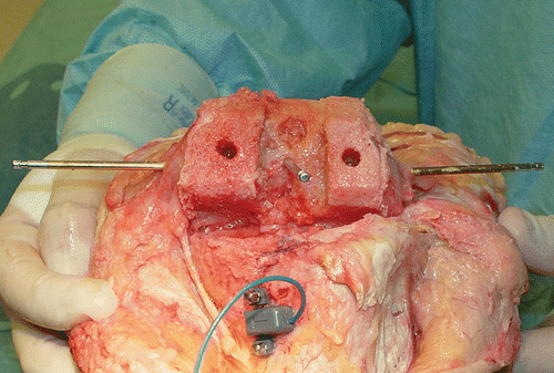
Four embalmed cadavers were evaluated for pin placement in various positions. It was found that the popliteal artery and vein consistently lie within 5 mm of the posterior cortical surface of the distal femur, beginning at a level of 10 mm above the knee joint line as these structures traverse Hunter's canal into the popliteal fossa. Moving distally, this location starts posterior medial and progresses to nearly midline posterior Citation[16]. It was found that placement of a pin from medial to lateral starting at the prominence of the medial epicondyle and attempting to parallel the transepicondylar axis placed the pin well posterior to the center of the intramedullary canal. A second pin could be placed 2 cm above the first from medial to lateral beginning at a point anterior to the supracondylar ridge of the distal femur. Distal pin placement was measured to be approximately 3.2 cm above the medial condylar surface and 2.5 cm above the lateral condyle. This allows for placement of intramedullary instrumentation into the distal femur, and will not interfere with bone cuts or implant placement (). Finally, it was noted that the popliteal vessels, the sciatic nerve, and the common peroneal nerve were more than 10 mm from the pins at all levels.
Figure 1. Anatomical specimen demonstrating pins placed medial and lateral in the transepicondylar axis, with a midline pin approximating the “femoral center” landmark which is referenced for the Medtronic StealthStation Treon system. Note the position of the intramedullary rod placement and that all bone cuts, including the femoral notch cut, are not interfered with by this pin placement.

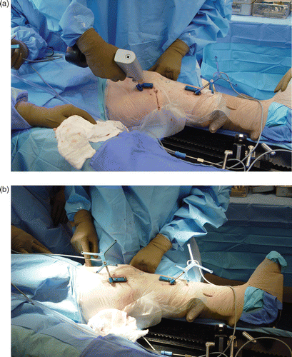
A group of 86 consecutive patients (33 males, 53 females) undergoing total knee arthroplasty (TKA) were identified as suitable for use of this new technique. The implants used were either the LCS mobile-bearing prosthesis or the LPS High Flex posterior stabilized implant. The surgical technique was a “tibia cut first” method in all cases, and flexion spacing was done using a tensioner device. Based on the anatomical study, pin placement was made percutaneously from medial to lateral by palpating and directing the pins into the medial epicondyle, approximating the transepicondylar axis. The second pin was placed 2 cm superior into the posterior third of the femoral shaft, just anterior to the supracondylar ridge. Both pins were inserted posterior to the vastus medialis (). The pins used were 3.2-mm pins threaded distally, with an overall length of 10 cm. The tracking arrays were oriented in the sagittal plane of the leg, and the line-of-site cameras were placed approximately 8 feet away, perpendicular to the longitudinal axis of the patient to the opposite side. Computer navigation was performed using the Medtronic StealthStation Treon system with the Universal Total Knee referencing protocol (Medtronic, Inc., Louisville, CO). A median parapatellar approach was used in 68 cases, while a lateral parapatellar approach was used in 18 valgus knees. In the valgus knees, the percutaneous pin placement was done parallel to the transepidcondylar axis, but from lateral to medial. Postoperative management was carried out in standard fashion.
Figure 2. (a) Intraoperative placement of the transepicondylar pins requires dissection of the medial synovial reflection to palpate the medial epicondyle and the supracondylar ridge just cephalad to this point. Pin insertion then parallels the transepicondylar axis and is perpendicular to the AP axis of Whiteside. (b) The femoral array is positioned in the medial side of the lower extremity.

Results
All procedures were successful for performing a navigation-assisted total knee replacement. Patient obesity was not a factor, nor did we find any loosening of the pin array during the procedure. One patient experienced early wound drainage from the percutaneous portal as stapling of the stab wound had inadvertently been neglected. There were no wound-healing problems in any patient. At one year follow-up, no patient could identify subjective symptoms related to either the medial epicondylar area or the stab wound portals. No direct neurovascular injuries were noted, and no patient developed a fracture of the femur related to the pin sites. Unrelated complications included one patient who developed a tourniquet palsy and a second patient who developed a tibial shaft stress fracture from one of the tibial array pin sites.
Discussion
Neurovascular injury posterior to the knee joint is a well-described potential complication of many surgical procedures in this region. Such injury may result from over-penetration of a drill, insertion of a reference pin beyond the far cortex of the distal femur, trauma from mechanical saws, or past-pointing of a depth gauge. Significant vessel pseudoaneurysm, or loss of structural integrity, may result from even a single-sided vessel wall trauma. It is therefore essential to have a clear understanding of the relationship between the neurovascular structures posterior to the knee joint when considering surgery in this area. Marchant et al. Citation[15] observed the existence of a definite, reproducible risk to the popliteal neurovascular structures when placing a bicortical reference pin. The popliteal artery and vein were observed to be most at risk due to their apparent intimate relationship to the posterior femoral cortex.
Several authors have attempted to accurately define this relationship through the use of imaging studies. Zaidi et al. Citation[14] found that at 90° of flexion the artery lay closer to the posterior tibial surface than in full extension. Shiomi et al. Citation[17] attempted to determine the angle of knee flexion which placed the popliteal artery at the least risk of injury during surgical procedures on the knee, finding that flexion increased the distance between the artery and posterior femoral cortical surface. Smith et al. Citation[18] studied the relationship of the popliteal artery at the level of knee replacement resection with the subjects in a supine position. They found that, even including the effect of gravity, knee flexion does not guarantee posterior movement of the neurovascular structures at the level of the knee.
Regarding the technique suggested in this paper for computer navigation tracker placement, there were a number of other reasons for developing this method. Most importantly, the author was seeking a way to avoid placing pins into the extensor mechanism in the anterior incision while allowing adequate room for placement of anterior knee instrumentation and minimizing the length of the incision. While studying the quadriceps-sparing approach in the laboratory, it became apparent that subcutaneous access to the medial epicondyle was simple and avoided placing the pin into the extensor mechanism. By inserting the two pins in the coronal plane, the frame of the dynamic reference base (DRB) could be placed such that the trackers were medial and in the sagittal plane of the lower extremity. This approach has virtually eliminated troublesome movement of the trackers arising from mechanical jolts or jostles that were typical with the anterior placement. Furthermore, the tracker is placed perpendicular to the optical camera which is normally viewing in this direction. Mihalko et al. have suggested that two-pin placement is optimal for DRB tracker anchorage, and found no difference when two pins were used in cortical or metaphyseal bone Citation[19]. Most importantly, Ossendorf et al. have published a case report of distal femoral stress fracture resulting from cortical placement of a larger single pin for knee navigation Citation[20].
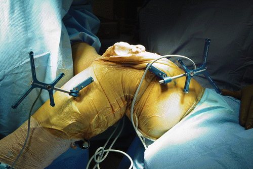
In conclusion, the author believes that the transepicondylar femoral pin placement is safe for placing the femoral DRB well away from neurovascular structures. Several other advantages accrue, including placement of the array posterior to the quadriceps muscle, avoiding muscle entrapment with range of motion, and allowing the array to become unobtrusive during the surgical procedure (). Accidental bumping or disturbance of the array becomes unlikely. Finally, extensive clinical experience has identified no inherent risks such as femoral fracture or wound-healing problems. This approach will be most useful for minimally invasive methods such as the subvastus or midvastus approach, as access to the distal femoral medial epicondyle is easily obtained through a very small anterior incision.
References
- Bolognesi MP, Hofmann AA. Computer navigation versus standard instrumentation for TKA: A single-surgeon experience. Clin Orthop Relat Res 2005; 440: 162–169
- Chauhan SK, Clark GW, Lloyd S, Scott RG, Breidahl W, Sikorski JM. Computer assisted total knee replacement. J Bone Joint Surg 2004; 86B: 818–823
- Decking R, Markmann Y, Fuchs J, Puhl W, Scharf HP. Leg axis after computer-navigated total knee arthroplasty: A prospective randomized trial comparing computer-navigated and manual implantation. J Arthroplasty 2005; 20: 282–288
- Haaker RG, Stockheim M, Kamp M, Proff G, Breitenfelder J, Ottersbach A. Computer-assisted navigation increases precision of component placement in total knee arthroplasty. Clin Orthop Relat Res 2005; 433: 152–159
- Krackow K. Total knee arthroplasty using the Stryker Knee Trac system. Navigation and MIS in Orthopaedic Surgery, JB Stiehl, W Konermann, R Hacker. Springer Verlag, Heidelberg 2006; 100–113
- Saragaglia D. Computer assisted implantation of a total knee prosthesis without preoperative imaging. The kinematic model. Navigation and MIS in Orthopaedic Surgery, JB Stiehl, W Konermann, R Hacker. Springer Verlag, Heidelberg 2006; 88–94
- Sparmann M, Wolke B, Czupalla H, Banzer D, Zink A. Positioning of total knee arthroplasty with and without navigation support. A prospective, randomised study. J Bone Joint Surg Br 2003; 85(6)830–835
- Stulberg SD, Loan P, Sarin V. Computer-assisted navigation in total knee replacement: Results of an initial experience of thirty five patients. J Bone Joint Surg 2002; 84A: 90–98
- Post WR, King SS. Neurovascular risk of bicortical tibial drilling for screw and spiked washer fixation of soft-tissue anterior cruciate ligament graft. J Arthroscopy 2001; 17(3)244–247
- Berger C, Anzbock W, Lange A, Winkler H, Klein G, Engel A. Arterial occlusion after total knee arthroplasty: Successful management of an uncommon complication by percutaneous thrombus aspiration. J Arthroplasty 2002; 17(2)227–229
- Mureebe L, Gahtan V, Kahn M, Kerstein MD, Roberts AB. Popliteal artery injury after total knee arthroplasty. American Surgeon 1996; 62: 366–368
- Roth JH, Bray RC. Popliteal artery injury during anterior cruciate ligament reconstruction. J Bone Joint Surg Br 1988; 70: 840
- Rush JH, Vidovich JD, Johnson MA. Arterial complications of total knee replacement: The Australian experience. J Bone Joint Surg Br 1987; 69(3)400–402
- Zaidi SH, Cobb AG, Bentley G. Danger to the popliteal artery in high tibial osteotomy. J Bone Joint Surg Br 1995; 77(3)384–386
- Marchant DC, Rimmington DP, Nusem I, Crawford RW. Safe femoral pin placement in knee navigation surgery. A cadaver study. Comput Aided Surg 2004; 9: 257–260
- Boileau JC. Grant's Atlas of Anatomy Sixth edition. Williams & Wilkins, Baltimore 1972, Figures 285–289 editor
- Shiomi J, Takahashi T, Imazato S, Yamamoto H. Flexion of the knee increases the distance between the popliteal artery and the proximal tibia: MRI measurements in 15 volunteers. Acta Orthop Scand 2001; 72(6)626–628
- Smith PN, Gelinas J, Kennedy K, Thain L, Rorabeck CH, Bourne RB. Popliteal vessels in knee surgery: A magnetic resonance imaging study. Clin Orthop 1999; 367: 158–164
- Mihalko WM, Duquin T, Axelrod JR, Bayers-Thering M, Krackow KA. Effect of one- and two-pin reference anchoring systems on marker stability during total knee arthroplasty computer navigation. Comput Aided Surg 2006; 11: 93–98
- Ossendorf C, Fuchs B, Koch P. Femoral stress fracture after computed navigated total knee arthroplasty. Knee 2006; 13(5)397–399
