Abstract
Objective: To assess the feasibility and accuracy of a drill template for the placement of a cervical pedicle screw in a single vertebral level.
Materials and Methods: A volumetric CT scan was performed on a cadaver cervical spine. Using computer software, a drill template with a predefined trajectory was constructed that was designed to match the posterior surface of the right side of the fifth cervical vertebra. A physical template was created from the computer model using a rapid prototyping machine. The drill template was used to guide drilling of a pilot hole, and a CT scan was performed to assess the accuracy of this hole. A 3.5-mm diameter pedicle screw was placed in the pilot hole. The spine was then dissected to separate the vertebrae and the trajectory of the screw was visually inspected.
Results: The feasibility of this patient-specific rapid prototyping technique was demonstrated. Imaging and visual inspection confirmed accurate placement of the pilot hole and cervical pedicle screw without cortical violation.
Conclusions: The potential use of drill templates to place cervical pedicle screws is promising. Our initial methodology appears to provide an accurate technique and trajectory for pedicle screw placement in the cervical spine.
Introduction
Despite advances in instrumentation techniques and intra-operative imaging, the successful implementation of posterior cervical instrumentation remains a challenge. The procedure generally considered to be the treatment of choice for stabilizing the cervical spine from the posterior approach employs bilateral placement of bone screws into the lateral masses of the cervical bodies in conjunction with plates or rods. The amount of bone available for purchase in the lateral mass is relatively small, raising concerns regarding inadequate fixation. In turn, this may prompt the surgeon to elect to perform additional fusion using an anterior approach to ensure stabilization. Lateral mass screw failure can result from screw fracture or, more commonly, from pullout with the resultant need for revision surgery. Using pedicle fixation of the vertebra, as commonly performed in the lumbar spine, would provide more bone for screw purchase and enable the use of longer screws, thereby increasing the likelihood of the screw staying in place until fusion occurs Citation[1]. Despite the perception that cervical pedicle fixation would provide superior holding strength, cervical pedicle screws have not been implemented routinely because the unique anatomic features of the cervical spine pose a challenge for accurate placement and there is potential for injury to the adjacent nerve roots and vertebral artery. The small diameter of the pedicle in the cervical spine provides little room for tolerance and requires accurate guidance, in contrast to the larger pedicle of the thoracolumbar portions of the spine.
Successful placement of pedicle screws requires an understanding of the individual patient's three-dimensional pedicle morphology and adjacent neurovascular structures, accurate identification of an appropriate entry point, and a safe and effective trajectory Citation[1–4]. Various methods have been explored for pedicle screw placement, including anatomic studies, image-guided techniques, and drill templates. Anatomic studies and image-guided techniques have several disadvantages, making patient-specific drill templates with pre-planned trajectories a promising alternative for pedicle screw placement. It is speculated that drill templates could shorten the surgical procedure and improve accuracy. Drill templates were initially demonstrated in the lumbar spine, hip, and knee Citation[5], but several studies have described their use in the cervical, thoracic, and lumbar spine Citation[6–8]. Using rapid-prototyping techniques, templates were created with three V-shaped knife edges to rest on the transverse and spinous processes Citation[6]. The ability to customize the placement of each screw based on the unique morphology of the cervical vertebra is an appealing concept.
This study explores the feasibility of creating rapid prototype custom-fit drill templates for pedicle screw placement in the cervical spine based on pre-procedural volumetric CT data sets and pre-selected trajectories. Our template design varies from those used in previous studies in that it is created to contact a greater surface area of the vertebra, and a custom template is designed for each trajectory.
Materials and methods
Specimen and Imaging
Formalin-fixed cervical spines were obtained with permission from the Anatomy Lab of the Department of Anatomy at the University of Iowa. The posterior surface of the specimens was cleared of soft tissue and then imaged with volumetric CT (SOMATOM Sensation 16, Siemens, Malvern, PA) with 0.75-mm slice thickness and 0.26-mm in-plane resolution. The images were stored in DICOM format.
Construction of drill templates
To develop the concept and process, one of the spines collected was selected and a plan developed for placing a right C5 pedicle screw. Software used for template construction included that of the StealthStation® 3D Spine module (Medtronic SNT, Louisville, CO) and Analyze (Mayo Clinic Biomedical Imaging Resource, Rochester, MN). The volumetric data set was transferred to the StealthStation® and a surgical plan was developed to accommodate a 3.5-mm diameter bone screw through the right C5 pedicle using the 3D spine operative planning software. The surgical plan was specifically developed so that the screw trajectory centered on the pedicle shaft passed into the vertebral body without violating the pedicle cortex and interacted with only the appropriate vertebral level. The beginning and end coordinates of the planned trajectory were recorded. Using Analyze, a template of the anatomic surface adjacent to the selected entry point was constructed. The template surface was created to be the inverse of the vertebral surface, thus potentially enabling a fit in a lock-and-key fashion similar to a physical casting of the vertebral surface, and specifically avoided overlap onto adjacent segments.
Image data was re-sampled using Analyze to yield cubic voxels with a size of 0.25 mm per side. The original 16-bit/pixel image data was remapped to 8 bits/pixel using an intensity window from −1000 Hu to + 1000 Hu. With a threshold of 140, bone segmentation was created in the image volume. The intensity of bone segmentation was inverted to create a cast of the spine. The drill template was created from this altered dataset.
Templates were designed to interface only with the appropriate side of the vertebrae at the appropriate level. This enabled the template to fit on the spine even if the spine was positioned differently than when scanned. Because each vertebral surface varies, it was difficult to develop the template so that it could be created in a single 3D step. In response to this challenge, individual transverse slices were altered, rather than the entire volume at once. Altering a set of adjacent slices had the same effect as altering the volume; however, it required more user input. Analyze enabled the following constraints to be implemented slice by slice.
All transverse slices before and after the desired vertebral level were set to zero, so that only the cast of the appropriate vertebrae remained. When choosing which slices to keep and which slices to set to zero, the coordinates from the StealthStation® were consulted to confirm that the trajectory would interact with or pass through the slices kept and the superior and inferior limits of the desired vertebrae were viewed. CT slices were obtained at an original thickness of 0.75 mm. Approximately 20 slices were used per template, resulting in a guide with dimensions of approximately 1.5 cm inferior-superior × 3 cm lateral × 2 cm anterior-posterior, which allowed coverage of the facet of interest and the majority of the adjacent lamina ().
Figure 1. (a) Posterior view of vertebra. Point a represents the superior limit of the lateral mass and point b represents the inferior limit of the lateral mass. The distance between a and b was made to be 1 cm or greater to provide stability. (b) Superior view of vertebra. Point a represents the maximum height of the template, corresponding to the inferior intersection of the laminas, and point b represents the minimum height of the template, corresponding to the superior intersection of the laminas. Point c represents the bottom surface of the template and is slightly anterior to the transverse process. Line d represents the lateral margin of the drill template.
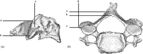
Anatomical landmarks on the vertebra were used to provide guidelines and maintain consistent template construction. The point at which the two laminas intersected superiorly on the vertebra was used for the minimum height of the template; the laminas’ inferior intersection was used for the maximum height of the template (). These minimum and maximum heights created sufficient template-bone interface and also limited the template to interfacing with the vertebral surface that can be most readily cleared of soft tissue. If the constructed template is higher than the inferior intersection of the lamina, the template interfaces with the spinous process, which is less readily cleared of soft tissue, and also has the potential to interact with spinous processes of adjacent vertebrae. The width of the template was set so as not to protrude past the midline of the vertebra, while extending slightly past the edge of the transverse process. The lower surface of the template was made to extend slightly anterior to the transverse process. This gave a hook-like aspect to the template, providing greater stability. On average the construction process created a template measuring 1.5 cm inferior-superior, 2 cm anterior-posterior, and 3 cm left-right.
The surgical plan from the StealthStation® was incorporated into the template through a unique intermediate computer code, written by the authors, linking the functions of the StealthStation® and Analyze together. This allowed a hollow cylinder of 9 mm diameter, based on the preplanned trajectory, to be added to the model to provide the trajectory for drilling. The inner diameter of the hollow cylinder was created to accommodate a 1.6-mm Kirschner wire (K-wire), commonly used as a drill guide for cannulated bone screws. The cylinder was constructed so that the distance from the start of the cylinder to the point where the template interfaced with the vertebral surface was 35 mm ().
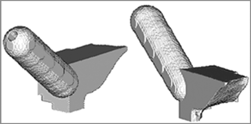
Figure 2. Two views of the computer model of the drill template. The lower surface of the template is the inverse or cast of the vertebral surface.
The computer model was then exported in STL format and converted into a physical template using a rapid prototyping machine (Stratasys, FDM 2000, Eden Prairie, MN). The rapid prototyping machine functions as a 3D “printer”, laying down consecutive layers of acrylonitrile butadiene styrene (ABS) plastic based on the computer model until a physical template is constructed. The resolution of the rapid prototyping machine is estimated to meet or exceed the 0.25-mm resolution of the template model and is not a limiting factor on the accuracy of reproduction.
Assessing the accuracy of the drill template
The constructed template was used to aid in drilling a 1.6-mm pilot hole using a K-wire. Since the drill template was created to be unique for the vertebra, it fitted securely on the vertebral surface. The template was easily and securely held in place by the surgeon's free hand. The K-wire was inserted through the cylinder of the drill template along the pre-planned trajectory and drilling took place until the vertebral body was penetrated. This pilot hole was then enlarged to a depth of approximately 3 cm with a 2.8-mm drill bit in order to accommodate a bone screw. The cervical spine was scanned intact on the same CT scanner as used initially, and the accuracy of the pilot hole was observed in the sagittal, coronal, and transverse planes. A 3.5 mm × 30 mm bone screw (Medtronic Sofamor Danek, Louisville, CO) was then placed freehand down the pilot hole. The accuracy of the screw placement was assessed by visual inspection, which involved completing the dissection of the spine so that the vertebrae were separated from one another and the soft tissue was cleared from the pedicle to allow direct inspection for cortical violation.
Results
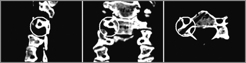
shows the computer-generated model of the template. Using this template, we demonstrated the ability to accurately place an initial K-wire followed by a 3.5-mm bone screw down the cervical pedicle. This was confirmed by CT imaging of the pilot hole and visual inspection of the screw placement after final placement (). The 3D image reconstruction showed no evidence of cortical violation. After imaging the intact specimen, dissection of the individual body confirmed that no cortical violation had occurred.
Figure 3. Sagittal, coronal and transverse CT slices of the pilot hole drilled with the aid of the template. The circles emphasize that the trajectory remained entirely within the pedicle.
Discussion
Despite careful surgical technique, image guidance and intra-operative imaging, prospective and retrospective studies have demonstrated the challenges of pedicle screw placement in the cervical spine, including the risk of vertebral artery and neurological injury Citation[9], Citation[10]. Each cervical vertebra has a unique morphology, making quantitative analysis challenging. It has been demonstrated that anatomic landmarks can assist in the placement of pedicle screws Citation[11]. However, due to the inherent variability and limited tolerance for error, anatomic landmarks alone are prone to inaccuracy in determining trajectory with resultant pedicle penetration. Various methods of screw placement have been described to improve pedicle screw accuracy, but perforations of the pedicle still occur Citation[12].
The use of image-guided techniques has been successful for pedicle screw placement in the thoracic and lumbar regions, and has been shown to increase the accuracy of screw placement when compared to the use of anatomic landmarks alone Citation[4], Citation[11], Citation[13–20]. To achieve the greatest accuracy, each vertebral body must be individually registered, as any movement of the individual segments relative to one another between the time of pre-operative imaging and surgery will be translated into decreased accuracy with simultaneous multi-level registration. Individual registration of each body during surgery is time-consuming and, due to the smaller size of the cervical vertebra, sometimes not possible. In addition, any movement of the registration frame between the time of registration and screw placement is problematic.
Encouraging results have been obtained in drill template studies. When additional supports were added to the V-shaped knife design described by Berry et al., the template was more stable and appeared to result in successful placement Citation[6]. However, with the four trajectories tested in the cervical spine, the surgeons were not always confident of the screw positioning. An advantage of the V-shaped knife design is that excessive soft-tissue dissection from the vertebra is not necessary. Our template design may provide greater stability because it contacts a greater surface area of the vertebra, but has the potential disadvantage of soft tissue interfering with a secure fit.
The challenges of pedicle screw placement make patient-specific drill templates attractive. Drill templates may add to the cost and time of the overall surgical process; however, once custom software is developed and the process streamlined, we estimate that each drill template could be created for $30–$50. Most bone screws used for applying spine instrumentation cost $80–$120 apiece and we believe the cost increase is potentially acceptable. This assumes only in-house production costs; commercial manufacturing would alter these estimates considerably.
We believe we have demonstrated a process that could improve the accuracy and safety of pedicle screw placement in the cervical spine. Our goal in this report was to describe our concept and to provide a framework for additional study. The utility of drill templates rests on the premise that pedicle screws provide superior fixation to lateral mass screws. This “proof of concept” report requires confirmation with additional studies, which are underway. The size of the template required to enable a secure/unique fit to the surface of the vertebra and the ability to place the template on the vertebral surface during the operation need to be addressed. In vitro, cervical spines are readily cleared of soft tissue; however, the clearing of soft tissue in vivo may be more problematic. The length of the guide tube built into the drill template may also be a consideration in order to minimize inaccuracy in the trajectory due to “wobble” from the K-wire. Following K-wire placement, the process of screw placement needs to be refined. As opposed to freehand placement, this process could incorporate either a fenestrated screw system to allow placement over the K-wire, or potentially an additional template with a larger cylinder allowing the pilot hole to be enlarged with a larger drill-bit to accommodate placement of the pedicle screw. These methods of screw placement could then be compared to the accuracy of the K-wire alone.
References
- Jones EL, Heller JG, Silcox DH, Hutton WC. Cervical pedicle screws versus lateral mass screws. Anatomic feasibility and biomechanical comparison. Spine 1997; 22(9)977–982
- Ludwig SC, Kramer DL, Vaccaro AR, Albert TJ. Transpedicle screw fixation of the cervical spine. Clin Orthop Relat Res 1999; 359: 77–88
- Rezcallah AT, Xu R, Ebraheim NA, Jackson T. Axial computed tomography of the pedicle in the lower cervical spine. Am J Orthop 2001; 30(1)59–61
- Steinmann JC, Herkowitz HN, el-Kommos H, Wesolowski DP. Spinal pedicle fixation. Confirmation of an image-based technique for screw placement. Spine 1993; 18(13)1856–1861
- Radermacher K, Portheine F, Anton M, Zimolong A, Kaspers G, Rau G, Staudte HW. Computer assisted orthopaedic surgery with image based individual templates. Clin Orthop Relat Res 1998; 354: 28–38
- Berry E, Cuppone M, Porada S, Millner PA, Rao A, Chiverton N, Seedhom BB. Personalised image-based templates for intra-operative guidance. Proc Inst Mech Eng [H] 2005; 219(2)111–118
- Goffin J, Van Brussel K, Martens K, Vander Sloten J, Van Audekercke R, Smet MH. Three-dimensional computed tomography-based, personalized drill guide for posterior cervical stabilization at C1-C2. Spine 2001; 26(12)1343–1347
- Mac-Thiong JM, Labelle H, Rooze M, Feipel V, Aubin CE. Evaluation of a transpedicular drill guide for pedicle screw placement in the thoracic spine. Eur Spine J 2003; 12(5)542–547
- Kast E, Mohr K, Richter HP, Borm W. Complications of transpedicular screw fixation in the cervical spine. Eur Spine J 2006; 15(3)327–334
- Yoshimoto H, Sato S, Hyakumachi T, Yanagibashi Y, Masuda T. Spinal reconstruction using a cervical pedicle screw system. Clin Orthop Relat Res 2005; 431: 111–119
- Karaikovic EE, Kunakornsawat S, Daubs MD, Madsen TW, Gaines RW, Jr. Surgical anatomy of the cervical pedicles: Landmarks for posterior cervical pedicle entrance localization. J Spinal Disord 2000; 13(1)63–72
- Karaikovic EE, Yingsakmongkol W, Gaines RW, Jr. Accuracy of cervical pedicle screw placement using the funnel technique. Spine 2001; 26(22)2456–2462
- Kamimura M, Ebara S, Itoh H, Tateiwa Y, Kinoshita T, Takaoka K. Accurate pedicle screw insertion under the control of a computer-assisted image guiding system: Laboratory test and clinical study. J Orthop Sci 1999; 4(3)197–206
- Girardi FP, Cammisa FP, Jr, Sandhu HS, Alvarez L. The placement of lumbar pedicle screws using computerised stereotactic guidance. J Bone Joint Surg 1999; 81(5)825–829
- Kotani Y, Abumi K, Ito M, Minami A. Improved accuracy of computer-assisted cervical pedicle screw insertion. J Neurosurg 2003; 99(3 Suppl)257–263
- Richter M, Mattes T, Cakir B. Computer-assisted posterior instrumentation of the cervical and cervico-thoracic spine. Eur Spine J 2004; 13(1)50–59, discussion: 60
- Kamimura M, Ebara S, Itoh H, Tateiwa Y, Kinoshita T, Takaoka K. Cervical pedicle screw insertion: Assessment of safety and accuracy with computer-assisted image guidance. J Spinal Disord 2000; 13(3)218–224
- Holly LT, Foley KT. Three-dimensional fluoroscopy-guided percutaneous thoracolumbar pedicle screw placement. Technical note. J Neurosurg 2003; 99(3 Suppl)324–329
- Ludwig SC, Kowalski JM, Edwards CC, 2nd, Heller JG. Cervical pedicle screws: Comparative accuracy of two insertion techniques. Spine 2000; 25(20)2675–2681
- Ludwig SC, Kramer DL, Balderston RA, Vaccaro AR, Foley KF, Albert TJ. Placement of pedicle screws in the human cadaveric cervical spine: Comparative accuracy of three techniques. Spine 2000; 25(13)1655–1667