Abstract
Proper rotational alignment of components is crucial for the success of total knee arthroplasty (TKA). The traditional reference guides for assessment of femoral rotation include the posterior condylar axis, the anteroposterior axis, also known as Whiteside's line, and the transepicondylar axis (TEA). The fixed-angle posterior referencing system recommends that the rotational femoral cut be made at 3° of external rotation. In a consecutive series of 100 patients undergoing TKA at our institution, the accuracy of these reference guides in determining the rotation of the femoral component was compared with that of a computerized navigation system. Although differences between the three reference methods were not statistically significant, the possibility of finding an outlier leading to excessive external or internal rotation of the femoral component when using a fixed posterior condyle reference guide mandates the use of other referencing methods to avoid this error. Using fixed posterior referencing, up to 17% of femoral components would have differed by more than 5° from the anatomic reference landmarks (TEA, Whiteside's line). This degree of rotational malalignment could lead to knee instability and early failure.
Introduction
Rotational alignment of the femoral component in total knee arthroplasty (TKA) has been shown to affect varus/valgus stability in flexion Citation[1], longevity of the implant Citation[2], and patellar tracking and other patellofemoral complications Citation[1], Citation[3–5]. Proper rotational alignment of the femoral component has traditionally been established using three reference guides: the posterior condylar axis Citation[6], the anteroposterior axis, also known as Whiteside's line Citation[7],Citation[8], and the transepicondylar axis (TEA) Citation[9]. None of these references have been shown to be consistently reliable in determining femoral component rotational alignment in valgus Citation[7], Citation[10–12] and varus Citation[13] knees.
Potentially compounding the inherent inaccuracy of posterior referencing surgical technique is the presence of limited or fixed 3° external rotational (ER) cutting guides used to set the degree of femoral component rotation Citation[11],Citation[13],Citation[14]. These limited and fixed posterior referencing systems may not be able to cover the broad range of anatomical variations in posterior condylar size and shape, and may thus lead to erroneous cuts with regard to rotation.
The purpose of the present study was to assess the accuracy of a fixed posterior condylar referencing method as compared to other traditionally used referencing systems for determining femoral component rotational alignment during primary TKA in patients with osteoarthritis.
Material and methods
From March 2005 to September 2005, we evaluated the femoral rotational alignment in 92 consecutive patients (100 knees: 72 in female patients, 28 in male patients) undergoing primary TKA for osteoarthritis. The average patient age at the time of surgery was 67.3 years (range: 45.9 to 97.2 years). Patients had a mean height of 165 cm (range: 145-185 cm) and mean weight of 86 kg (range: 41-150 kg), corresponding to a mean Body Mass Index (BMI) of 31.6 kg/m2 (range: 19.5-43.7 kg/m2). With regard to ethnicity, 87 knees were in Caucasian patients, 10 in African-American patients, 2 in Hispanic patients, and 1 in an Asian patient. All 100 knees had a diagnosis of osteoarthritis. Eight patients underwent a simultaneous bilateral procedure (16% of cases).
All 100 knees were operated by a single surgeon (W.J.H.) using an image-free computer assisted Stryker Knee Navigation System (Stryker Navigation, Kalamazoo, MI). The intra-operative measurements for mechanical axis and range of motion, prior to any bone cut, revealed 78 knees to be in varus alignment at a mean of 7.4° (range: 0° to 17° varus) and 22 knees to be in valgus alignment at a mean of 4.9° (range: 0° to 16° valgus). The knees had a mean extension of 6.4° (range: 10° hyperextension to 30° flexion contracture) and passive flexion of 117° (range: 93° to 136°), corresponding to an arc of motion of 110.6° (range: 63° to 141°).
The TEA was assessed by identifying the lateral epicondyle, then the medial epicondyle Citation[15],Citation[16] and Whiteside's line were assessed by identifying the trochlear sulcus so that the navigation system could find the perpendicular to it Citation[8],Citation[17]. To assess the accuracy of a fixed posterior referencing guide for the given system, the guide was set at 3° of external rotation and placed on the distal femoral resection cut prior to performing the proximal tibial cut. Great care was taken to make sure the skids of the guide were flush against the posterior condyles of the femur. A navigated tracker was coupled to this fixed posterior referencing guide () and the degree of rotation created by the guide was measured and compared to the previously registered values for the TEA and Whiteside's line. The exact degree of difference was measured using the computer navigation system and recorded in degrees of external or internal rotation relative to the other two landmarks. The remainder of the procedure was then continued as per routine.
Figure 1. Posterior reference guide set at 3° of external rotation. A navigated tracker has been coupled to the posterior referencing guide (arrow); the degree of rotation created by the guide was measured and compared to the previously registered values for the TEA and Whiteside's line. [Color version available online.]
![Figure 1. Posterior reference guide set at 3° of external rotation. A navigated tracker has been coupled to the posterior referencing guide (arrow); the degree of rotation created by the guide was measured and compared to the previously registered values for the TEA and Whiteside's line. [Color version available online.]](/cms/asset/d87ba0f5-0560-4cbb-8b70-193d73d86bb6/icsu_a_304732_f0001_b.gif)
Data analysis including chi-square, Student's t-test, and linear regression techniques was performed to identify significant differences among the variables such as age, gender, race, weight, height, body mass index (BMI), degree of flexion, degree of extension and total range of motion, as well as degree of valgus or varus alignment.
Results
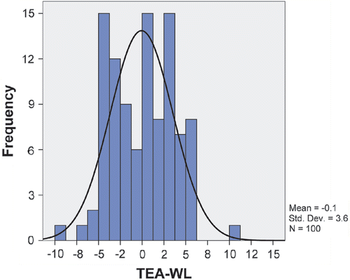
When analyzed as an average, the fixed 3° ER guide matched up very well with the TEA and Whiteside's line. Compared to the TEA, the fixed 3° guide placed the femoral component with a mean of 0.07° internal rotation (IR) (range: 9° internal rotation to 9.5° external rotation), a median rotation of 0° with a standard deviation of 3.4°, and a normal distribution curve. The mean rotation of the femoral component as determined by the 3° fixed guide and Whiteside's line matched perfectly with a mean of 0° (range: 8.5° internal rotation to 11° external rotation), a median of 0.5° IR with a standard deviation of 3.9°, and a normal distribution curve. The difference between these two comparative groups (TEA - Whiteside's line) had a mean value of -0.1° (TEA–Whiteside's line = -0.1), with a 3.6° standard deviation, and a normal distribution (). No difference was found between the two reference systems with regard to femoral component rotation (p = 0.892). Only 2% of the knees were outside two standard deviations (−7.1° to + 7.3°). This validates the commonly held belief that a fixed 3° external rotation cutting guide will consistently match the TEA or Whiteside's line.
Figure 2. Normal distribution curve on transepicondylar axis (TEA) - Whiteside's line with standard deviation value.
Assessing variability within each group showed that the fixed 3° external rotation guide was considerably less accurate. The accuracy was determined by assuming that the registered TEA (or Whiteside's line) was the correct rotation for that particular femoral component. The percentage of cases in which the fixed guide placed the femoral cut within a specific degree with respect to the TEA (or Whiteside's line) thus reflected the accuracy of the guide. If ±1° was the desired endpoint, then the fixed guide was 35% accurate when using the TEA as the reference and only 24% accurate when using Whiteside's line. Looking at this another way, the fixed 3° external rotation guide set a femoral rotational cut differing by more than 1° from the TEA in 65% of knees and by more than 1° from Whiteside's line in 76% of cases. By incrementing the acceptable range to ±2°, only 57% of the knees matched the TEA reference value, and only 49% matched the Whiteside's line reference value. By incrementing the range to ±3°, 71% of the knees matched the TEA reference value, and 62% matched the Whiteside's line reference value. When the range was incremented to ±4°, 80% of the knees matched the TEA reference value, and 75% matched the Whiteside's line reference value. Finally, an increment in the range up to ±5° showed that 86% of the knees matched the TEA reference value, and 83% matched the Whiteside's line reference value (see b). Again, looking at this another way, the fixed 3° external rotation guide set a femoral rotational cut differing by more than 5° from the TEA in 14% of knees and by more than 5° from Whiteside's line in 17% of cases.
Figure 3. (a) The rotational alignment as determined using the posterior reference guide set at 3° of external rotation compared to the transepicondylar axis (TEA). The actual reading for each patient is represented by a histogram. (b) The rotational alignment as determined using the posterior reference guide set at 3° of external rotation compared to Whiteside's line. The actual reading for each patient is represented by a histogram. [Color version available online.]
![Figure 3. (a) The rotational alignment as determined using the posterior reference guide set at 3° of external rotation compared to the transepicondylar axis (TEA). The actual reading for each patient is represented by a histogram. (b) The rotational alignment as determined using the posterior reference guide set at 3° of external rotation compared to Whiteside's line. The actual reading for each patient is represented by a histogram. [Color version available online.]](/cms/asset/9c30b4b0-a73f-45bf-ac9d-fcd3e0544f74/icsu_a_304732_f0003_b.gif)
Variables such as age, gender, race, weight, height, BMI, degree of flexion, degree of extension, and total range of motion, as well as degree of pre-operative valgus or varus alignment, did not influence the results with regard to the femoral rotation registered by these three reference systems.
Discussion
The proper rotational alignment of components during total knee arthroplasty is crucial to the success of this procedure. Various reference guides are currently used by reconstructive surgeons to position the femoral component appropriately. Almost all knee systems using a posterior referencing guide recommend that the femoral cut be performed in 3° of external rotation. Although the reasoning behind this can be comprehended, it is not known if this common practice is likely to apply to all patients. It is conceivable that the femoral component may be rotationally malpositioned in some cases. Furthermore, the same issue may apply to two other commonly used rotational referencing guides, namely the transepicondylar axis (TEA) and Whiteside's line. One of the main questions that remains, and to our knowledge had not yet been investigated, is the degree of correlation between the fixed 3° external rotation guide and the other two referencing guides. The present study found that, on average, the fixed 3° external rotation cutting guide matched the TEA and Whiteside's line reference values as registered using the navigation system. Furthermore, the study found that there was minimal difference between the TEA and Whiteside's line in determining the rotational alignment of the femur. This is important, as some authors have advocated the use of the TEA during surgery Citation[15],Citation[16], whilst others have found the use of Whiteside's line to be a more accurate method for the establishment of rotational alignment Citation[8],Citation[17]. However, a recently published study on cadavers found no difference between the results obtained using either method Citation[18].
A somewhat more critical question is whether a fixed posterior referencing system is applicable to all patients. Based on the findings of this study, the use of fixed-angle instrumentation as the sole guide for a femoral rotational cut could have given rise to rotational malpositioning in a substantial number of patients. Rotational malpositioning, particularly placing the femoral component in internal rotation, could in turn lead to patellofemoral complications and suboptimal outcome of the knee arthroplasty Citation[4]. Another problem with rotational malalignment relates to soft tissue balancing and the potential for flexion instability Citation[19]. Increased external rotation has been reported to affect the medial flexion gap and to lead to symptomatic flexion instability Citation[17]. While malrotation is believed to result in early mechanical failure due to the theoretical increase in stress forces in the femoral and patellar components, little is known about the relationship between the degree of malrotation of the femoral component and the rate of failure Citation[5]. We observed that up to 17% of femoral components could have been placed ±5° outside the acceptable rotational alignment range, highlighting the fact that universal application of a fixed referencing guide may not be appropriate in all cases and some degree of individualization may be needed.
To enable the rotational alignment to be “customized”, some knee designs use posterior referencing systems that allow “dialing in” of a variable degree of external rotation. Based on the findings of this study, we strongly believe that a combination of referencing systems must be employed to minimize the potential for rotational malalignment. While the authors have adopted navigated instrumentation for use in TKA, standard marking of the TEA and Whiteside's line is also carried out to assess rotational alignment before committing to bone cuts. Detailed scrutiny of the rotational alignment of the femur, particularly in patients with “abnormal” anatomy, is indicated to minimize rotational malalignment.
We attempted to identify patients who may be “outliers” and in whom the use of standard fixed-angle devices is precluded. Our results demonstrated no correlation between the examined variables and the rotational status of the distal femur. This was also true for the mechanical axis of the extremity and the pre-operative deformity. In other words, patients with severe valgus or varus deformity were no more likely to exhibit variation in rotational alignment that those without any deformities. It is, however, important to point out that we did not encounter any patients with anatomical aberrations such as hypoplastic condyles.
Limitations
Inter-observer measurements were not obtained in this study due to its nature, being performed during actual surgical procedures and not as a cadaver study, which would render less bias with respect to anatomical landmark identification and its consequent navigational input for the TEA and Whiteside's line.
Conclusion
This study demonstrated a close correlation in accuracy between the three commonly used referencing systems for determining the rotational alignment of the femoral component during total knee arthroplasty. The study also found that the use of a fixed posterior referencing guide alone could lead to femoral rotational malalignment in a significant number of cases. While it has not been established how much variability in femoral rotational alignment can be tolerated for a successful total knee arthroplasty, our recommendation is to take advantage of the anatomical references (TEA, Whiteside's line) to avoid malalignment problems with respect to the femoral component.
References
- Anouchi YS, Whiteside LA, Kaiser AD, Milliano MT. The effects of axial alignment of the femoral component on knee stability and patellar tracking in total knee arthroplasty demonstrated on autopsy specimens. Clin Orthop Relat Res 1993; 287: 170–177
- Oswald MH, Jakob RP, Schneider E, Hoogewoud HM. Radiological analysis of normal axial alignment of femur and tibia in view of total knee arthroplasty. J Arthroplasty 1993; 84: 419–426
- Barrack RL, Schrader T, Bertot AJ, Wolfe MW, Myers L. Component rotation and anterior knee pain after total knee arthroplasty. Clin Orthop Relat Res 2001; 392: 46–55
- Berger RA, Crossett LS, Jacobs JJ, Rubash HE. Malrotation causing patellofemoral complications after total knee arthroplasty. Clin Orthop Relat Res 1998; 356: 144–153
- Miller MC, Berger RA, Petrella AJ, Karmas A, Rubash HE. Optimizing femoral component rotation in total knee arthroplasty. Clin Orthop Relat Res 2001; 392: 38–45
- Hungerford DS, Krackow KA. Total joint arthroplasty of the knee. Clin Orthop Relat Res 1985; 192: 23–33
- Whiteside LA, Arima J. The anteroposterior axis for femoral rotational alignment in valgus total knee arthroplasty. Clin Orthop Relat Res 1995; 321: 168–172
- Arima J, Whiteside LA, McCarthy DS, White SE. Femoral rotational alignment, based on the anteroposterior axis in total knee arthroplasty in a valgus knee. A technical note. J Bone Joint Surg Am 1995; 779: 1331–1334
- Berger RA, Rubash HE, Seel MJ, Thompson WH, Crossett LS. Determining the rotational alignment of the femoral component in total knee arthroplasty using the epicondylar axis. Clin Orthop Relat Res 1993; 286: 40–47
- Griffin FM, Insall JN, Scuderi GR. The posterior condylar angle in osteoarthritic knees. J Arthroplasty 1998; 137: 812–815
- Nagamine R, Miura H, Inoue Y, Urabe K, Matsuda S, Okamoto Y, Nishizawa M, Iwamoto Y. Reliability of the anteroposterior axis and the posterior condylar axis for determining rotational alignment of the femoral component in total knee arthroplasty. J Orthop Sci 1998; 34: 194–198
- Olcott CW, Scott RD. A comparison of 4 intraoperative methods to determine femoral component rotation during total knee arthroplasty. J Arthroplasty 2000; 151: 22–26
- Pagnano MW, Hanssen AD. Varus tibial joint line obliquity: a potential cause of femoral component malrotation. Clin Orthop Relat Res 2001; 92: 68–74
- Hungerford DS, Kenna RV. Preliminary experience with a total knee prosthesis with porous coating used without cement. Clin Orthop Relat Res 1983; 176: 95–107
- Olcott CW, Scott RD. The Ranawat Award. Femoral component rotation during total knee arthroplasty. Clin Orthop Relat Res 1999; 367: 39–42
- Poilvache PL, Insall JN, Scuderi GR, Font-Rodriguez DE. Rotational landmarks and sizing of the distal femur in total knee arthroplasty. Clin Orthop Relat Res 1996; 331: 35–46
- Katz MA, Beck TD, Silber JS, Seldes RM, Lotke PA. Determining femoral rotational alignment in total knee arthroplasty: reliability of techniques. J Arthroplasty 2001; 163: 301–305
- Siston RA, Patel JJ, Goodman SB, Delp SL, Giori NJ. The variability of femoral rotational alignment in total knee arthroplasty. J Bone Joint Surg Am 2005; 8710: 2276–2280
- Krackow KA, Mihalko WM. The effect of medial release on flexion and extension gaps in cadaveric knees: implications for soft-tissue balancing in total knee arthroplasty. Am J Knee Surg 1999; 124: 222–228