Abstract
Objective: To demonstrate the feasibility of an integer programming model to assist in pre-operative planning for open reduction and internal fixation of a distal humerus fracture.
Materials and Methods: We describe an integer programming model based on the objective of maximizing the reward for screws placed while satisfying the requirements for sound internal fixation. The model maximizes the number of bicortical screws placed while avoiding screw collision and favoring screws of greater length that cross multiple fracture planes.
Results: The model was tested on three types of total articular fractures of the distal humerus. Solutions were generated using 5, 9, 21 and 33 possible screw orientations per hole. Solutions generated using 33 possible screw orientations per hole and five screw lengths resulted in the most clinically relevant fixation plan and required the calculation of 1,191,975 pairs of screws that resulted in collision. At this level of complexity, the pre-processor took 104 seconds to generate the constraints for the solver, and a solution was generated in under one minute in all three cases.
Conclusion: Despite the large size of this problem, it can be solved in a reasonable amount of time, making use of the model practical in pre-surgical planning.
Introduction
Distal humeral fractures account for less than 1% of adult fractures. The majority of such fractures result from high-energy trauma, and these types of fractures are among the most difficult to treat. Their management is further complicated by their rarity: outside of trauma centers, most orthopaedic surgeons encounter relatively few of these complex fractures, and are thus unable to accumulate sufficient personal experience in their treatment Citation[1-3]. Therefore, there is a need for surgeons who treat these fractures infrequently to have access to advanced decision-support tools to assist in decision making.
The current standard of treatment for distal humeral fractures is operative with open reduction and stable internal fixation using plates and screws. While not indicated for every fracture of the distal humerus, CT scans and 3D reconstructions are helpful in the pre-operative planning for complex fractures Citation[4]. The outcome of treatment, as evaluated by elbow function, has been observed to be most closely related to the stability of fixation and early range of motion with prompt initiation of physical therapy Citation[5]. Therefore, the objective of treatment with internal fixation is to provide sufficient stability to allow the patient to undergo early post-operative rehabilitation and to provide an environment for bone healing to occur Citation[6]. For this reason, the goal of the surgeon is to maximize the stability of the surgical construct. Failure of fixation most often results from a lack of secure fixation of the plates at the time of surgery due to the use of an inadequate number of screws Citation[2].
Linear programming, which is a type of mathematical programming, is a mathematical model in which a linear function is maximized or minimized subject to a set of linear constraints. Applications of this technique range from models that optimize hospital function, assisting with the management of scarce resources such as operating room time Citation[7],Citation[8] and vaccinations Citation[9], to tools that can assist in treatment planning. The use of linear programming is well established in radiation oncology for the optimization of treatment plans in intensity-modulated radiation therapy (IMRT) Citation[10–15].
Mathematical programming has also been used in surgical planning. Joskowicz and Taylor described an algorithm using integer programming, a subtype of mathematical programming, for computing an interference-free insertion path for a body into a cavity, and demonstrated its application in insertability analyses of orthopaedic hip implants Citation[16]. Computational methods have also been developed to assist surgeons in fracture fixation. Tockus et al. described a system in which 3D bone models generated from pre-operative CT images are used to improve the positioning accuracy of bone segments during closed long bone fracture reduction Citation[17]. This system has also been used to measure periaxial rotation and provide a quantitative measure to the surgeon while aligning the distal and proximal femur segments Citation[18]. However, no work has been published to date describing a system for computer-aided planning of distal humerus fracture fixation.
The purpose of our work was to demonstrate the feasibility of an integer programming model to assist in pre-operative planning for open reduction and internal fixation of a distal humerus fracture. Our model will generate an optimal solution for screw placement while taking into consideration areas of bone that provide better fixation and other areas which must be avoided.
Materials and methods
Surgical anatomy of the distal humerus
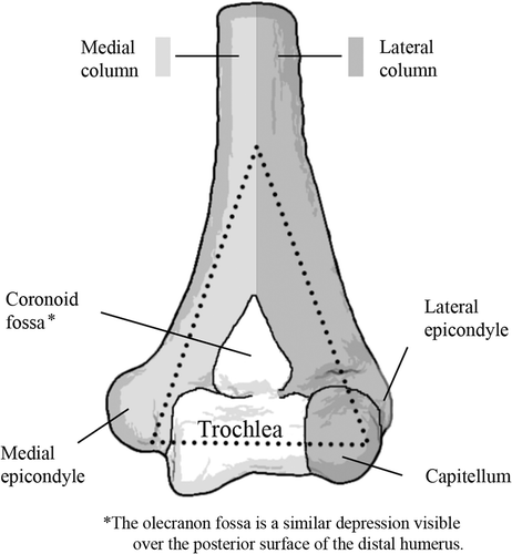
The elbow is a hinge joint with a single axis of rotation formed by the articulation of the humerus and ulna. The shape of the humerus can be represented by two columns that diverge in a wishbone fashion near the distal end of the bone. The trochlea, which resembles a spool, is supported between the columns of the distal humerus. The proximal portion of the ulna articulates around the trochlea, forming the central axis of this joint Citation[2]. The distal humerus also articulates with the radius at the capitellum, a rounded protuberance at the end of the lateral column.
The distal portion of the humerus, formed by the interconnection of the divergent columns, most resembles a triangle in shape (). Disruption of any one of the arms of this triangle weakens the construct considerably Citation[19]. This mechanical property helps to explain the instability of the joint after internal fixation unless all three arms of the triangle are effectively stabilized Citation[2].
On viewing the humerus from a posterior aspect, the olecranon fossa, a triangular depression, is appreciated. This fossa accommodates the proximal tip of the olecranon when the elbow is in full extension Citation[2]. It is essential that the fossa be clear of obstruction to minimize any restriction of movement.
The internal structure of long bones such as the humerus must be considered when planning fixation for fractures. Cortical bone, which forms the outer layer of long bones, is far denser than the cancellous bone that occupies the interior. The increased density of cortical bone allows for better screw purchase and stronger fixation. This structure has significant implications for the effectiveness of screws placed in the distal humerus.
Classification of fractures of the distal humerus
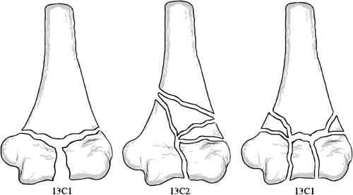
The AO/ASIF (Association for the Study of Internal Fixation) classification for long bone fractures uses an alpha-numeric coding system in which each fracture is provided with a five-character designation. The first number is used to designate the bone in which the fracture has occurred. Each long bone is divided into proximal, diaphyseal and distal segments, and this provides the second number. Fractures within the proximal and distal segments are either extra-articular (type A), partial articular (type B), or total articular (type C) fractures Citation[20]. Total articular fractures are further classified into three subtypes: The C1 fracture is a simple fracture of the articular surface and the metaphysis; the C2 fracture is a simple fracture of the articular surface with comminution of the metaphysis; and the C3 fracture is a multifragmentary fracture of the articular surface and metaphysis Citation[3]. Each of these fractures is further divided into three categories which we did not consider in testing our model. The 13C fracture patterns are illustrated in .
Figure 2. OTA classification of complete articular fractures of the distal humerus (13C): (1) articular simple, metaphyseal simple; (2) articular simple, metaphyseal multifragmentary; and (3) multifragmentary.
Although total articular fractures represent a small percentage of all fractures, they are by far the most common type of distal humerus fracture and the most difficult to treat. These fractures disrupt each component of the distal humeral triangle, resulting in the disassociation of the columns from each other and from the proximal humerus. Treatment of these fractures is further complicated by small fracture fragments and a limited amount of sub-chondral bone, which is often osteopenic Citation[2],Citation[5].
Technique of internal fixation for fractures of the distal humerus
The principle of fixation is to surgically recreate the anatomy of the distal humerus by securing the sides of the humeral triangle to each other and to the more proximal columns. The complex anatomy of the humerus can make this a difficult task for several reasons. One major consideration is that the fragments in these fractures are often small, limiting the number of screws that can be placed. The distal fragments are also composed of cancellous bone, making it difficult to gain adequate purchase with screws. It is also essential that hardware be placed taking care to avoid articular surfaces and the fossae so the patient may regain maximum range of motion.
The periarticular location of the distal humerus prevents the use of standard fixation techniques in repairing these fractures. Upper extremity shaft fractures (humerus, radius, ulna) are typically considered to require a minimum of three screws for fixation on each side of the fracture, but it is not unusual to be unable to achieve three-screw fixation on each side of distal humeral fractures. Surgeons will usually attempt to place as many screws as possible into the articular segment. Long “home run” screws placed up the medial and lateral columns are believed to provide increased fracture stability. However, placement of these screws often affects the ability to place additional screws into the articular segment. Therefore, screw placement involves selecting the optimal set of screw locations given the physical restrictions imposed by the requirement that screws not intersect.
Biomechanical studies have suggested that the ideal construct for plate application is a posterolateral plate and medial plate placed at 90° to one another Citation[21]. However, there are currently no clear guidelines on the placement of screws to avoid hardware impingement and to maximize fixation strength given the complex anatomy of the distal humerus.
Mathematical formulation
In the screw placement problem, the objective is to maximize the reward for screws placed while satisfying the requirements of sound internal fixation. Our model obtains a solution that maximizes the number of bicortical screws that do not collide with each other, favoring screws of greater length that cross multiple fracture planes. The model is also designed to strongly select against screws that penetrate the olecranon fossa.
We start by defining the I holes of the fixation plates i = 1, 2, …, I, K discrete screw lengths k = 1, 2, …, K, and J discrete screw orientations per hole j = 1, 2, …, J. The binary decision variable Xijk defines a screw of length k inserted into hole i at orientation j:The region of space surrounding the humerus is divided into M equal-sized cubes indexed by m=1, 2, …, M. δijkm represents the intersection of screw Xijk with cube m. Let rim be the reward to be gained if a screw from hole i passes through cube m. Let cijk be the combined reward for screw Xijk having passed through cubes each associated with individual rewards rim.
We can formulate the screw placement (SP) problem as a nonlinear integer programming formulation as follows:subject to
where M = {indices of 6 most distal holes of medial plate}
where L = {indices of 5 most distal holes of lateral plate}
where T = {indices of 5 most proximal holes of both plates}
The objective function (2) maximizes the total reward for placed screws. Constraint (3) ensures that at most one screw will be placed in each hole. Constraint (4) ensures that any two screws that intersect will not both be chosen as part of the solution. Constraints (5) and (6) ensure that at least two screws are placed in the distal six and five holes of the medial and lateral plate, respectively. Constraint (7) ensures that at least three screws are placed in the proximal portion of the plates, and constraint (8) sets an upper limit on the number of screws placed.
A pre-processor is necessary to generate the constraints imposed by screw interference. The pre-processor iterates through every combination of orientation and length for every combination of two screws, determining if there is screw interference. Any combination of screws that intersect is added to the constraint matrix barring that pair from the solution using equation (4).
The pre-processor is also responsible for calculating the reward associated with each possible screw placement. The reward associated with each cube is calculated based on the number of fracture planes crossed by a line from the hole under consideration to the cube.
Implementation
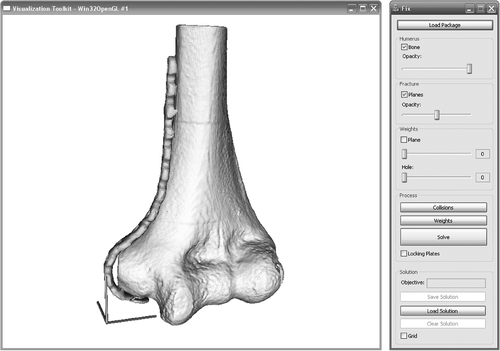
We have implemented our model using the C++ programming language. The graphical user interface was implanted using Microsoft Foundation Classes and the Visualization Toolkit (VTK, Kitware, Inc., Clifton Park, NY). An open source physics library (Open Dynamics Engine by Russell Smith) is used for accurate collision detection. The solutions were generated using an integer program solver (CPLEX, ILOG, Mountain View, CA). The cases were processed on a laptop with a 1.83-GHz Intel Core 2 Duo processor and 1 GB of random access memory. The graphical user interface used to interactively define a fracture pattern and solve for an optimal solution is shown in .
Figure 3. The graphical user interface (GUI) of the system developed for surgical planning. The controls on the right allow the user to load a fracture configuration and generate a solution. Controls are also provided to view the weighting scheme used to generate the solutions.
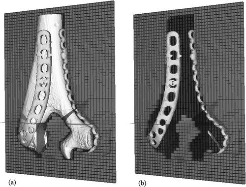
Our implementation requires, as input, a mesh representing the distal humerus, a mesh representing the plates, vectors defining the position of each hole in the plate, and vectors defining the orientations of the fracture planes. These mesh files were created from CT scans by standard segmentation and surface reconstruction methods. The fracture planes were manually defined based on stereotypical fracture patterns of increasing complexity. The plates used in our testing were a 14-hole, pre-contoured, medial, distal humeral plate and a 13-hole, pre-contoured, posterior/lateral, distal humeral plate (Zimmer, Inc., Warsaw, IN).
The 10 cm × 6 cm × 14 cm region of space surrounding the humerus was divided into 53,760 cubes of 2.5 mm on each side for use in reward calculation. One plane of this grid displaying a weighting scheme is shown in .
Results
In this section, we illustrate the practical applicability and computational behavior of this model by using our model to develop a fracture fixation plan for the sub-classes of the total articular fracture of the distal humerus (13C). For each fracture pattern, we quantify the complexity of the model and the time needed to obtain optimal solutions with varying degrees of freedom in choosing screw orientations for each hole.
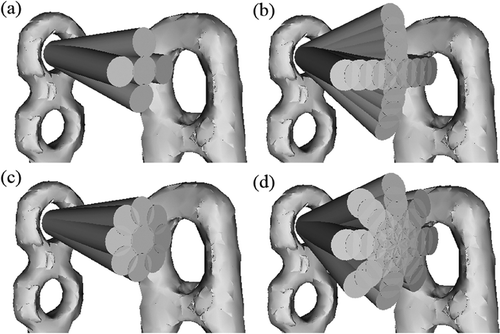
We obtained the optimal solutions for each fracture pattern with varying numbers of discrete screw orientations per hole. The initial implementation of our model allowed for screws to deviate from the neutral position by 10° along four and eight planes. The implementation was revised to allow for more freedom in screw placement by allowing screws to deviate by 5°, 10°, 15° and 20°. These configurations are shown in .
Figure 5. Possible screw orientations in each configuration: (a) 1 deviation, 4 planes; (b) 1 deviation, 8 planes; (c) 4 deviations, 4 planes; (d) 4 deviations, 8 planes.
The solutions generated with each configuration for the 13C1 fracture are shown in . The solutions for 13C2 and 13C3 fractures generated using eight planes and four deviations are also shown in . The pre-operative plan with screw hole and orientation choices for the 13C3 fracture generated with eight planes and four deviations is shown in . Orientation is expressed in degrees of clockwise rotation, with 0° pointing towards the proximal end of the plate. The size of each problem and the execution time for the pre-processor is shown in . The processing time required by the solver to attain an optimal solution for each fracture pattern is shown in . The fossa was not penetrated by a screw in any of the solutions.
Figure 6. Solutions generated with varying degrees of freedom in screw orientation: (a) 13C1, 4 orientations, 1 deviation; (b) 13C1, 4 orientations, 4 deviations; (c) 13C1, 8 orientations, 1 deviation; (d) 13C1, 8 orientations, 4 deviations; (e) 13C2, 8 orientations, 4 deviations; (f)13C3, 8 orientations, 4 deviations.
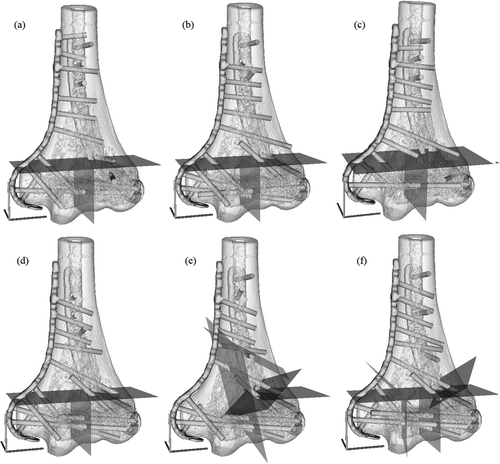
Table I. Pre-operative plan for 13C3 fracture generated with 4 deviations and 8 planes.
Table II. Pre-processor problem size and processing time in seconds.
Table III. Solver processing time in seconds.
Discussion
We demonstrated the application of integer programming in planning distal humerus fracture fixation. Our model implements the principles of internal fixation to generate screw placement plans for a complex fixation problem. As implemented, our model can generate a pre-operative plan when provided with a definition of the fracture to be treated.
Optimizing screw orientation and placement in fracture fixation shares many similarities with optimizing beam orientation in IMRT. However, a fundamental difference between these tasks lies in the value associated with the intersection of the spaces occupied by two beams versus two screws. These techniques also share some of the same limitations, which are overcome using similar methods.
A limitation of applying integer programming in planning surgical fixation is the discretization of possible screw orientations. In reality, a screw may be placed into a hole at any angle within the physical constraints of the plate. It is not feasible to allow this in our model. This problem is similar to one encountered in IMRT planning, where the number of possible configurations of a multi-leaf collimator must be limited to obtain solutions in reasonable time. This limitation was overcome by finding a balance between providing an adequate number of possible screw orientations such that a reasonable solution could be found, and limiting this number sufficiently to allow a solution to be generated in a reasonable amount of time. We succeeded in generating acceptable solutions to the problem in a reasonable time by providing 33 orientations per hole. When collisions are calculated based on 33 orientations, approximately 20 million combinations of two screws are tested for collision to find approximately 1.4 million combinations of screws which collide and must be added to the constraint matrix. Adding one more degree of deviation to the four currently used would increase the number of orientations to 41 and require the pre-processor to determine screw interference for an additional 10 million screw combinations.
The most obvious opportunity for model improvement would be the incorporation of biomechanical considerations into the model. For example, the objective could be modified to include stiffness of the distal humerus construct based on a finite element analysis. This formulation was considered but discarded early on because of the extreme computational burden associated with solving a FEM model for each possible combination of screw placements. Future work will explore improving binary integer programming model solutions methods to allow for FEA subproblems. Another option for improvement of our model is further refinement of the cube-weighting algorithm. The design of our system is such that the only element that needs to be modified to change the solutions generated is the weighting algorithm. The remaining elements of this system are mostly supporting functions, performing straightforward calculations of collision detection and generating a constraint matrix. The solver generates a solution based on the weights associated with each possible screw orientation constrained by collision information. The current implementation of our weighting scheme is relatively simple, being based only on the number of fracture planes that a screw will cross, and was designed to emulate current practices in distal humerus fracture fixation. The weighting scheme can be altered in future revisions to consider positions and sizes of the fragment, awarding higher weights to fragments that are likely to be penetrated by fewer possible screws.
Our initial results show that this problem, while large, can be solved in a reasonable amount of time, making use of the model practical in pre-surgical planning. Further testing necessary to demonstrate the validity of the solutions generated by our model is planned for the future.
Acknowledgments
Funding for this research was provided by the University of Michigan Department of Orthopaedic Surgery. We also thank Jeffrey Meganck for scanning plates for us and Steve Goldstein for providing access to the MicroCT scanner.
References
- Jupiter JB, Neff U, Holzach P, Allgower M. Intercondylar fractures of the humerus. An operative approach. J Bone Joint Surg Am 1985; 67(2)226–239
- McKee MD, Mehne DK, Jupiter JB. Fractures of the distal humerus. Skeletal Trauma: Basic Science, Management, and Reconstruction, Second, BD Browner, JB Jupiter, AM Levine, PG Trafton. Saunders, Philadelphia 1998; 1483–1522
- Müller ME, Allgöwer M. Arbeitsgemeinschaft für Osteosynthesefragen. Manual of Internal Fixation: Techniques Recommended by the AO-ASIF Group. Springer-Verlag, Berlin 1991
- Doornberg J, Lindenhovius A, Kloen P, van Dijk CN, Zurakowski D, Ring D. Two and three-dimensional computed tomography for the classification and management of distal humeral fractures. Evaluation of reliability and diagnostic accuracy. J Bone Joint Surg Am 2006; 88(8)1795–1801
- Gupta R, Khanchandani P. Intercondylar fractures of the distal humerus in adults: a critical analysis of 55 cases. Injury 2002; 33(6)511–515
- Crenshaw AH. Fractures of shoulder girdle, arm, and forearm. Campbell's Operative Orthopaedics, 9th, ST Canale. Mosby, St. Louis 1998; 2281–2362
- Kuo PC, Schroeder RA, Mahaffey S, Bollinger RR. Optimization of operating room allocation using linear programming techniques. J Am Coll Surg 2003; 197(6)889–895
- Mulholland MW, Abrahamse P, Bahl V. Linear programming to optimize performance in a department of surgery. J Am Coll Surg 2005; 200(6)861–868
- Sheldon HJ, Edward CS, Robert D, Bruce GW. An integer programming model for vaccine procurement and delivery for childhood immunization: a pilot study. Health Care Management Science 1999; 2(1)1–9
- Bednarz G, Michalski D, Houser C, Huq MS, Xiao Y, Anne PR, Galvin JM. The use of mixed-integer programming for inverse treatment planning with pre-defined field segments. Phys Med Biol 2002; 47(13)2235–2245
- D'Souza WD, Meyer RR, Shi L. Selection of beam orientations in intensity-modulated radiation therapy using single-beam indices and integer programming. Phys Med Biol 2004; 49(15)3465–3481
- D'Souza WD, Meyer RR, Thomadsen BR, Ferris MC. An iterative sequential mixed-integer approach to automated prostate brachytherapy treatment plan optimization. Phys Med Biol 2001; 46(2)297–322
- Lee EK, Fox T, Crocker I. Simultaneous beam geometry and intensity map optimization in intensity-modulated radiation therapy. Int J Radiat Oncol Biol Phys 2006; 64(1)301–320
- Romeijn HE, Ahuja RK, Dempsey JF, Kumar A, Li JG. A novel linear programming approach to fluence map optimization for intensity modulated radiation therapy treatment planning. Phys Med Biol 2003; 48(21)3521–3542
- Yang R, Dai J, Yang Y, Hu Y. Beam orientation optimization for intensity-modulated radiation therapy using mixed integer programming. Phys Med Biol 2006; 51(15)3653–3666
- Joskowicz L, Taylor RH. Interference-free insertion of a solid body into a cavity: An algorithm and a medical application. Int J Robotics Res 1996; 15(3)211–229
- Tockus L, Joskowicz L, Simkin A, Milgrom C (1998) Computer-aided image-guided bone fracture surgery: modeling, visualization, and preoperative planning. Proceedings of the First International Conference on Medical Image Computing and Computer-Assisted Intervention (MICCAI ’98), Berlin, October, 1998, WM Wells, A Colchester, S Delp. Springer, Cambridge, MA, 29–38, Lecture Notes in Computer Science 1496
- Ron O, Joskowicz L, Simkin A, Milgrom C. Computer-based periaxial rotation measurement for aligning fractured femur fragments: method and preliminary results. Computer Assisted Radiology and Surgery, HU Lemke, MW Vannier, K Inamura, AG Farman, K Doi. Elsevier, Berlin, Amsterdam 2001; 295–300, Proceedings of the 15th International Congress and Exhibition (CARS 2001), June 2001.
- Mehne DK, Matta JK. Bicolumn fractures of the adult humerus. Proceedings of the 53rd Annual Meeting of the American Academy of Orthopaedic Surgeons, New Orleans, LA, February, 1986
- Martin JS, Marsh JL. Current classification of fractures. Rationale and utility. Radiol Clin North Am 1997; 35(3)491–506
- Helfet DL, Hotchkiss RN. Internal fixation of the distal humerus: a biomechanical comparison of methods. J Orthop Trauma 1990; 4(3)260–264
