Abstract
While navigation is now recognized as an efficient tool for improving femoro-tibial alignment of primary knee prostheses, its use in revision surgery has not yet been fully evaluated. We describe a procedure based on a bone morphing acquisition performed on the surface of the original implants, followed by a dependant bone cut sequence (tibia first). Using the current system, a preoperative CT-scan measurement of the original femoral component was required. Knee balancing was achieved using spacer blocks, with the trial tibial component and the original femoral component still in place. Preliminary experience from 19 cases, some with severe bone loss requiring reconstruction, is reported. A retrospective comparison to 10 non-navigated revision cases performed concomitantly by the same operating surgeon was carried out. Although there was no significant difference in the number of outliers for the two series, navigation appeared to be a valuable aid in reconstructing both bone extremities, while controlling the level of the joint line. However, definitive validation requires further prospective and comparative investigations in larger series.
Introduction
Numerous studies have shown navigation to be a valuable tool for improving femoro-tibial alignment in primary knee arthroplasty Citation[1], Citation[2] by reducing the number of outliers outside a 3° interval around the neutral position. However, the use of navigation in revision cases, in which osseous landmarks are often missing due to severe bone loss, remains the subject of investigation. In revision total knee arthroplasty, surgeons face several challenges, including the restoration of the joint line level and ensuring optimal patellar tracking. Because the position of the initial implant may not be satisfactory, it cannot be assumed to be a dependable anatomical reference for the rest of the procedure. In fact, the first step consists of deciding whether the joint level and the femoral rotation of the original prosthesis need to be changed. This decision relies on several parameters such as the ligamentous status, the severity of the bone destruction, and the reason for the failure of the original implant. A preliminary experiment was carried out to evaluate how navigation would help in applying the initial planning. To date, few papers have been published on navigated revisions, some of them dealing with computer-guided bone reconstruction of cavitary defects Citation[3]. Other recent preliminary reports agree that CT-free navigation improves leg alignment in revision surgery Citation[3–5].
We describe an imageless computer-aided procedure for revision total knee replacement, based on the bone morphing technique followed by a dependant bone cut (tibia first) sequence. Because no revision-dedicated software was available, the present study was carried out using a system designed for primary surgery.
Nineteen computer-aided revisions were performed, five of which were necessitated by failed unicompartmental arthroplasties with moderate bone loss and 14 by failed total knee prostheses, some with severe femoral bone loss.
An attempt was made to answer three questions: 1) Does navigation allow a better femoro-tibial alignment, by retrospective comparison to a non-navigated series from the same operator?; 2) Is navigation an efficient tool only in those simple cases with no or only moderate bone loss (revision of uniarthroplasties), or is it also useful in more difficult cases requiring bone reconstruction of one or both extremities?; and 3) Is a system designed for primary surgery also adequate for revision surgery? In trying to answer these questions, these very short-term clinical and radiological results were examined with special regard to the position of each component and to leg alignment.
Materials and methods
Materials
Nineteen patients had their knee prosthesis revised using a navigation system (PRAXIM Medivision, La Tronche, France) designed for primary implantation of the PFC total knee prosthesis (Depuy, Warsaw, IN). The average age of the patients was 66 years (range: 20–80 years) and they comprised 6 men and 13 women.
Seven patients had previously undergone multiple operations (tibial osteotomies, drainages due to infection), and three knees had been replaced two or more times. Five revisions were justified by the failure of medial unicompartmental prostheses: loosening of the femoral component in one case, loosening of the tibial component in two cases, and unexplained pain without loosening in two cases. In the other 14 patients, revision was required following the failure of total knee prostheses. Six of these revisions were related to the polyethylene insert: in one case this was loosened from the tibial baseplate, while in the other five cases the insert was worn through. In all six cases, the femoral component had rubbed against the metallic baseplate, provoking a severe metallosis with irreversible damage to both components, and an associated severe osteolysis below the tibial baseplate in one patient. Three patients were revised due to aseptic loosening of one or both components (the tibial component in one case, the femoral component in another case, and both components in the third case), and two other patients were revised due to severe instability, having a severe malunion of a supra-condylar fracture, combining varus, internal rotation and recurvatum. Finally, three patients had a chronic infection: In these patients, a two-stage revision was carried out with temporary implantation of cemented spacers that remained in place for 3, 8 and 36 months, respectively.
All revisions were performed by the same operating surgeon (P.M.), who also performed 82 navigated primary cases over the same period of time (2005 to 2007). All but one patient with failed unicompartmental prostheses were revised using standard knee prostheses. The other 15 patients were revised using a TC3 postero-stabilized implant with metallic augments and stems (Depuy, Warsaw, IN). Severe bone segmental defects of the femoral condyles were present in 10 cases, requiring metallic augments in 10 cases and reconstruction using a femoral head bulk allograft in one case. There were major cavitary tibial defects in three cases, which were filled using compacted allografts in two cases and a metallic conical sleeve in one case.
A control group was retrospectively constituted, including 10 revisions performed without navigation by the same operator over the same period of time. The demographic data are shown in .
Table I. Main characteristics of the navigated and non-navigated groups.
Methods
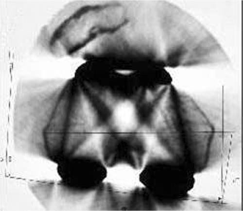
The height of the joint line relative to the head of the fibula was measured on both sides on 100% magnification AP X-rays centered on the upper tibia. Preoperative determination of the femoral component rotation was obtained using a CT-scan coronal view of the lower femur at the level of the most prominent points of both femoral epicondyles (). Comparative bilateral measurements of the posterior condylar angle were performed. In the absence of a healthy contralateral knee, the decision regarding changing the level of the joint line and/or the femoral component rotation was based on the presence of a patellar tilt and on the patellar height relative to the joint line.
Figure 1. A preoperative CT scan of the lower femur was required to determine the posterior condylar angle with the original femoral component in place. In this example, the femoral component was implanted in internal rotation with a subluxated patella.
Knees were exposed without tourniquet through a medial subvastus (15 cases) or a medial parapatellar (4 cases) approach. An osteotomy of the tibial anterior tubercle was required in 4 cases to optimize exposure and knee flexion.
The femoral pins supporting the beacons were placed medially as close as possible to the joint () and the tibial pins were placed distally, 20 cm from the joint line, to prevent impingement with the stems ().
Figure 2. a) Peroperative view showing severe destruction of both condyles. b) The femoral pins are placed medially, so as not interfere with the preparation of the medullary canal. Two hemi-femoral head allografts were required to reconstruct the femoral condyles. [Color version available online.]
![Figure 2. a) Peroperative view showing severe destruction of both condyles. b) The femoral pins are placed medially, so as not interfere with the preparation of the medullary canal. Two hemi-femoral head allografts were required to reconstruct the femoral condyles. [Color version available online.]](/cms/asset/ceb3a999-4e66-4737-9c44-97f8912e6d46/icsu_a_323251_f0002_b.gif)
Figure 3. Operative view showing the placement of the beacons on the tibia and femur. [Color version available online.]
![Figure 3. Operative view showing the placement of the beacons on the tibia and femur. [Color version available online.]](/cms/asset/300b663d-6068-4b4b-8cbe-4a6aecbba83e/icsu_a_323251_f0003_b.gif)
Bone morphing was performed on the surface of the original prosthesis. It allowed determination of the rotational position of the femoral component by palpation of the posterior bicondylar axis, and fixation of the level of the joint line for the rest of the operation. When the preoperative planning suggested lowering the joint line, bone morphing was performed on the surface of the tibial baseplate after removal of the polyethylene insert. The initial level of the joint line served as a reference for balancing the knee and calculating the size of the bone defects. In two cases, the femoral components were grossly loosened and mobile and had to be removed immediately. Provisional implants of approximately the same size were implanted in the same rotation as the original component, and were stabilized using an intramedullary stem of adequate size and temporary screws. Bone morphing was then performed on the surface of this trial implant. In one case, in which there was a major deformation due to malunion of a supracondylar fracture, a metaphyseal osteotomy of the lower femur was first performed and then stabilized using the stem of the trial femoral component before proceeding with bone morphing.
The tibial component was then removed, and an (as thin as possible) navigated tibial cut was made to expose the healthy bone (). The computer indicated the difference in height between the real cut and the ideal level (defined as being 10 mm below the joint line), thus giving an indication to eventually augment the thickness of the tibial component (). After preparing the medullary canal, the diameter and size of the tibial stem were determined and the tibial component of the largest possible size was assembled, with its stems and eventual metallic augments, and a 10-mm trial insert.
Figure 4. Operative view showing the navigation of the tibial cut. [Color version available online.]
![Figure 4. Operative view showing the navigation of the tibial cut. [Color version available online.]](/cms/asset/a6bb9906-5b5c-4449-84ce-6ab1844568cc/icsu_a_323251_f0004_b.gif)
Figure 5. Screenshot showing the difference in height between the ideal cut (with reference to the level of the joint line) (blue line) and the actual bone cut through the remaining bone (yellow line). In this example, the bone defect was measured at 10 mm. [Color version available online.]
![Figure 5. Screenshot showing the difference in height between the ideal cut (with reference to the level of the joint line) (blue line) and the actual bone cut through the remaining bone (yellow line). In this example, the bone defect was measured at 10 mm. [Color version available online.]](/cms/asset/a3434899-6a06-4a7f-83a9-500f8c0cdda1/icsu_a_323251_f0005_b.gif)
Balancing the knee was achieved using spacer blocks with the provisional tibial component and the original femoral component still in place, or using a stable trial femoral component in two cases, as described above. The extension gap was balanced first, then the flexion gap, thus determining the size and rotation of the definitive femoral component. The effective rotation of the femoral component was deduced from its preoperative value (as determined on the preoperative CT scan) augmented by the peroperative variation in rotation displayed by the computer. If these goals were not attained despite adequate sizing and rotation of the femoral component, the polyethylene thickness was increased until the flexion gap was stabilized. This raised the level of the joint line by a certain number of millimeters that was also automatically applied to the extension gap. In those cases, in which an osteotomy of the anterior tibial tuberosity was required, care was taken at the end of the operation to fix the tuberosity in a higher position to compensate for the joint line elevation and to prevent patella bara.
The ideal bone cut level was memorized by the computer and the femoral component was removed. The femoral bone cuts were navigated, placing the saw guide on an intramedullary jig (). This allowed selection of the adequate femoral angle, while performing the distal cuts at the level of the remaining healthy bone, if present. As on the tibial side, the computer indicated the difference in height between the ideal and actual bone cuts, thus allowing determination of the thickness of the distal, lateral, medial and, if necessary, posterior augments. Stems with a 5-mm posterior offset were used in five cases. Distal femoral augments 5 to 16 mm thick were required in 10 cases, while tibial augments were used in only three cases. In one case, a massive femoral allograft was used to reconstruct a major femoral bone loss ().
Figure 6. Preoperative view showing the navigation of the distal femoral cut, with the cutting guide mounted on an intra-medullary jig. In the present system, two femoral angles were proposed (5° and 7°). If one of these angles was adequate, canal-filling femoral stems could be used; if neither of them was adequate, a thinner stem was used, allowing some degree of tilting until the correct angle was obtained. [Color version available online.]
![Figure 6. Preoperative view showing the navigation of the distal femoral cut, with the cutting guide mounted on an intra-medullary jig. In the present system, two femoral angles were proposed (5° and 7°). If one of these angles was adequate, canal-filling femoral stems could be used; if neither of them was adequate, a thinner stem was used, allowing some degree of tilting until the correct angle was obtained. [Color version available online.]](/cms/asset/b0234fba-e662-434b-9160-662ed4602ddb/icsu_a_323251_f0006_b.gif)
The definitive trial femoral component was finally assembled with its stem and augments and a reduction was performed to check the final alignment and stability. If satisfactory, the definitive components were inserted, being cemented only in their proximal part, whereas the canal-filling stems were left uncemented.
In the control group, metallic femoral augments were required in 4 of the 10 cases. Standard implants were used in only three cases on the femoral side and two cases on the tibial side. In the other cases, the same implants were used as in the navigated group, except for one case with gross instability which required a hinge prosthesis. The level of the joint line was determined according to the preoperative planning, and was measured relative to a metallic marker inserted in the tibial metaphysis at the start of the operation.
Patients were evaluated clinically and radiologically at 3, 6 and 12 months after the operation using the IKS score and based on non-weight-bearing anteroposterior and lateral X-rays of the operated knee, as well as on a skyline view and long-leg standing radiographs. The distribution of the HKA angles, the operative time, and the rate of complications in the navigated series and control group were compared using the Wilcoxon 2-sample test with a 0.05 level of significance.
Results
The average operation time for the navigated procedures was 3 hours and 20 minutes (120–300 min). With a 6-month minimum follow-up and an 11-month average follow-up, there were three postoperative infections, two of them in patients who had been revised for chronic infection. A joint drainage was performed in association with adapted and prolonged antibiotherapy. There was no recurrence at 11, 12 and 26 months, respectively, after the joint drainage.
Postoperative HKA angles ranged from 176 to 185°. For a (−3°; +3°) target interval, there were three outliers (176°, 185°, 185°), as measured on long-leg standing radiographs, while the values obtained from the navigation report indicated 179°, 180° and 182°, respectively. One outlier (176°) occurred in a uni revision, while the other two occurred in revisions of total knee prostheses with severe bone loss. When they were analyzed using the Cusum plot Citation[6] and the Cusum test, as described by Nizard et al. Citation[7], the femoro-tibial axes were contained within the decision interval (−3.2°; +3.2°) if the accepted error was 3° around the neutral axis ().
Figure 7. Cusum test showing that the values of the femoro-tibial axis were inside the decision interval (−3.2; +3.2). [Color version available online.]
![Figure 7. Cusum test showing that the values of the femoro-tibial axis were inside the decision interval (−3.2; +3.2). [Color version available online.]](/cms/asset/e3f48ae6-8dd9-4340-9672-c98cb47c358a/icsu_a_323251_f0007_b.gif)
Due to laxity in flexion, seven patients received PE inserts of thickness greater than 12 mm (including one 20-mm-thick and two 25-mm-thick inserts). One patient with a 25-mm-thick PE and major patella bara complained of instability. The patella was laterally dislocated. In the other patients, the patella appeared centered on the skyline view except in three cases in which it was slightly lateralized.
In accordance with the preoperative estimation and the correction that was applied during surgery, the postoperative femoral rotation was always contained within a 0–5° interval of external rotation. In five cases, the femoral component had to be externally rotated by 5, 5, 5, 10 and 10°, respectively, to correct a preoperative malposition in internal rotation, while the femoral rotation was not changed in the other cases. The tibial slopes were measured at 0° in 15 cases, 3° in three cases, and 4° in one case.
In the overall series, the average 6-month flexion at last follow-up was 105°, with an average gain of 4° (−20°; +30°). The IKS knee scores were satisfactory (> 85) in 16 cases, fair in one case (IKS 75), and poor in two cases (IKS 28 and 56).
In the control group, the average operation duration was 3 hours (145–210 min). There were five complications, including two recurrent loosenings at 20 and 24 months, respectively, after re-implantation, one of them in a patient who had undergone four previous knee replacements. Another patient without loosening with a patella bara was re-revised due to unexplained pain. One patient complained of instability and was revised successfully by implanting a thicker polyethylene insert. Finally, there were two infections that required joint debridement and drainage with apparent success after more than two years of follow-up.
The average postoperative HKA angle was 181° (180°–185°). The femoral rotation was not measured postoperatively; however, all patellae were centered on the skyline view except for one with a slight lateral translation.
The PE thickness exceeded 12 mm in 5 cases (15–20 mm). The tibial slope was 0° in five cases, 3° in three cases, and respectively 4° and 6° in the last two cases. With an average follow-up of 18 months (range: 6–24 months), the IKS scores were satisfactory in all but two cases. The average flexion at last review was 107° (range: 90–120°) and the average gain of flexion was −3° (−20°; +20°).
There was no significant difference between the two groups with respect to the HKA angle (p = 0.76), the rate of complications (p = 0.8), or the duration of the procedure (p = 0.52), although the latter was longer, at an average of 20 min, in the navigated group.
Results for both groups are summarized in .
Table II. Position of the components as measured on the postoperative X-rays. Mean and standard deviation (SD) are given for each measurement in the navigated and non-navigated groups. The patellar tilt was measured on a skyline view: It is the angle between a line connecting the anterior limits of the condyles and a line drawn through the prosthesis bone interface. If the angle is positive, the patellar angle is considered normal; if the angle is zero or reversed (luxation), patellar tilt is considered abnormal. The medial tibial angle was measured on an AP view of the upper tibia: It is the medial angle between the line drawn though the tibial baseplate and the tibial anatomical axis. The lateral femoral angle was measured on an AP view of the lower femur: It is the lateral angle between the line connecting the distal limits of the condyles and the femoral anatomical axis. The femoral slope was measured by the angle between the femoral anatomical axis and the line perpendicular to the tangent of the femoral component on a lateral view of the lower femur.
Discussion
The present results must be considered as preliminary and report only the very short-term outcome following navigated revision of a failed knee arthroplasty. Therefore, such data are insufficient to validate the method, but are intended to describe and demonstrate the feasibility of the procedure that should now be evaluated prospectively and comparatively. However, a fair comparison with a controlled non-navigated group is likely to be very difficult in revision surgery, which involves so many parameters, such as the severity of the bone defects, the laxity, and the reasons for revision. Although it was designed retrospectively, the present study involved two groups of patients who were comparable with regard to the severity of the femoral bone destruction and the presence of a laxity in flexion, and who were operated on by the same surgeon with the same sequence using the same implants.
Navigation did not improve the postoperative HKA angle. In fact, some outliers were identified on long-leg standing radiographs even though the navigation reports were satisfactory. Thus, it is probable that navigation ignored some degree of deformity due to residual laxity that would only appear in weight-bearing situations. Although the present report failed to show any improvement in the short-term results for navigated revision surgery, it is the authors’ opinion that navigation contributed valuable information throughout the procedure, at almost no extra time expense. While navigation required careful preoperative planning, it helped the surgeon to keep the reference joint level in mind throughout the operation. This is important in those knees in which the flexion gap balance may be difficult to achieve due to significant preoperative frontal laxity. In the case of major laxity in flexion, navigation showed the exact amount of joint line elevation required to stabilize the joint. Thus, navigation can be a helpful decision-making tool for choosing more constrained implants in these cases, in which the amount of joint elevation exceeds 1 cm relative to the ideal level. In particular, it is noteworthy that the poor clinical results in this series were obtained in patients with severe patella bara. Retrospectively, it is the authors’ opinion that these patients might have benefited from a hinge prosthesis, allowing for a lower joint level that would have optimized patellar tracking and extensor mechanism function. The system was of particular assistance in those cases with severe bone destruction because it helped calculate the height of bone to be reconstructed. In particular, it restored a satisfactory anatomy in the three infected knees that had a cemented spacer, as already described by Perlick et al. Citation[8]. The case requiring femoral reconstruction using a bulk allograft did not present any major difficulties, because navigation allowed the bone cuts to be performed through the allograft at the level that had been memorized at the start of the procedure, just like in a primary case.
Finally, the system designed for the primary procedure proved efficient in revision cases as well. Its most important features were the efficiency of the bone morphing software that allowed a model to be obtained regardless of the shape of the initial implant, and the measurement of the difference in height between the ideal and actual bone cuts.
In conclusion, although the retrospective comparison with a non-navigated series showed no significant difference, navigation is considered to be a valuable aid in guiding reconstruction of both extremities, while controlling the level of the joint line. It proved useful in simple as well as more complicated cases. The navigation system designed for primary surgery appeared to be convenient for performing revision surgery. The method requires careful preoperative planning, including a CT scan to measure the preoperative rotation of the femoral component. Further prospective evaluations are necessary.
References
- Bauwens K, Matthes G, Wich M, Gebhard F, Hanson B, Ekkernkamp A, Stengel D. Navigated total knee replacement. A meta-analysis. J Bone Joint Surg Am 2007; 89: 261–269
- Bäthis H, Shafizadeh S, Paffrath T, Simanski C, Grifka J, Lüring C. [Are computer assisted total knee replacements more accurately placed? A meta-analysis of comparative studies]. Orthopade 2006; 35: 1056–1065
- Sikorski JM. Computer-assisted revision total knee replacement. J Bone Joint Surg Br 2004; 86: 510–514
- Perlick L, Bäthis H, Perlick C, Lüring C, Tingart M, Grifka J. Revision total knee arthroplasty: a comparison of postoperative leg alignment after computer-assisted implantation versus the conventional technique. Knee Surg Sports Traumatol Arthrosc 2005; 13: 167–173
- Thielemann FW, Clemens U, Hadjicostas PT. Computer-assisted surgery in revision total knee arthroplasty: early experience with 46 patients. Orthopedics 2007; 30: S132–S135
- Nix AB, Rowlands RJ, Kemp KW, Wilson DW, Griffiths K. Internal quality control in clinical chemistry: a teaching review. Stat Med 1987; 6: 425–440
- Nizard RS, Porcher R, Ravaud P, Vangaver E, Hannouche D, Bizot P, Sedel L. Use of the Cusum technique for evaluation of a CT-based navigation system for total knee replacement. Clin Orthop Relat Res 2004; 425: 180–188
- Perlick L, Bäthis H, Tingart M, Grifka J. Implementation of a CT-based navigation system in two-stage reimplantation for infected total knee arthroplasty. Acta Orthop Belg 2003; 69: 355–360