Abstract
Objective: During computer navigated hip arthroplasty, impingement of soft tissues on reference markers (RMs) may cause errors in the navigation process. The aim of this study was to investigate the effect of such soft tissue impingement during limb movement on the stability of different RMs.
Materials and Methods: The stability of one- and two-screw RM systems inserted using three different levels of soft tissue dissection was analyzed in fresh cadaver lower limbs. All tests were done with RMs inserted in both the distal-anterior femur and the distal-lateral femur.
Results: Rotations of less than 0.15° and translations of less than 0.4 mm occurred in most test combinations. The combination that showed the greatest instability was when a stab incision was used to insert a screw in the distal-lateral femur (translation: 0.73 ± 0.05 mm; rotation: 0.25 ± 0.05°) (p < 0.001). This instability occurred in both single- and double-screw RMs (p = 0.21).
Conclusions: Reference markers can be placed in the anterior distal femur through stab incisions without resulting in significant impingement during limb movement. When RMs are placed in the lateral distal femur, impingement may occur from the fascia lata. Release of the fascia lata 1 cm either side of the screw prevents significant impingement. Wide skin incision is unnecessary in any location.
Introduction
Computer aided orthopaedic surgery (CAOS) has become widely available for a broad range of orthopaedic interventions including hip Citation[1], Citation[2] and knee Citation[3] arthroplasty, spinal surgery Citation[4], trauma surgery Citation[5], Citation[6] and high tibial osteotomy Citation[7]. The use of CAOS can undoubtedly reduce the variability of implant placement Citation[3], and may reduce the complication rate by increasing the accuracy of percutaneous screw placement Citation[8]. It is postulated that the improved accuracy of implant placement will lead to improved long-term survival rates, but this has not been proven.
CAOS relies on spatial information provided by reference marker arrays attached rigidly to bone. These arrays are visualized in space by a binocular camera system and the data is integrated by computer, allowing calculation of all necessary variables determining bone position, axis and joint centers. Therefore, CAOS is only as accurate as the information it receives from the reference markers, and any instability in these markers will lead to inaccuracies in the navigation process and a substandard outcome. Reference markers are inserted either within the surgical field, free from soft tissue impingement, or percutaneously at sites distant from the exposed areas. Movement of the limb may cause soft tissue impingement and consequently disturb these percutaneously inserted reference markers, causing registration errors which might not be detected by the operating surgeon intraoperatively. In particular, the limb movements required during total hip replacement, e.g., the figure-of-four position, knee flexion and hip adduction, all create tension in the rectus femoris and fascia lata, potentially impinging on the RM. The purpose of this study was therefore to investigate the effect of different degrees of soft tissue dissection on the stability of reference markers in the femur during simulated limb movements such as occur during total hip replacement.
Materials and methods
The experiments were performed on fresh intact cadavers placed on a mobile operating table. Cadavers had no previous lower limb surgery or trauma. The Hannover Medical School review board approved this human subject study before any tests were performed.
We evaluated both single-screw () and double-screw () minimally invasive reference arrays (MIRA system, BrainLAB, Heimstetten, Germany) in two different positions on the femur. The positions used were as follows:
15 cm proximal to the superior pole of the patella in the sagittal plane (“Anterior”).
10 cm proximal to the lateral femoral epicondyle in the coronal plane (“Lateral”).
1-cm stab incision in the skin with no further dissection.
3-cm skin incision with dissection through muscle/fascia.
As in B, but with additional complete freeing of muscle from the bone surrounding the screw.
Maximum external then internal hip rotation in neutral hip flexion.
Flexion of hip to 90° and maximal knee flexion.
Flexion of hip and knee to 90° and maximum internal/external hip rotation.
Flexion of hip to 45° and maximum hip adduction.
Figure 1. Single-screw MIRA system.
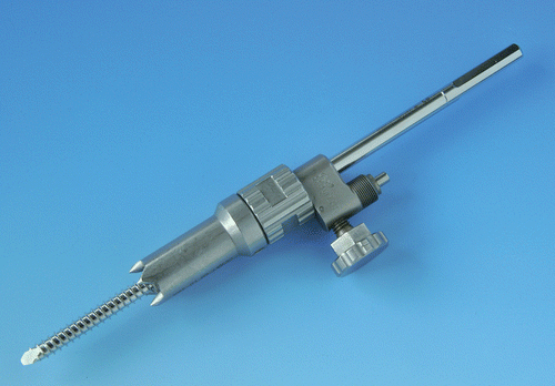
Figure 2. Double-screw MIRA system.
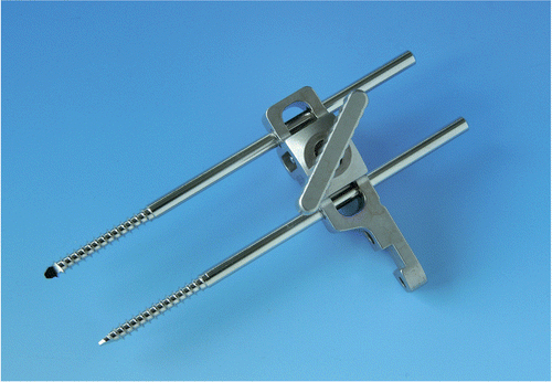
Figure 3. (A) Two-pin RM inserted via stab incisions. (B) Two-pin RM inserted via larger incision and soft tissue dissection. (C) Two-pin RM inserted with full dissection of muscle from bone. (D) Single-pin RM inserted with full dissection of muscle from bone.
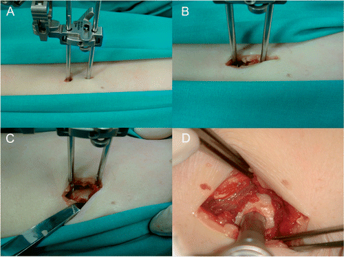
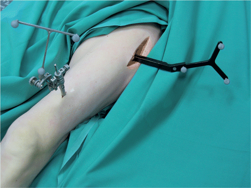
Figure 4. The super-stable reference array (right) and the percutaneously inserted single-pin MIRA reference array in the lateral position.
All tests were accomplished on unfixed cadaver limbs in order to reproduce similar tissue rigidities to those in live patients. The tests were performed on four limbs, with two observers repeating each test ten times.
Statistical analysis was performed using SPSS software. For our evaluation, the average value including the standard deviation of each test series was calculated. Student's T-test was used to assess the difference between means, with the significance level set at 0.05.
Results
The tests showed that defined movements of the lower extremity caused measurable deviations between the test reference array and the super-stable array, indicating relative movement between the test array and the femur. No complications occurred during the test series.
In test series I (maximum external and internal hip rotation in neutral hip flexion), the mean translational and rotational movements for both one- and two-screw RMs inserted in the anterior femur with all dissection levels were 0.33 mm (SD = 0.03) and 0.11° (SD = 0.02), respectively. For one- and two-screw RMs inserted into the lateral femur through a stab incision, the mean translational and rotational movements were 1.56 mm (SD = 0.11) and 0.40° (SD = 0.06). When further dissection was carried out with release of the fascia lata, the movements were reduced to 0.33 mm (SD = 0.03) and 0.12° (SD = 0.03). These differences were significant (p < 0.01).
Test series II (maximum flexion of the hip and knee) gave similar results for the anteriorly placed RMs (one- and two-screw in all dissection levels) of 0.39 mm (SD = 0.03) and 0.13° (SD = 0.02). Laterally placed RMs showed the greatest instability when placed through a stab incision [2.23 mm (SD = 0.10); 0.51° (SD = 0.05)]. Greater dissection again resulted in reduced movements [0.42 mm (SD = 0.02); 0.13° (SD = 0.02)] (p < 0.01).
Test series III (hip flexion to 90° and maximum internal/external rotation) produced minimal displacements in the anteriorly placed RMs [0.39 mm (SD = 0.03); 0.12° (SD = 0.02)]. Laterally placed RMs again showed the greatest movement when inserted through a stab incision [2.15 mm (SD = 0.07); 0.42° (SD = 0.07)]. Release of the fascia lata reduced the impingement resulting in less movement [0.37 mm (SD = 0.03); 0.12° (SD = 0.02)] (p < 0.01).
Test series IV (flexion of the hip to 45° followed by maximal adduction) confirmed the previously observed results. Movements for anterior screws remained similar [0.39 mm (SD = 0.03); 0.13° (SD = 0.03)]. Laterally inserted screws again showed the highest movement when inserted percutaneously [2.19 mm (SD = 0.08); 0.56° (SD = 0.03)] compared to more invasive dissection [0.35 mm (SD = 0.04); 0.12° (SD = 0.02)] (p < 0.01).
Comparing RM types showed mean translation and rotation of 0.44 mm (SD = 0.03) and 0.14° (SD = 0.02) for the single-screw RM for all site/dissection combinations and movements of 0.41 mm (SD = 0.03) and 0.14° (SD = 0.03) for the double-screw RM. The differences were not significant (p = 0.51 for translation and p = 0.73 for rotation).
summarizes all the results in detail, showing relative translational and rotational movements for all RM/soft tissue dissection combination and movement series. Mean values ± one standard deviation are shown.
Figure 5. Graphs showing (A) mean relative translation (mm ± SD) and (B) mean relative rotation (degrees ± SD) of RM during movement series I-IV. A: Single-pin, anterior femur, stab incision. B: Single-pin, anterior femur, muscle release. C: Single-pin, anterior femur, complete dissection. D: Double-pin, anterior femur, stab incision. E: Double-pin, anterior femur, muscle release. F: Double-pin, anterior femur, complete dissection. G: Single-pin, lateral femur, stab incision. H: Single-pin, lateral femur, muscle release. I: Single-pin, lateral femur, complete dissection. J: Double-pin, lateral femur, stab incision. K: Double-pin, lateral femur, muscle release. L: Double-pin, lateral femur, complete dissection. [Color version available online.]
![Figure 5. Graphs showing (A) mean relative translation (mm ± SD) and (B) mean relative rotation (degrees ± SD) of RM during movement series I-IV. A: Single-pin, anterior femur, stab incision. B: Single-pin, anterior femur, muscle release. C: Single-pin, anterior femur, complete dissection. D: Double-pin, anterior femur, stab incision. E: Double-pin, anterior femur, muscle release. F: Double-pin, anterior femur, complete dissection. G: Single-pin, lateral femur, stab incision. H: Single-pin, lateral femur, muscle release. I: Single-pin, lateral femur, complete dissection. J: Double-pin, lateral femur, stab incision. K: Double-pin, lateral femur, muscle release. L: Double-pin, lateral femur, complete dissection. [Color version available online.]](/cms/asset/dce7af46-6d83-4533-ad0a-f31a6b89bca8/icsu_a_326951_f0005_b.gif)
Discussion
Rigid fixation of a reference marker to the surgical extremity or trunk remains a crucial step in navigated orthopaedic procedures Citation[5], Citation[9]. Any relative movement between the marker and the bone will result in inaccurate navigation with potential inaccuracy in the surgical technique. Recent work has suggested that non-invasive reference markers do not achieve adequate stability during limb movement Citation[10]. Desired accuracy for most navigated procedures should be within 1 mm and 1° Citation[4], Citation[9], Citation[11], Citation[12]. Users should be able to transfer these values into their measurements of the leg axis, soft tissue balancing or implant placements Citation[1], Citation[3], Citation[7], Citation[13], Citation[14]. Many computer navigated techniques allow RM fixation in areas with little soft tissue movement, one such example being navigated pedicle screw placement Citation[15], Citation[16]. However, in total hip arthroplasty the marker must be placed in the femur distal to the proposed stem position, where significant soft tissue movement occurs during handling of the limb throughout the surgical procedure. The choice of surgical approach will dictate the positioning of the marker: the posterior approach necessitates fixation in the lateral femur, whereas the lateral or trans-trochanteric approach allows for fixation in the anterior femur.
Previous studies have investigated the stability of RMs during limb movement but have not assessed the effect of soft tissue dissection Citation[17]. We have shown that RM screws can be safely inserted in the anterior distal femur through a 1-cm stab incision without any detrimental effect on stability during limb movement when compared to the same RM inserted following complete soft tissue dissection. However, when the lateral distal femur is used for RM screw insertion, the use of a stab incision alone is associated with significant translational and rotational movements during limb movement. Increasing the length of the skin incision to 3 cm and releasing the fascia lata is sufficient to ablate this excess movement. Further dissection including complete release of the underlying musculature from the bone around the screw has no additional effect. When appropriate soft tissue dissection is performed, equal stability is achieved in both the lateral and anterior positions. This is in contrast to the findings of Mayr et al. Citation[17], who suggested that the lateral position was superior to the anterior position. The difference in our findings is probably related to the RM type used in this study, which is a very stable design. This is demonstrated by the relative differences in stability between the two studies. Our observations show that most translational movements were less than 0.4 mm, whereas the lowest mean translation in the report by Mayr et al. was 1.8 mm. Similarly, our rotational movements of approximately 0.15° compare favorably with the 1.5° to 2.5° observed by Mayr et al.
In comparing one- and two-screw RM devices we found no significant differences in observed stability. Little data is available in the literature to confirm our findings. One previous study showed superior stability in two-screw fixators when tested with incremental loads in a material testing machine Citation[18]. Whilst this is an important finding, and the biomechanics of a two-screw device will always be inherently superior, we have shown that in the clinical situation the one-screw device is working within what can be termed its window of stability. We therefore have no evidence to suggest that the added potential morbidity of a two-screw device is justified.
We found little difference in RM stability between the different limb movement series in the stable situations. When significant instability was seen, then the movement that generated the least instability was rotation in hip extension. Flexion of the hip and knee and adduction both tighten the fascia lata, which explains why the instability was seen in movement series II, III and IV. These findings are clinically relevant, as it is these movements that occur during hip arthroplasty.
The limitations of our study arise from the limited number of cadavers representing specific soft tissue conditions. Specimens with higher muscle mass and obesity might lead to even higher deviations of the reference array positions relative to the femur.
In conclusion, anterior percutaneous fixation of reference markers to the distal femur does not result in significant soft tissue impingement during limb movement. When markers are inserted laterally, care should be taken to release the fascia lata from around the screw to alleviate any soft tissue impingement. One-screw fixation systems can be as stable as two-screw systems provided they are designed and inserted appropriately.
References
- Nogler M, Kessler O, Prassl A, Donnelly B, Streicher R, Sledge JB, Krismer M. Reduced variability of acetabular cup positioning with use of an imageless navigation system. Clin Orthop Relat Res 2004; (426): 159–163
- Wixson RL, MacDonald MA. Total hip arthroplasty through a minimal posterior approach using imageless computer-assisted hip navigation. J Arthroplasty 2005; 20(7 Suppl 3)51–56
- Sparmann M, Wolke B, Czupalla H, Banzer D, Zink A. Positioning of total knee arthroplasty with and without navigation support. A prospective, randomised study. J Bone Joint Surg Br 2003; 85(6)830–835
- Hüfner T, Gebhard F, Grützner PA, Messmer P, Stöckle U, Krettek C. Which navigation when?. Injury 2004; 35(Suppl 1)S-A30–S-A34
- Hüfner T, Geerling J, Kfuri M Jr, Gansslen A, Citak M, Kirchhoff T, Sott AH, Krettek C. Computer assisted pelvic surgery: registration based on a modified external fixator. Comput Aided Surg 2003; 8(4)192–197
- Hüfner T, Kfuri M Jr, Kendoff D, Richter M, Geerling J, Krettek C. Navigated osteosynthesis of the proximal femur. An experimental study. Unfallchirurg 2003; 106(11)975–979
- Keppler P, Gebhard F, Grützner PA, Wang G, Zheng G, Hüfner T, Hankemeier S, Nolte LP. Computer aided high tibial open wedge osteotomy. Injury 2004; 35(Suppl 1)S-A68–S-A78
- Stöckle U, König B, Dahne M, Raschke M, Haas NP. [Computer assisted pelvic and acetabular surgery. Clinical experiences and indications]. Unfallchirurg 2002; 105(10)886–892
- Gebhard F, Krettek C, Hüfner T. Computer aided orthopedic surgery (CAOS)–a rapidly evolving technology. Injury 2004; 35(Suppl 1)S–A1
- Kendoff D, Bogojevic A, Citak M, Citak M, Maier C, Maier G, Krettek C, Hüfner T. Experimental validation of noninvasive referencing in navigated procedures on long bones. J Orthop Res 2007; 25(2)201–207
- Li Q, Zamorano L, Jiang Z, Gong JX, Pandya A, Perez R, Diaz F. Effect of optical digitizer selection on the application accuracy of a surgical localization system–a quantitative comparison between the OPTOTRAK and FlashPoint tracking systems. Comput Aided Surg 1999; 4(6)314–321
- Khadem R, Yeh CC, Sadeghi-Tehrani M, Bax MR, Johnson JA, Welch JN, Wilkinson EP, Shahidi R. Comparative tracking error analysis of five different optical tracking systems. Comput Aided Surg 2000; 5(2)98–107
- Kinzl L, Gebhard F, Keppler P. Total knee arthroplasty–navigation as the standard. Chirurg 2004; 75(10)976–981
- Lüring C, Hüfner T, Kendoff D, Perlick L, Bäthis H, Grifka J, Krettek C. What is the influence of the approach to intraoperative measurement of the knee joint on total knee arthroplasty? A navigation-controlled study on a cadaver knee. Unfallchirurg 2005; 108(4)274–278
- Richter M, Cakir B, Schmidt R. Cervical pedicle screws: conventional versus computer-assisted placement of cannulated screws. Spine 2005; 30(20)2280–2287
- Seller K, Wild A, Urselmann L, Krauspe R. [Prospective screw misplacement analysis after conventional and navigated pedicle screw implantation]. Biomed Tech (Berl) 2005; 50(9)287–292
- Mayr E, Moctezuma de la Berrera JL, Eller G, Bach C, Nogler M. The effect of fixation and location on the stability of the markers in navigated total hip arthroplasty. A cadaver study. J Bone Joint Surg Br 2006; 88-B(2)168–172
- Mihalko WM, Duquin T, Axelrod JR, Bayers-Thering M, Krackow KA. Effects of one- and two-pin reference anchoring systems on marker stability during total knee arthroplasty computer navigation. Comput Aided Surg 2006; 11(2)93–98