Abstract
Computer-assisted navigation in total knee arthroplasty has been shown to improve implant positioning and may lead to improved patient outcomes. The purpose of this study was to assess differences in time and accuracy using a navigation system with either a conventional (20 knees) or specially designed (20 knees) system of cutting blocks. The time needed for fixing and positioning the specially designed blocks averaged 2.9 minutes, compared to 6.4 minutes for the conventional blocks (p < 0.001). In the coronal plane, the mean angular difference between the instrument slot and the resected bone was smaller for the specially designed blocks than for the conventional blocks. This difference was significant for both the femoral (p = 0.007) and tibial (p = 0.028) cuts. These encouraging results show the need for further study of navigation in total knee arthroplasty.
Introduction
In total knee replacement (TKR), precise alignment is critical for the long-term survival of an implant. Prior research has shown that it is possible to more accurately position a prosthesis using an implant navigation system Citation[1–4].
Conventional cutting blocks can be difficult to position correctly and fix to the bone, particularly when using a navigation system. This is due to the lack of opportunity for a provisional fixation followed by navigated fine adjustment. It must be taken into account that, during the fixation of a conventional cutting block by a pin or screw, shear stress and agitation can easily lead to a deviation from the desired navigated position.
In TKR without navigation the importance of this effect is limited by the tolerance of the mechanical guides used for correct adjustment of the cutting blocks. However, in navigated TKR the surgeon aims for millimetric accuracy in the fixation of the cutting blocks as planned and guided by the navigational system.
The process required to achieve such accuracy with conventional cutting blocks can be time-consuming and may influence cutting accuracy. This study compared two different types of cutting blocks: 1) conventional blocks specific to each device; and 2) a block designed specifically for use with a navigation system (Pivotal™; Stryker Orthopaedics, Mahwah, NJ). Unlike conventional blocks, the structure of the Pivotal™ block allows for three-dimensional adjustment after primary fixation on the bone ( and ). The purpose of the study was to compare the positioning time and the accuracy of bone cuts made using either type of block in combination with a navigation system, to determine if any measurable benefit was provided by the Pivotal™ block.
Figure 1. Femoral Pivotal™ cutting block. 1 = block tracker; 2 = femoral tracker; 3 to 5 = screws for spatial adjustment (3 is for depth of resection, 4 is for varus/valgus, and 5 is for flexion/extension); 6 = pins for temporary fixation; 7 = hole for variable angle pin; 8 = holes for pins; 9 = tracker socket; 10 = temporary block holder; 11 = release button for resection block. [Color version available online.]
![Figure 1. Femoral Pivotal™ cutting block. 1 = block tracker; 2 = femoral tracker; 3 to 5 = screws for spatial adjustment (3 is for depth of resection, 4 is for varus/valgus, and 5 is for flexion/extension); 6 = pins for temporary fixation; 7 = hole for variable angle pin; 8 = holes for pins; 9 = tracker socket; 10 = temporary block holder; 11 = release button for resection block. [Color version available online.]](/cms/asset/462a635a-3bed-4022-bc3e-cca098488066/icsu_a_327020_f0001_b.gif)
Materials and methods
This prospective study involved 40 patients (23 females, 17 males) randomly assigned to total knee replacement (TKR) surgery using either conventional (n = 20) or Pivotal™ (n = 20) cutting blocks. The mean patient age was 67.3 years for the conventional group and 69.1 years for the Pivotal™ group. The primary indication for surgical treatment was osteoarthritis (n = 36), followed by rheumatoid arthritis (n = 3) and secondary post-traumatic gonarthrosis after tibial plateau fracture (n = 1).
Figure 2. Tibial Pivotal™ cutting block. 1 = block tracker; 2 = tibial tracker; 3 to 5 = screws for spatial adjustment (3 is for depth of resection, 4 is for varus/valgus and 5 is for flexion/extension). [Color version available online.]
![Figure 2. Tibial Pivotal™ cutting block. 1 = block tracker; 2 = tibial tracker; 3 to 5 = screws for spatial adjustment (3 is for depth of resection, 4 is for varus/valgus and 5 is for flexion/extension). [Color version available online.]](/cms/asset/b76001ca-4175-412b-a5d7-b4e6f60d2d36/icsu_a_327020_f0002_b.gif)
The navigation system used for each surgery was an imageless system (Stryker Instruments, Kalamazoo, MI; software version 2.0) that did not require acquisition of X-ray images or CT scans during surgery Citation[4]. The navigation system displayed axis changes in 0.5° increments and resection height in millimeter increments.
In all cases, a posterior stabilized system with a standard patellar component was implanted. The surgical approach used was either a medial parapatellar (n = 8) or a midvastus (n = 32) approach. The implants used included Scorpio® PS (7 cases) and Scorpio® Flex (20 cases), both manufactured by Stryker Orthopaedics (Mahwah, NJ), and the NexGen® LPS (10 cases) and LPS Flex mobile bearing systems (4 cases), both manufactured by Zimmer, Inc. (Warsaw, IN).
With the Scorpio® (), the system-specific, proximal tibial and distal femoral conventional cutting blocks were fixed to the bones using pins. With the NexGen® (), the system-specific proximal tibial and distal femoral conventional cutting blocks were fixed to the bones using pins and screws. For precise positioning of the conventional cutting blocks using the navigation system, a 1.3-mm resection plane probe was placed into the resection slot of the block. The surgeon used the navigation system to finalize the block position. The anterior femoral cut was performed with the system-specific 4-in-1 cutting block and was dependent on the extension and flexion gaps.
Figure 3. Conventional cutting block for the femur after navigated positioning. 1 = tracker; 2 = resection plane probe; 3 = 4-in-1 cutting block (Scorpio®). Inset is a screenshot from the navigation system showing the internal/external rotational alignment of the cutting block. [Color version available online.]
![Figure 3. Conventional cutting block for the femur after navigated positioning. 1 = tracker; 2 = resection plane probe; 3 = 4-in-1 cutting block (Scorpio®). Inset is a screenshot from the navigation system showing the internal/external rotational alignment of the cutting block. [Color version available online.]](/cms/asset/ff58ac57-d4b3-4ad9-9224-d0cc3b454dd0/icsu_a_327020_f0003_b.gif)
Figure 4. Conventional cutting block for the tibial cut after navigated positioning. 1 = tracker; 2 = resection plane probe; 3 = resection block (NexGen®). [Color version available online.]
![Figure 4. Conventional cutting block for the tibial cut after navigated positioning. 1 = tracker; 2 = resection plane probe; 3 = resection block (NexGen®). [Color version available online.]](/cms/asset/44a8bfd4-ecde-4479-aa83-2fd265c52f5c/icsu_a_327020_f0004_b.gif)
When the Pivotal™ cutting block was used, it was first fixed to the tibia (), and then to the distal femur (). After fixation, final adjustments were made using the navigation system. The exact position of the Pivotal™ cutting block was controlled with a system-associated removable LED tracker. Positioning of the block was accomplished using three adjustment screws for varus/valgus, flexion/extension or anterior/posterior slope, and resection height, respectively.
The adjustment and fixation of the cutting blocks and the bone cuts themselves were performed by the first author (S. K.). All cuts were made with a Stryker System 5 oscillating saw, using a new saw blade (thickness: 1.27 mm; length: 90 mm) for each surgery. In all cases, the accuracy of the bone cuts was checked with the aid of the navigation system and the resection plane probe. The quality of the bone cut was assessed using the probe to compare the bone resection plane with that of the cutting block slot. The total time elapsed was measured from the time the block was placed on the bone until the final fixed position for cutting was attained.
Postoperatively, standard AP and lateral radiographs were obtained to check the overall radiographic alignment of the final implant positioning.
Statistical differences in time and cut angles were evaluated using the Mann-Whitney test (two-tailed; SPSS® for Windows, Version 11.5). The significance level was set at p ≤ 0.05 for all analyses.
Results
The time required to adjust the saw blades and to perform the proximal tibial and the anterior and femoral resections using the Pivotal™ blocks was approximately half that required when using the conventional blocks (, ). In most cases, the largest amount of time was consumed when using the conventional blocks to make the distal femoral cut.
Figure 5. The time required to adjust the blocks and to perform the proximal tibial, anterior, and distal femoral resections using the Pivotal™ blocks was approximately half that required for the conventional blocks.
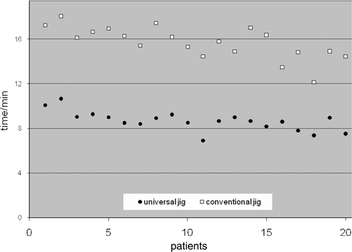
Table I. Mean leg axis in preoperative control by navigation.
The overall radiographic alignment of the final implant positioning for both instrument systems investigated was within a total limit of 3° of aberration in accordance with the planning using the navigational system. In addition, the location of the implant axis for all knees studied was within ±3° of the mechanical axis given by the navigational system.
The angular differences between the instrument slots and the resultant bone cuts for the Pivotal™ and conventional blocks were statistically significant in the frontal plane, but not in the sagittal plane (). Generally, not only did using the Pivotal™ guides result in fewer angular differences, but they also virtually eliminated angular differences greater than 1°. The distribution of the angular differences for the Pivotal™ and conventional blocks is illustrated in and .
Figure 6. The distribution of the difference in angle between the slot of the positioned block and the bone resection for the proximal tibial cut.
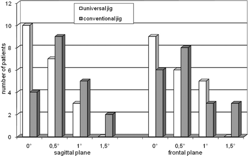
Figure 7. The distribution of the difference in angle between the slot of the positioned block and the bone resection for the distal femoral cut.
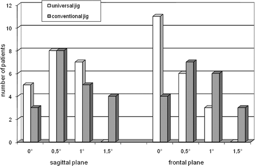
Table II. Time (in minutes) for positioning the cutting blocks in the precise position.
Table III. Differences between the accuracy of the cutting block and the cut.
Discussion
Precise positioning of cutting guides is critical to the successful outcome of a total knee arthroplasty procedure and the long-term survival of the prosthesis. Quicker, more precise cuts may improve the efficiency of this procedure. Our data support the hypothesis that the use of Pivotal™ cutting blocks would result in more accurate bone cuts made in a more time-efficient manner as compared to conventional blocks.
Rand and Coventry Citation[5] reported that frontal plane angular bone-cut deviations greater than 4° resulted in decreased long-term survival for knee implants. Over a period of 10 years, the survival rate in the group with a frontal deviation of more than 4° was only 73%, compared to a 90% survival rate when the frontal deviation was less than 4°. Additionally, aseptic loosening has been reported in nearly 25% of patients with more than 3° of frontal deviation, compared to only 3% in those with less than 3° deviation Citation[6], Citation[7]. In our series, all patients were within 3° of the ideal mechanical axis as calculated by the navigational system.
In our study, the time required for positioning and adjustment was reduced by nearly 50% when using Pivotal™ adjustable cutting blocks. The use of navigation allowed initial positioning to be achieved in approximately 5–10 seconds and, once fixed, the block could be adjusted in all three dimensions without removing any pins; this was not possible with the conventional blocks. In addition, the conventional blocks were associated with some motion during insertion of the pins at an angle to the cortical bone. This type of motion often occurs at the medial aspect of the tibia when conventional blocks are used.
Our study does present some limitations, including the measurement accuracy of the navigation system itself (±0.5°) and the possibility of non-optimal statistical power. Additionally, two different surgical approaches were used with four different implants. However, this appeared to have no impact on the respective time outcomes for bone cuts made using either the Pivotal™ or conventional cutting blocks ().
As shown by other studies, computer-assisted navigation can improve implant positioning with respect to the mechanical leg axis because the risk of extensive misalignment can be reduced Citation[2–4], Citation[8], Citation[9–11]. The use of navigation, in combination with universal and adjustable cutting blocks, can lead to improved operative technique. When differences between the conventional block and Pivotal™ block techniques were within the error range of the method, we found that the two techniques yielded similar accuracy. However, when compared to the conventional cutting blocks, using the Pivotal™ adjustable blocks reduced operating time considerably.
The Pivotal™ block allows the surgeon to leverage the real-time information from the navigation system and quickly and easily adapt, thereby saving time without sacrificing cutting accuracy, and potentially resulting in cost-savings in terms of reduced operating time. Although the results of this study are promising, the value of computer-assisted navigation in total knee arthroplasty requires further evaluation.
Declaration of interest: The authors report no conflicts of interest. The authors alone are responsible for the content and writing of the article.
References
- Jenny JY, Boeri C. Computer-assisted implantation of a total knee arthroplasty: a case-controlled study in comparison with classical instrumentation. Rev Chir Orthop Reparatrice Appar Mot 2001; 87(7)645–652
- Mielke RK, Clemens U, Jens JH, Kershally S. Navigation in knee endoprosthesis implantation - preliminary experiences and prospective comparative study with conventional implantation technique. Z Orthop Ihre Grenzgeb 2001; 139(2)109–116
- Perlick L, Bäthis H, Tingart M, Perlick C, Grifka J. Navigation in total-knee arthroplasty: CT-based implantation compared with the conventional technique. Acta Orthop Scand 2004; 75(4)464–470
- Sparmann M, Wolke B, Czupalla H, Banzer D, Zink A. Positioning of total knee arthroplasty with and without navigation support. A prospective, randomised study. J Bone Joint Surg Br 2003; 85(6)830–835
- Rand JA, Coventry MB. Ten-year evaluation of geometric total knee arthroplasty. Clin Orthop Relat Res 1988; 232: 168–173
- Jeffery RS, Morris RW, Denham RA. Coronal alignment after total knee replacement. J Bone Joint Surg Br 1991; 73(5)709–714
- Ritter MA, Faris PM, Keating EM, Meding JB. Postoperative alignment of total knee replacement. Its effect on survival. Clin Orthop Relat Res 1994; 299: 153–156
- Bäthis H, Perlick L, Tingart M, Perlick C, Lüring C, Grifka J. Intraoperative cutting errors in total knee arthroplasty. Arch Orthop Trauma Surg 2005; 125(1)16–20
- Clemens U, Konermann WH, Kohler S, Kiefer H, Jenny JY, Mielke RK. Computer-assisted navigation with the OrthoPilot system using the Search evolution TKR prosthesis. Results of a multi-centre study. Navigation and Robotics in Total Joint and Spine Surgery, JB Stiehl, WH Konemann, RG Haaker. Springer, Berlin 2003; 234–241
- Oberst M, Bertsch C, Wurstlin S, Holz U. CT analysis of leg alignment after conventional vs. navigated total knee prosthesis implantation. Initial results of a controlled, prospective and randomized study. Unfallchirurg 2003; 106(11)941–948
- Saragaglia D, Picard F, Chaussard C, Montbarbon E, Leitner F, Cinquin P. Computer-assisted knee arthroplasty: comparison with a conventional procedure. Results of 50 cases in a prospective randomized study. Rev Chir Orthop Reparatrice Appar Mot. 2001; 87(1)18–28