Abstract
Introduction: The removal of metal shrapnel in the sub-acute phase of casualty treatment requires the utmost accuracy in detection and removal, especially when there is proximity to major neurovascular structures. Inability to successfully locate and remove retained fragments may lead to a variety of complications due to fragment migration. In this study we prove the feasibility of a new technique which uses metal detector technology combined with a surgical navigation system, resulting in improved accuracy and decreased operating time.
Methods: In each of the experiments, 6 metal nuts were inserted into a dummy leg to simulate shrapnel wounds. Two major experiments were then conducted. Experiment 1 was a comparison of two methods: (a) localization of the nuts using surgical navigation alone, and (b) localization by means of metal detector technology combined with a surgical navigation system (StealthStation® TREON® plus). Experiment 2 employed the same two methods, but this time migration of the metal fragments was introduced. The localization time was measured from incision of the dummy skin to the moment the metal fragment was touched by the searching device.
Results: In experiment 1 the results showed no significant differences between the two approaches. In experiment 2 the new technique was found to significantly decrease the mean fragment localization time, taking 9.6 seconds (±7.2 seconds) as compared to 26.4 seconds (±13.8 seconds) when using the regular technique.
Conclusion: Combining a metal detector probe and a surgical navigation system was found to significantly decrease operating time and increase the surgeon's confidence, especially in cases where migration of the metal fragment occurred during searching and extraction.
Introduction
As the frequency of terrorist attacks increases, so does the number of civilian victims presenting at hospitals with gunshot wounds, stab wounds, blast injuries, burns and non-typical injuries caused by the penetration of nails, nuts, metal balls or other sharp objects which terrorists add to their bombs to maximize casualties and damage Citation[1–5]. The most common metal fragments used in homemade bombs are ball-bearings of radius 2–8 mm or metal nuts with an average size of 5–10 mm.
Some metal fragments can be removed at the primary stage of wound treatment Citation[6]; however, fragments which are retained in the body and require removal in the sub-acute phase necessitate the utmost accuracy in detection and extraction. Inability to successfully localize and remove the retained fragments may lead to a variety of problems such as pain, neurological symptoms, vascular compression, sterile abscesses or granulomas, infections, lead synovitis and systemic lead intoxication, and can also result in complications due to natural migration Citation[7] or migration forced by external magnetic fields such as those associated with MR imaging Citation[8],Citation[9].
The conventional method for locating and extracting metal fragments involves almost continuous use of fluoroscopy to generate the real-time dual-plane location of the extraction tool and metal fragment Citation[10],Citation[11]. Some reduction in the use of fluoroscopy during metal fragment extraction can be achieved by using surgical navigation systems Citation[12]. Theoretically, the surgical navigation system can guide an extraction tool to the exact location in 3D space using two fluoroscopic images. Practically, however, in order to achieve the desired accuracy with a surgical navigation system, two conditions must be met: (1) the point in space to be reached should be associated with a continuous rigid body; and (2) the point or object to be reached should not change its position relative to the reference frame of the surgical navigation system after the fluoroscopic images have been acquired. In the case of extraction procedures for metal fragments, these two conditions are usually not met. A metal fragment penetrating soft tissue may collide with the bone but remain free to move if external forces are applied to it. In the case of an attempt to extract a small fragment, the forces applied to the object with the extraction tool may result in migration of the fragment. The possibility of such migration allows the surgical navigation system to act only as a coarse locater; once migration has occurred, the navigation system loses its efficiency as a tool for locating and extracting small metal fragments in a soft tissue environment.
In this study we demonstrate the feasibility of a new technique using metal detector technology Citation[13–15] combined with a surgical navigation system (StealthStation® TREON® plus, Medtronic, Louisville, CO). The addition of a metal detector probe, defined within the surgical navigation system, takes advantage of the navigation system as a coarse and quick locater of the metal fragment, but also adds the ability to perform precise location of the fragment despite any migration that may occur.
The combination of the two technologies assists the surgeon in detecting and extracting small metal fragments with improved accuracy and decreased operating time, especially in cases were the fragments tend to migrate.
Materials and methods
The development of the device was based on known models of metal detector Citation[16] which were suited in terms of their size and function to the specific purpose of detecting metal fragments in the human body. Different detection methods were investigated, and the Beat Frequency Oscillator (BFO) technology was the model chosen. The generator frequency was 250 kHz and the audible beats were within human hearing range. The probe's magnetic field of view has been designed to detect a metal particle up to 1.5 cm from the tip of the probe with a spatial angle of 30° (). In general, metal detector technology is capable of detecting any object made of a conductive material, though the size of the object and the type of metal involved may necessitate calibration of the system. Since the goal of this study was to investigate the feasibility of the concept, the metal detector probe was not designed to meet sterilization requirements or to function in a liquid environment (). A plastic cylinder was used for the construction of the probe, and its dimensions were designed to permit the insertion of a surgical extractor through it. Thus, a tube 14 cm long and 1.2 cm in diameter was used. All major components of the metal detector were constructed in-house.
Figure 1. Graphical output of a preliminary test intended to estimate the geometrical range of the metal detector. The z axis indicates frequency change, which is easily distinguished by the human ear.
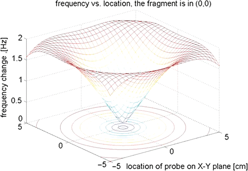
Figure 2. A prototype of the metal detector. The apparatus included a plastic tube, a coil wound at the tip, and a tracking array which was used by the navigation system (StealthStation® TREON® plus).
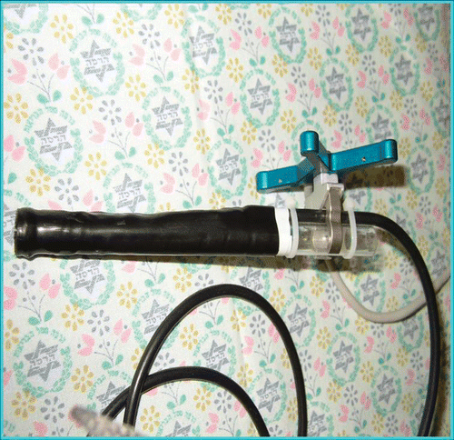
The StealthStation® TREON® plus navigation system (Medtronic, Louisville, CO) was used in this study. A tracking array (Medtronic SureTrak II) was installed on the metal detector probe, enabling definition of the probe tip in the navigation system. The navigation system is infra-red-based and no electromagnetic interference is expected.
The dummy leg model used in this study was a foam-coated right knee with intact femur and tibia (SYNBONE AG, Malans, Switzerland).
Two major experiments were conducted:
Experiment 1
Six steel nuts were inserted into a dummy leg and fixated (). A surgical navigation reference frame was attached (), and two fluoroscopic images were acquired. The physician had to locate the six nuts first using only the surgical navigation system (Method A) and then using the metal detector probe combined with the navigation system (Method B). The six nuts were repositioned between the two attempts to prevent development of a learning curve for their locations. Extraction time was measured from incision of the dummy skin to the moment the steel fragment was touched by the searching device ( and ).
Figure 3. Metal nuts placed in the dummy leg. The physician had no knowledge of their exact locations.
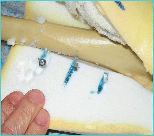
Figure 4. The dummy leg ready for the operational procedure. The reference frame is fixed to the leg. Movement of the leg during the procedure does not spoil the navigation process as long as there is no relative movement between the reference frame and the dummy leg.
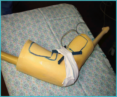
Figure 5. The physician searches for the metal nut with the navigated metal detector.
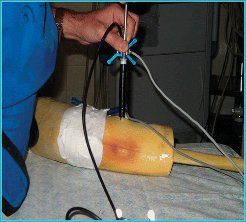
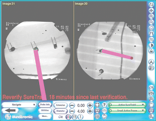
Figure 6. A screenshot of the surgical navigation system displaying the location of the metal detector. This visual guidance is supplemented by the audio indications heard by the surgeon.
Experiment 2
Up to the image-acquisition stage, experiment 2 was identical to experiment 1. After the images were acquired, the nuts were released from their fixation and their locations were moderately changed. The release and re-positioning of the nuts mimicked a situation in which metal fragments migrate during the extraction procedure. The whole process was first carried out with surgical navigation alone, and then using the metal detector combined with the navigation system. In each of these experiments, the surgeon had no previous knowledge of the metal fragments' locations. Again, extraction time was measured from incision of the dummy skin to the moment the steel nut was touched by the searching device.
Results
summarizes the results of experiment 1, in which a comparison was made between localizations performed by methods A and B. The results of this experiment showed no significant difference between the methods with regard to the time taken for the surgeon to reach the fragment within the dummy leg.
Table I. Time required for extraction of metal nuts from the dummy leg in experiment 1. The results were not significant.
summarizes the results of experiment 2, which compared localization of the fragment in the dummy leg following a simulated migration using the same two methods as in experiment 1. The mean localization time was 1 min 26.4 seconds (standard deviation: 1 min 13.8 seconds) for Method A, and 9.6 seconds (standard deviation: 7.2 seconds) for Method B (P = 0.048786).
Table II. Time required for extraction of metal nuts from the dummy leg in experiment 2. Results with the two methods are significantly different, with an average of 1 min 26.4 seconds required for Method A and an average of 9.6 seconds for Method B.
Discussion
The main objective of this study was to prove the feasibility of using an operative invasive tool (a metal detector probe) to assist the surgeon in the detection and extraction of small metal fragments, especially in cases where the fragments undergo migration during the extraction process. For this purpose, the probe was constructed and two experiments were conducted as described in the preceding sections.
The first experiment included a real shrapnel detection and extraction procedure performed in a dummy leg using methods A and B, in order to prove the feasibility of the concept. The duration of the procedure was measured from the instant the surgeon cut the dummy's “skin” to the instant the metal fragment was reached by the tip of the searching tool. The second experiment was similar to the first, except for one change. Here, the time measured was that taken by the surgeon to touch the nut.
We concluded that using the metal detector probe in combination with the navigation system is beneficial, especially in cases where migration of the metal fragment has occurred. In other cases, where no migration occurred, we found no advantage in using Method B. We assume that in medical centers where navigation systems are not in use, a metal detector extraction tool may be of assistance to surgeons attempting to remove metal fragments by helping them locate the metal shards more efficiently. However, we have not yet studied this issue.
Limitations of the study
Actual extraction time was not studied since the metal detector probe did not include an extraction mechanism, thus the time recorded relates to detection of the metal target by touching it, but without subsequent physical removal. The dummy leg contraption, along with the prototype of the metal detector specially prepared for this experiment, made maneuvering more difficult than would be expected when using a well designed and commercially manufactured device in an in vivo environment.
References
- Mayo A, Kluger Y. Terrorist bombing. World Journal of Emergency Surgery 2006; 1: 33, doi:10.1186/1749-7922-1-33
- Peleg K, Aharonson-Daniel L, Stein M, Michaelson M, Kluger Y, Simon D, Noji E-K. Gunshot and explosion injuries: Characteristics, outcomes, and implications for care of terror-related injuries in Israel. Ann Surgery 2004; 239(3)311–318
- Amir LD, Aharonson-Daniel L, Peleg K, Waisman Y. the Israel Trauma Group. The severity of injury in children resulting from acts against civilian populations. Ann Surgery. 2005; 241(4)666–670
- Marcus NA, Blair WF, Shuck JM, Omer GE, Jr. Low velocity gunshot wounds to the extremities. J Trauma 1980; 20(12)1061–1064
- Weil Y, Mosheiff R, Liebergall M. Blast and penetrating fragment injuries to the extremities. J Am Acad Orthop Surgeons 2006; 14(10 suppl)S136–S139
- Peyser A, Khoury A, Liebergall M. Shrapnel management. J Am Acad Orthop Surgeons 2006; 14(10 Suppl)S66–S70
- Eylon S, Mosheiff R, Liebergall M, Wolf E, Brocke L, Peyser A. Delayed reaction to shrapnel retained in soft tissue. Injury 2005; 36(2)275–281
- Teitelbaum GP. Metallic ballistic fragment: MR imaging safety and artifacts (letter). Radiology 1990; 177: 883
- Shellock FG. MR imaging of metallic implants and materials: A compilation of the literature. AJR 1988; 151: 811–814
- Rhee JM, Martin R. The management of retained bullets in the limbs. Injury 1997; 28(Suppl 3)S-C23–S-C28
- Williams MS, Hutcheson RL, Miller AR. A new technique for removal of intraarticular bullet fragments from the femoral head. Bull Hosp Joint Dis (United States) 1997; 56(2)107–110
- Mosheiff R, Weil Y, Khoury A, Liebergall M. The use of computerized navigation in the treatment of gunshot and shrapnel injury. Comput Aided Surg 2004; 9(1–2)39–43
- Cakir B, Akan M, Yildirim S, Akoz T. Localization and removal of ferromagnetic foreign bodies by magnet. Ann Plast Surg 2002; 49(5)541–544
- Veselko M, Trobec R. Intraoperative localization of retained metallic fragments in missile wounds. J Trauma 2000; 49(6)1052–1058
- Trobec R, Avbelj V, Veselko M, Demsar F. Metal implant localizers: Frontiers and diagnostic feasibility. J Med Eng Technol 1996; 20(3)134–140
- Yamazaki S, Nakane H, Tanaka A. Basic analysis of a metal detector. IEEE Trans Instrum Meas 2002; 51(4)810–814