Abstract
A laboratory study was conducted to compare the accuracy with which a LISS plate could be placed on the distal metaphysis of a model femur using both a fluoroscopy-based computer assisted technique and the conventional fluoroscopic technique. A significant difference was found between outcomes with the two approaches with respect to the maximum distance from the plate to the diaphysis of the bone, but there was no significant difference in the maximum distance to the condylar area. There was also no difference with respect to the number of holes that required re-drilling for adjustment of the plate placement or screws with poor purchase in bone. There were, however, significant differences between the two techniques in terms of duration of the procedure and radiation exposure.
Introduction
The potential applications for computer assisted surgery in orthopedics are expanding. Current clinical usage of this technology is mainly in arthroplasty, in which the computer is used to predict the appropriate orientation of a bone cut, or in the placement of pedicle screws for spinal fusion Citation[1]; however, computer assistance has also proven useful in localizing tumors and planning osteotomies Citation[2],Citation[3].
Intraoperative conventional fluoroscopy is currently the most common method of providing visual feedback to surgeons during fracture reduction and hardware placement in orthopedic trauma surgery. The disadvantages of this imaging modality include the radiation dosage to the patient and surgical team, as well as the cumbersome positioning of the C-arm that is necessary to obtain orthogonal intraoperative views. As familiarity with computer assisted techniques has matured, expansion of their application in the trauma field has ensued Citation[4],Citation[5]. The most common application of computerized fluoroscopy-based navigation in an orthopedic trauma context has been in intramedullary nailing of the femur Citation[4],Citation[5].
Open reduction and internal fixation of periarticular fractures can be a very challenging procedure involving a significant learning curve and a high risk of intraoperative complications or failure to achieve an acceptable reduction and stable fixation. The patient and surgical team are also exposed to an untoward amount of ionizing radiation during fluoroscopy. The application of computer planning and navigation technology to this surgical problem offers the potential for improved accuracy and reliability, as well as reduced radiation exposure.
In a small series of cases, a previous study Citation[6] demonstrated the feasibility of using computer assisted surgery technology to achieve fracture reduction and accurate placement of a LISS (Less Invasive Stabilization System) plate in proximal tibial fractures.
The purpose of the present study, which was performed in a laboratory setting, was to compare the accuracy with which a LISS plate could be placed on the distal metaphysis of a model femur using both a fluoroscopy-based computer assisted technique and the conventional fluoroscopic technique.
Materials and methods
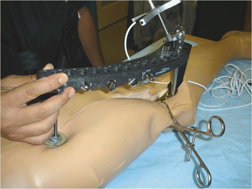
Two surgeons (a clinical fellow and a senior orthopedic resident) performed 32 procedures in which a 9-hole LISS plate (Synthes, West Chester, PA) was placed on an intact Sawbones femur model (Pacific Research Laboratories, Vashon, WA) which was masked with simulated soft tissue to reproduce the in vivo visibility constraints (). In randomly assigned order, each of the two surgeons performed 8 conventional procedures using a standard C-arm and 8 computer assisted fluoroscopy-guided trials. In addition to randomization, efforts were made prior to the study to minimize bias and learning-curve effects by having each surgeon perform five procedures of each type.
Figure 1. The model femur covered with simulated soft tissue.
Prior to the trials, a 3D virtual model of the LISS plate was created using a standard CT scan. The data from the scan was converted into a virtual model using computer guidance system software (VSS, IGO Technologies, Kingston, Ontario, Canada). The result was a virtual LISS plate that could be moved in 3D virtual space on a computer screen.
For the computer assisted trials, an optical marker array was mounted on the anterior aspect of the femur, and a second optical marker array was mounted on the LISS plate drill guide using a custom-designed mounting interface. A C-arm was fitted with an optical electronic tracking drum (Traxtal Technologies, Toronto, Ontario, Canada) for position monitoring during image acquisition, then anteroposterior (AP) and lateral C-arm images of the femur were acquired with tracking arrays in place (). These images were fed into the computer and provided the basis of the feedback to the surgeon. While an optical tracking system (Polaris, Northern Digital Inc., Waterloo, Canada) tracked the femur and LISS plate in real time, a computer guidance system (VSS, IGO Technologies) was used to overlay a virtual image of the LISS plate on the fluoroscopic images in real time and display them on a computer monitor (). The procedure was performed in accordance with the implant's technique manual. For the conventional technique, C-arm fluoroscopy was used to apply the LISS plate in the standard fashion.
Figure 2. AP and lateral views were acquired with the tracking marker arrays in place.
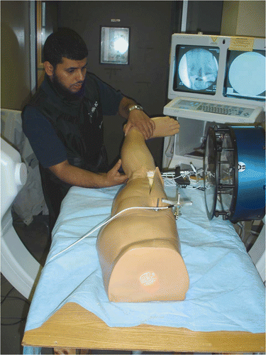
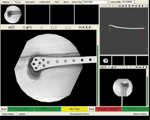
Figure 3. The LISS plate superimposed on the fluoroscopic image of the model femur.
The data obtained from each procedure was used to evaluate three aspects: the efficiency of the trial, the accuracy and reliability of the plate placement, and the radiation exposure. The efficiency of the procedure was measured by timing the trial and by counting the number of drill holes that required re-drilling to facilitate adjustment of the plate. The accuracy was measured in several ways: The maximum distance between the plate and the bone was measured at both the condylar area and the diaphysis of the bone, while an objective measurement was used to provide a binary data point as to whether a plate was completely within a subjectively defined zone of acceptability on the bone. The number of screws that had poor fixation in bone was also recorded. Following the trial, the femur and LISS plate were registered to 3D virtual models generated from CT images, and the registration was used to obtain the coordinates of the LISS plate holes. To assess reliability, an analysis of the variance in screw placement at each individual screw position on the LISS plate for all 32 trials was performed. To measure radiation exposure, the number of C-arm fluoroscopy shots was recorded for each trial. Descriptive statistics including measures of variability were assessed for each parameter. Independent sample Student's t-tests were used to determine statistical significance between the computer navigated and conventional groups. A p-value of less than or equal to 0.05, for a two-tailed test, was considered statistically significant.
Results
In terms of efficiency, the number of holes requiring re-drilling to enable adjustment of the plate was five for the conventional group and zero for the navigated group. When examining accuracy, we found a significant difference between the two groups when measuring the maximum distance from the LISS plate to the diaphysis of the bone, but not when measuring the maximum distance to the condylar area. There was no difference between the two procedures with respect to the number of holes that required re-drilling for plate adjustment or the number of screws with poor purchase in the bone. There were, however, significant differences between the two techniques in terms of duration of the procedure and radiation exposure. Specific data for the conventional and navigated groups are presented in .
Table I. Comparison of results for conventional and navigated procedures.
Discussion
Intraoperative fluoroscopy is a valuable tool for visualizing underlying bone and surgical instrument positions in orthopedic procedures Citation[7], but it exposes both the surgical team and the patient to multiple doses of X-ray radiation Citation[8]. We have used LISS plate placement on a distal femur model to show that the computer assisted technique can be as accurate as the conventional technique in the positioning of orthopedic hardware on bone. We have also shown in our laboratory setting that this technique has the added benefit of reduced radiation exposure for the surgical team and the simulated patient.
A learning curve is to be expected when beginning to apply computer navigation to a surgical technique. To minimize learning effects, both surgeons performed practice cases prior to the study. The randomized study design also helped to offset the learning-curve effect.
There were a few weaknesses in the study that should be mentioned. Besides being an in vitro investigation, the study involved re-using the same plate and phantom. In addition, only two surgeons were involved in the trials. However, using the same model and phantom and having a small number of surgeons involved helped to minimize the variance and bias of the results.
Based on our findings, we believe that an in vivo study should be conducted on real fractured femora to investigate whether our results translate to real bones.
Acknowledgments
The authors wish to thank Heather Grant for her help with the statistical analyses and for reviewing the manuscript. Two 9-hole LISS plates, along with the associated screws and drill guide, were provided by Synthes for use in this project.
References
- John PS, James C, Antony J, Tessamma T, Ananda R, Dinesh K. A novel computer-assisted technique for pedicle screw insertion. Int J Med Robot 2007; 3: 59–63
- Athwal GS, Pichora DR, Ellis RE, Rudan JF. A computer-assisted guidance technique for the localization and excision of osteoid osteoma. Orthopedics 2004; 27(2)195–197
- Croitoru H, Ellis RE, Prihar R, Small CF, Pichora DR. Fixation-based surgery: A new technique for distal radius osteotomy. Comput Aided Surg 2001; 6(3)160–169
- Khoury A, Liebergall M, Weil Y, Mosheiff R. Computerized fluoroscopic-based navigation-assisted intramedullary nailing. Am J Orthop 2007; 36(11)582–585
- Weil YA, Gardner MJ, Helfet DL, Pearle AD. Computer navigation allows for accurate reduction of femoral fractures. Clin Orthop Relat Res 2007; 460: 185–191
- Grützner PA, Langlotz F, Zheng G, von Recum J, Keil C, Nolte LP, Wentzensen A, Wendl K. Computer-assisted LISS plate osteosynthesis of proximal tibia fractures: Feasibility study and first clinical results. Comput Aided Surg 2005; 10(3)141–149
- Hofstetter R, Slomczykowski M, Sati M, Nolte LP. Fluoroscopy as an imaging means for computer-assisted surgical navigation. Comput Aided Surg 1999; 4(2)65–76
- Suhm N, Jacob AL, Nolte LP, Regazzoni P, Messmer P. Surgical navigation based on fluoroscopy - clinical application for computer-assisted distal locking of intramedullary implants. Comput Aided Surg 2000; 5(6)391–400