Abstract
There are many published reports demonstrating the accuracy of CT-based navigation systems. However, the use of such systems often subjects patients to a high level of radiation exposure. CT scans acquired using thinner slices are considered to lead to more accurate results, but also increase radiation exposure. We took the postoperative CT scans for 56 cases of total hip arthroplasty performed using a CT-based navigation system and analyzed the accuracy of the cup and stem positioning. Of these cases, 41 were performed using 3-mm CT slices and 15 were performed using 1-mm slices, enabling us to compare the accuracy of the system and the radiation exposure using the different slice thicknesses.
CT-based navigation appears to be very accurate with regard to cup anteversion and leg length, but inaccurate with regard to stem anteversion. As for the varus/valgus angle of the stem, the navigated approach seems to be very accurate in terms of the numerical value, but this does not satisfy us: Stem anteversion is still inaccurate with this system, while cup inclination is sufficiently accurate with both navigation and manual methods.
Use of 1-mm CT slices results in twice the radiation exposure associated with 3-mm CT slices, but there is little difference with respect to accuracy. It is therefore recommended to use a CT-based navigation system with 3-mm CT slices for accurate and safe total hip arthroplasty.
Introduction
It has been proven that malpositioning of implants in total hip arthroplasty (THA) is one of the main causes of postoperative dislocation, limited range of motion, leg length discrepancy and early polyethylene wear. However, the majority of osteoarthritic hips encountered in Japan originate from developmental acetabular dysplasia with a high hip center Citation[1], and it is therefore difficult to place the implant precisely in the proper position. Furthermore, as a standard THA procedure is performed with the patient in the decubitus position, it is difficult to check the implant position properly on the table. Therefore, numerous types of navigation system, including imageless, fluoroscopy-guided and CT-based systems, have been developed and improved to enable proper positioning in THA Citation[2–6].
Because Japanese patients often present with severe deformity and even a dislocated hip, CT-based navigation is considered to be the most useful approach and is therefore the most frequently used option in Japan. However, while CT-based navigation is regarded as the most accurate system available at present Citation[5], it also has several disadvantages: it is expensive, time-consuming, and involves significant radiation exposure for the patient, while imposing limitations on the choice of implant design. The degree of radiation exposure for the patient Citation[2], Citation[6] when using CT-based navigation depends on the pitch of CT employed. The use of thicker-pitched CT entails lower radiation exposure; however, accuracy of navigation is suspected to be worse.
The purpose of this study was two-fold: First, to compare the results for implant positioning accuracy as determined using the standard manual technique without navigation and with a CT-based navigation system; and second, to compare the results for implant positioning accuracy obtained using 3-mm and 1-mm CT slices for navigated implantation of the cup and stem.
Patients and methods
Navigation system
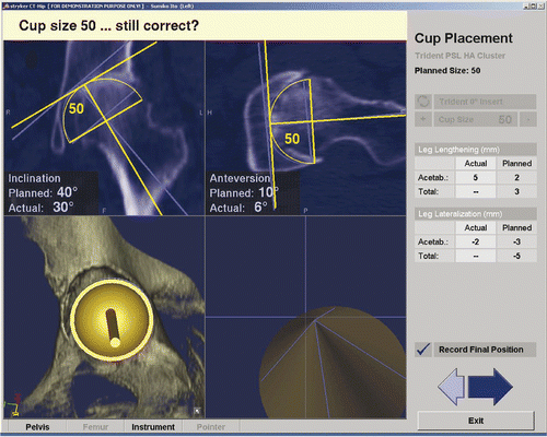
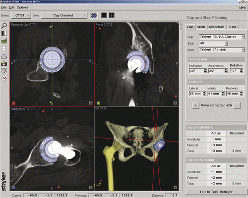
The Stryker Hip Navigation System is a CT-based system that is able to display the three-dimensional shape of implants and bony hip structure on the screen (). Use of an infrared tracker reduces error and enables the leg length to be checked and compared with the contralateral side. However, the system cannot avoid radiological exposure for the patient, is one of the most expensive machines of its type, and is also one of the most time-consuming systems for hip navigation. Cup anteversion and inclination are determined by the pelvic plane, which is defined by the bilateral anterior superior iliac spine (ASIS) and the pubic tubercle and bilateral end of the ischium. Stem anteversion is determined by the axial neck angle and the line that links the medial and lateral malleolar points of the knee. Leg length is determined by the longitudinal distance between each knee center and the bilateral ASIS line. These landmark anatomical points are registered at the outset of the navigation procedure.
Patients
Patients who underwent navigated THA from October 2004 to May 2008 were included in the study. During this period, 66 cases of THA were performed using the CT-based navigation system (Stryker Hip Navigation System, version 1.0/21). The first 10 joints to be operated on were excluded to avoid the effect of the learning curve. A total of 56 joints (in 53 patients: 50 females and 3 males) were analyzed, comprising 53 hips with osteoarthritis caused by developmental acetabular dysplasia and 3 with osteonecrosis of the femoral head. Prior to October 2006, all patients were operated on using the navigation system with 3-mm CT slices (41 cases); thereafter, all operations were performed with 1-mm CT slices (15 cases). The inclination and anteversion of the cup, anteversion and varus/valgus angle of the stem, and leg length compared with the contralateral side were all measured intraoperatively by the navigation system ().
In all cases, the inclination and anteversion of the cup, as well as the anteversion of the stem, were also measured by the manual method, using a protractor to measure the angles intraoperatively. Cup inclination is measured as the angle between the shaft of the cup inserter and the horizontal plane reflected to the patient's coronal plane. Cup anteversion is measured as the angle between the shaft of the stem inserter and a vertical line reflected to the axial plane. Stem anteversion is measured as the angle between the direction of the broach handle and the lower leg line. At present, we have no useful method for measuring the varus/valgus angle of the stem and leg length compared with the contralateral side, and were therefore unable to obtain these values intraoperatively.
Postoperative CT scans were acquired 3 months after surgery. Differences in each angle as well as in leg length were evaluated, comparing values measured by the navigation system or manually during the THA procedure and those measured on the postoperative CT (). In each case, the postoperative CT scans were acquired with the same slice thickness (3 mm or 1 mm) used in the CT-based navigated THA. Each value for the cup angle is referenced to the radiological angle as defined by Murray Citation[7]. Each position was evaluated by importing the postoperative CT into the navigation DICOM software and superimposing the CAD model onto the implant position.
All CT scans were acquired using a Toshiba Aquilion 64 scanner, and we could easily estimate the radiation dose of our scanning protocol, which covered a region from the top of the pelvis to approximately 10 cm below the knee joint. The radiation dose was calculated by the CT software and displayed on the CT screen after each scan. The DLP (Dose Length Product) was used in the study; this is generally used by the ICRP (International Commission for Radiological Protection) and is transcribed as mGy·cm.
Each patient was implanted with a TriAD acetabular cup (Stryker) and a Cent-Pillar anatomical stem (Stryker). The operations were performed by two well-trained surgeons (M.M., N.H). It should be noted that our target acetabular component position was not always in the safe zone as defined by Lewinnek et al. Citation[8]. Japanese patients have varying degrees of femoral neck anteversion, and the cup and stem were therefore implanted so that the sum of the cup anteversion and stem anteversion was 40° Citation[9]. For example, in a patient with 30° of neck anteversion, the cup might be implanted with 10° of anteversion. An MIS (Minimally Invasive Surgery) anterolateral subgluteal approach Citation[10] was employed in every case.
Statistical analysis
All data are presented as the mean and standard deviation. Statistical comparisons between measurements obtained with navigated THA and those obtained with conventional manual methods, as well as between results obtained with 1-mm and 3-mm CT slices, were performed using the Mann-Whitney U test. The level of significance was set at a probability value of <0.05.
Results
Navigated versus manual measurements
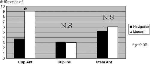
The mean difference between measurements of cup inclination obtained intraoperatively using the navigation system and the corresponding measurements on the postoperative CT was 3.2° (range: 0 to 11°; SD: 2.7), while the mean difference between manual intraoperative measurements and measurements on the postoperative CT was 3.1° (range: 3 to 11°; SD: 2.5). The mean difference between measurements of cup anteversion obtained intraoperatively with navigation and those from the postoperative CT was 3.8° (range: 0 to 12°; SD: 3.4), while the mean difference between manual intraoperative measurements and measurements on the postoperative CT was 9.0° (range 0 to 29°; SD: 7.0). The mean difference between measurements of stem anteversion obtained intraoperatively with navigation and those from the postoperative CT was 5.2° (range: 0 to 17°; SD: 4.8), while the mean difference between manual intraoperative measurements and measurements on the postoperative CT was 6.1° (range: 2 to 26°; SD: 5.4) ().
Figure 3. Mean deviation of cup anteversion, inclination and stem anteversion using navigation and manual methods. There is only a significant difference for cup anteversion.
The mean difference between measurements of leg length compared with the contralateral side obtained intraoperatively with navigation and the corresponding measurements on the postoperative CT was 2.2 mm (range: 0 to 9°; SD: 1.8). The mean difference between measurements of the varus angle of the stem obtained intraoperatively with navigation and the corresponding measurements on the postoperative CT was 2.2° (range: 0 to 9°; SD: 2.0).
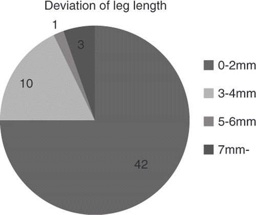
Thus, only with respect to cup anteversion was the difference between the measurements obtained intraoperatively with navigation and the corresponding measurements on the postoperative CT significantly smaller than that between the manual intraoperative measurements and the measurements on the postoperative CT. With respect to leg length compared with the contralateral side, 52 of the 56 THA procedures were accomplished with less than 5 mm difference using navigation ().
Effect of slice thickness
Changing the CT slice thickness results in a difference between measurements of leg length obtained intraoperatively with navigation and those obtained from the postoperative CT. The results are presented in . The mean difference between measurements of cup inclination obtained intraoperatively with navigation and those obtained from the postoperative CT was 3.1° (range: 0 to 11°; SD: 2.8) when using 3-mm slices and 3.2° (range: 0 to 11°; SD: 2.7) when using 1-mm slices. The mean difference between measurements of cup anteversion obtained intraoperatively with navigation and those obtained from the postoperative CT was 3.8° (range: 0 to 16°; SD: 3.8) when using 3-mm slices and 3.7° (range: 0 to 7°; SD: 2.3) when using 1-mm slices. The mean difference in measurements of stem anteversion obtained intraoperatively with navigation and those obtained from the postoperative CT was 5.3° (range: 0 to 16°; SD: 4.8) when using 3-mm slices and 5.0° (range: 0 to 16°; SD: 4.8) when using 1-mm slices. The mean difference between measurements of leg length compared with the contralateral side obtained intraoperatively with navigation and those obtained from the postoperative CT was 2.3 mm (range: 0 to 9°; SD: 1.8) when using 3-mm slices and 1.8 mm (range: 0 to 7°; SD: 1.7) when using 1-mm slices.
Figure 5. Mean deviation of cup inclination, cup anteversion and leg length using 3-mm and 1-mm CT slices. The deviation of the two cup parameters showed no significant difference.
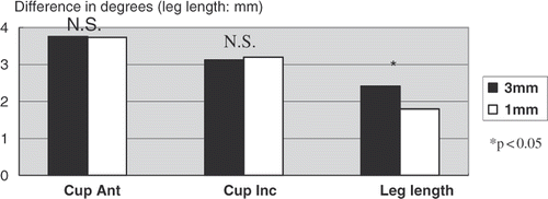
Thus, except in the case of leg length measurements, there was no significant difference between results obtained using 3-mm CT slices and 1-mm slices. However, the mean radiation dose associated with a one-time examination was 2404 mGy·cm for a CT scan performed using 1-mm slice thickness, as compared to only 1276 mGy·cm for a scan performed using 3-mm slices.
Discussion
Use of navigation significantly reduces deviation in cup anteversion, but makes no difference to the deviation in cup inclination and stem anteversion. It is not too difficult to measure the inclination of the cup intraoperatively, but it is difficult to recognize the sagittal pelvic plane with the patient in the decubitus position, with the result that accuracy in controlling cup anteversion without navigation is inferior to that when using navigation. Many imageless navigation systems can calculate the anatomical angle of the pelvis, but this has been reported to be inaccurate for cases of severe deformity in dysplasic hip patients Citation[6]. As mentioned previously, the majority of Japanese hip osteoarthritis cases result from developmental dysplasia, and many of these patients exhibit severe deformity. Therefore, CT-based navigation is thought to be the most appropriate system for measuring cup anteversion accurately in hip arthritis in Japanese patients.
There was no difference in the deviation of stem anteversion with or without navigation, and the accuracy of measurements for stem anteversion was unsatisfactory in both cases. In the navigation system employed, the computer measures the anteversion of the stem by using the neck line of the stem and the bilateral epicondyle line of the knee. However, it is difficult to locate the bilateral epicondyle line exactly on a CT image. Therefore, it is supposed that the difference between the measurement obtained during navigation and that obtained from the postoperative CT may result from the difficulty of identifying the bilateral epicondyle line correctly every time.
The deviation in leg length is thought to be accurate. Previously, a number of articles have explained how to equalize leg length following THA Citation[11], Citation[12]; however, they have been unable to achieve a mean discrepancy of less than 2.5 mm. Therefore, the results of the current study were satisfactory to us as accurate measurements.
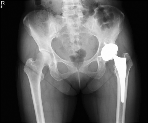
The deviation in stem varus/valgus alignment was very accurate in terms of the numerical value. However, leading surgeons consider 2° of stem varus to be unacceptable in X-ray evaluation (), so the varus/valgus angle was not thought to be satisfactorily controlled in this study. As cementless stems are anatomical and short, it is difficult to insert them appropriately, and more accurate systems are required.
Figure 6. Although the varus angle of this stem is accurate to 3°, it appears to be ill-shapen on the X-ray. This demonstrates that accurate numerical values for the varus/valgus angle do not guarantee good results.
In the current study, navigation using 1-mm CT slices was found to be more accurate than with 3-mm slices with respect to leg length. However, the difference between the measurements is considered to be negligible in a clinical context. Sugano et al. indicated that a combination of 3-mm slice thickness and 1-mm reconstruction pitch may be the optimal trade-off, as investigated using a bone model Citation[13]. For a patient in our hospital, the normal radiation dose for a plain brain CT (10 mm slice thickness) is approximately 1500 mGy·cm, and for an abdominal CT (10-mm slice thickness) it is approximately 800 mGy·cm. The mean radiation exposure using 1-mm CT slices (2404 mGy·cm in our study) is considered to be too high. At present, there is no defined upper limit for the radiation dose related to CT scanning, but a proposed reference dose for routine pelvic osseous CT of 514 mGy·cm has been reported in EUR 16262 (European Guidelines on Quality Criteria for Diagnostic Radiographic Images) Citation[14]. The surgeon has to evaluate the trade-off between CT-scan radiation exposure and procedural accuracy. A safer approach is to choose the lowest radiation dose that still gives acceptable precision.
Although the mean difference in operative time with and without navigation is only approximately 10 min, an additional 20 to 40 min is required for preoperative planning in each case, and it might be difficult for busy surgeons to accept such a time-consuming procedure.
There are many papers in the literature concerning hip navigation, but most of them evaluated only cup anteversion and inclination; few addressed stem alignment. However, to avoid postoperative hip dislocation, accurate positioning of both the cup and the stem are necessary Citation[9]. In the current study, the navigation system was significantly better than the conventional method with respect to cup anteversion and leg length.
The limitation of this study is that it only relates to a very small number of cases. The time-consuming nature of the procedure is the key factor preventing us from using the system in a larger number of patients. Development of a simpler and more accurate system is therefore desirable.
Conclusion
CT-based navigation is a useful tool for accurate positioning of implants in total hip arthroplasty. A CT slice thickness of 3 mm is an optimal compromise with regard to radiation exposure and accuracy.
References
- Takeyama A, Naito M, Shiramizu K, Kiyama T. Prevalence of femoroacetabular impingement in Asian patients with osteoarthritis of the hip. Int Orthop 2009; 33(5)1229–1232
- Widmer K, Grützner PA. Joint replacement – total hip replacement with CT based navigation. Injury Int J Care Injured 2004; 35: S-A84–S-A89
- Kalteis T, Handel M, Bäthis H, Perlick L, Tingert M, Grifka J. Imageless navigation for insertion of the acetabular component in total hip arthroplasty. J Bone Joint Surg 2006; 88-B(2)163–167
- Grützner PA, Zheng G, Langlotz U, von Recum J, Nolte LP, Wentzensen A, Widmer KH, Wendl K. C-arm based navigation in total hip arthroplasty – background and clinical experience. Injury Int J Care Injured 2004; 35: S-A90–S-A95
- Hube R, Birke A, Hein W, Klima S. CT-based and fluoroscopy-based navigation for cup implantation in total hip arthroplasty. Surg Technol Int 2003; 11: 273–279
- Tannast M, Langlotz F, Kubiak-Langer M, Langlotz U, Siebenrock KA. Accuracy and potential pitfalls of fluoroscopy-guided acetabular cup placement. Comput Aided Surg 2005; 10: 329–336
- Murray DW. The definition and measurement of acetabular orientation. J Bone Joint Surg Br 1993; 75-B: 228–232
- Lewinnek GE, Lewis JL, Tarr R, Compere CL, Zimmerman JR. Dislocation after total hip replacement arthroplasties. J Bone Joint Surg Am 1978; 60: 217–220
- Widmer KH, Zurfluh B. Compliant positioning of total hip components for optimal range of motion. J Orthop Res 2004; 22(4)815–821
- Röttinger H. The MIS anterolateral approach for THA. Orthopade 2006; 35(7)708, 710–715
- Bose WJ. Accurate limb-length equalization during total hip arthroplasty. Orthopedics 2000; 23(5)433–436
- Shiramizu K, Naito M, Shitama T, Nakamura Y, Shitama H. L-shaped caliper for limb length measurement during total hip arthroplasty. J Bone Joint Surg Br 2004; 86(7)966–969
- Sugano N, Sasama T, Sato Y, Nakajima Y, Nishii T, Yonenobu K, Tamura S, Ochi T. Accuracy evaluation of surface-based registration methods in a computer navigation system for hip surgery performed through a posterolateral approach. Comput Aided Surg 2001; 6: 195–203
- Shrimpton PC, Hillier MC, Lewis MA, Dunn M. Dose from Computed Tomography (CT) Examinations in the UK – 2003 Review. NRPB-W67. Chilton: National Radiological Protection Board; 2005.
