Abstract
Objective: Multilevel Oblique Corpectomy (MOC) is an emerging technique for surgical treatment of multi-segmental cervical spondylotic myelopathy (CSM) featuring extensive ossification of the posterior longitudinal ligament (OPLL). However, the use of an oblique drilling plane is unfamiliar to most surgeons and there is no anatomical landmark present on the posterior portion of the vertebral body. To overcome these difficulties, the authors used intraoperative C-arm-based image guided navigation (IGN), and this study was conducted to evaluate the efficacy of IGN in MOC.
Methods: Following the introduction of IGN for MOC, 24 patients underwent MOC procedures at our institution. Two patients who had undergone previous cervical operations were excluded from the present study. Of the remaining 22 patients, 11 underwent MOC with IGN, and 11 underwent MOC without IGN support. The completeness of MOC (CMOC) is measured as the sum of the bilateral remaining posterior body minus the remaining approach-side anterior body in millimeters at the most compressive level. For each patient, the preoperative Japanese Orthopaedic Association Score (JOAS) and postoperative 5th day JOAS were collected as well as several other perioperative parameters.
Results: The mean CMOC was 0.89 mm for the IGN group and 5.9 mm for the control group. The mean change in JOAS was 5.58 for the IGN group and 3.34 for the control group at 1-year follow-up. In the control group, two patients underwent re-exploration due to remaining OPLL. Despite the intraoperative IGN set-up time, the mean operation time for the IGN group was shorter than that for the control group (248 min versus 259 min). Mean treated levels were 3.55 for the IGN group and 3.36 for the control group.
Conclusion: Through the use of image guided navigation, it was possible to accomplish faster and more complete MOC.
Introduction
Traditionally, anterior or posterior approaches have been used for the treatment of cervical spondylotic myelopathy (CSM) Citation[1–7]. Since the early 1990s, however, surgeons have developed alternatives to anterior median corpectomy and fusion; these new techniques are referred to as multilevel oblique corpectomy (MOC) or antero-lateral partial vertebrectomy Citation[8], Citation[9].
In 1966, Verbiest et al. Citation[10] described the concept of the oblique transcorporeal approach for CSM, and in 1992, George and colleagues Citation[8] named this approach MOC without fusion. In 1994, Ohara et al. Citation[9] reported the use of MOC in treating ossification of the posterior longitudinal ligament (OPLL) of the cervical spine.
As the names imply, trapezoid bony resection in these procedures provides a window for wide anterior decompression of the spinal cord while preserving the anterior portion of the vertebral body and the anterior longitudinal ligament. Fusion is therefore unnecessary and the number of target levels is not restricted. As a result, patients with CSM at more than three disc levels can be treated without anterior or posterior fusion.
Despite a relatively long history and several inherent advantages of these procedures, the use of MOC has not been as widespread as that of mid-line corpectomy with fusion Citation[11–19]. One reason for this might be the oblique plane of drilling from the anterior cortical bone to the contralateral pedicle area, which is unfamiliar to most surgeons, compounded by the absence of a definite anatomical landmark on the posterior portion of the vertebral body Citation[12]. Also, depth perception via the narrow window in the pathologic condition is troublesome, a factor that prevents free selection of these techniques.
To overcome these difficulties, the authors have introduced IGN into MOC, using an intraoperative C-arm-based image guided navigation (IGN) system: the StealthStation ION™ Fluoroscopic Navigation System (Medtronic SNT, Louisville, CO) with FluoroNav™ virtual fluoroscopy software.
Materials and methods
MOC has been performed in our institute since 2004 and IGN has been applied to this procedure since May 2007. From May to December 2007, 24 patients underwent MOC. Two patients were excluded from the present study because of previous cervical operations. Of the remaining 22 patients, 11 underwent MOC in conjunction with IGN, and 11 underwent MOC without IGN support. The assignment of patients to the IGN group was based on the surgeon's choice of whether to use IGN: Two surgeons (H.Y.L, O.K.B) opted to use IGN during the procedure while the other two surgeons did not use IGN. The patients’ demographic data, preoperative Japanese Orthopedic Association Scores (JOAS), postoperative 5th day and 1-year JOAS, and operation times are presented in .
Table I. Demographic and clinical data of patients. For mean values, ranges are in parentheses.
Surgical procedures
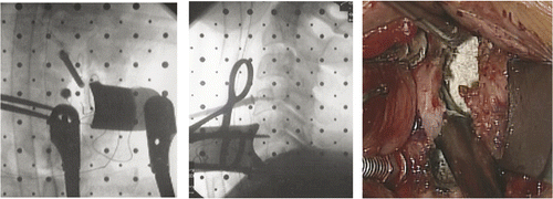
For each patient, the head and shoulders were fixed to the operating table and the cervical spine was exposed through a transverse skin incision using the standard Smith-Robinson approach. After confirmation of the correct level with intraoperative lateral X-rays, 14-mm distraction screws were inserted in the center of the bodies as a guide to the midline and also to serve as medial retractors. The approach-side longus colli muscle was dissected and retracted laterally to expose the medial portion of the transverse process. A blade was then positioned to retract the longus colli muscle laterally, and the other arm of the retractor was fixed to one of the central screws ().
Figure 1. The longus colli muscle was retracted by a lateral blade connected to one of the central screws. Left: Anterior-posterior view with C-arm. Center: Lateral view with C-arm. Right: Microscopic view during contralateral-side drilling.
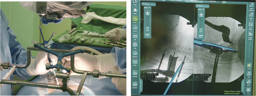
From this point on, the standard procedures for MOC were continued in the control group. In the IGN group, a dynamic reference frame was attached to the operating table with an external frame and C-arm views were acquired for navigation using the StealthStation ION system ().
Figure 2. Left: A dynamic navigation frame was attached to the operating table with an external frame. In this configuration, navigation can be implemented while maintaining the microscopic view. Right: IGN is useful in the axial plane and also in the sagittal plane.
As an initial registration step was not present in the ION system, IGN was introduced after the cervical retractor to obtain the highest accuracy. In other words, two-dimensional IGN was used rather than three-dimensional IGN, which latter resulted in a significant metal artifact.
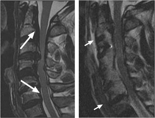
In this configuration, IGN could be set up without removing the microscope from the operating field. This step required 20 to 30 minutes. IGN was usually applied to determine the position of the bilateral posterior cortical bone near the uncovertebral joints and the sagittal margins of drilling, under microscopic view. IGN guidance made oblique drilling possible not only in the axial plane, but also the sagittal plane. Therefore, the anterior portion of the disc was not even touched at its upper and/or lower end segments ().
Figure 3. Preoperative and postoperative sagittal T2 images of a 62-year-old male with OPLL. Left: The upper and lower arrows indicate the direction of drilling. Right: The anterior portion of the upper and lower end segments was preserved.
Once a sufficient trapezoid cavity was created, the epidural compressive lesion was usually floated. At this point, the accuracy of IGN was of no further value and the remaining procedures were carried out in the same manner as for the control group.
Completeness of MOC (CMOC)
Three radiographic parameters were measured to calculate the completeness of MOC (CMOC). Component A is defined as the horizontal length of the remnant OPLL or posterior body from the ipsilateral posterior margin of the surgically created furrow to the ipsilateral medial margin of the pedicle on an axial CT scan. Component B is defined as the horizontal length of the remnant OPLL or posterior body from the contralateral posterior margin of the surgically created furrow to the contralateral medial margin of the pedicle on an axial CT scan. Component C is defined as the horizontal length of the remnant anterior body from the midline to the contralateral anterior margin of the surgically created furrow on an axial CT scan. CMOC is defined as the sum of A plus B minus C in millimeters at the pedicle level of the most compressive segment ().
Figure 4. Left: The arrow indicates the center of the vertebral body. The two lines represent the ideal trapezoid bony resection. Right: A is the remaining posterior body in millimeters from the ipsilateral medial margin of the pedicle; B is the same portion from the opposite side; and C is the remaining body from the center.
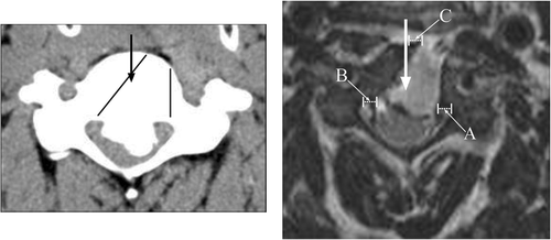
In the completed trapezoid trough, CMOC should be a negative value; a positive value indicates an incomplete posterior bony resection or a violation of the anterior central portion.
Results
In all 22 patients, an MR image of the cervical spine was acquired postoperatively and used to determine the CMOC. The mean CMOC was 0.89 mm (range: −3 to 5.3 mm) for the IGN group and 5.9 mm (range: −1 to 18 mm) for the control group. This difference had no statistical significance. However, the P-value for component B (contralateral remaining posterior body) of CMOC was determined to be 0.034 by an Independent Samples T-test. The mean value for component B was 4.1 mm (range: 0.6 to 7.6 mm) for the IGN group and 6.4 mm (range: 3.13 to 8.79 mm) for the control group ().
Table II. Completeness of multilevel oblique corpectomy (CMOC): mean values with ranges in parentheses (all measurements are in mm).
In the control group, two patients who underwent re-exploration due to remaining OPLL had CMOCs of 4.9 mm and 18 mm, respectively. A nearly quadrangular median bony resection was seen in the 18 mm CMOC case and a fusion procedure was added. In the IGN group, two patients had to be revised because of hematoma and their CMOCs were −1 mm. CSF leakage occurred in 3 cases in the control group and in 1 case in the IGN group; all four of these cases were managed by cervical epidural drainage without further complication.
The mean change in JOAS between preoperative and postoperative 5th day was 4.27 (range: 2 to 6) in the IGN group and 2.91 (range: −1 to 6) in the control group. This improvement in JOAS further increased to 5.58 (range: 2 to 11) for the former and 3.34 (range: −8.5 to 10) for the latter group at 1-year follow-up. The mean number of treated levels was 3.55 in the IGN group and 3.36 in the control group: their dispersion is summarized in . In the IGN group, anterior discs were spared at the lower end segment in all 11 patients and at the upper end segment in 7 patients.
Table III. Dispersion of treated levels.
The mean operation time was 248 min (range: 195 to 410 min) in the IGN group, and 259 minutes (range: 165 to 360 min) in the control group. This difference in operation time between groups is not statistically significant, despite the additional intraoperative IGN set-up time.
Discussion
The application of IGN to anterior spinal surgery has been restricted to thoracolumbar segments or upper cervical areas (the craniovertebral junction) for technical reasons and because of tiresome registration requirements Citation[20–25]. IGN has also been used in screw placement, but its use has not been considered in simple anterior plate-screw systems on the subaxial cervical spine Citation[26–28]. An initial registration step is needed with preoperative image-based navigation systems, and this has prevented the practical use of IGN in anterior cervical surgery. However, there is no need for such a registration step in navigation systems supported by intraoperative imaging modalities, so the authors were able to apply IGN to anterior surgery of the subaxial cervical spine Citation[27–30].
This application did not result in prolongation of the operation for the IGN group. In fact, the mean operation time was shorter than that for the control group. In addition, the contralateral body resection was more complete when maintaining an oblique plane than in the controls. These results demonstrate the theoretical advantages of IGN application in MOC, such as the ability to provide information about the depth and angle of drilling in real time.
The clinical outcomes seemed better in the IGN group than in the control group. The long-term effect has not yet been evaluated, but in the IGN group it appeared that the entire anterior portion of the upper and/or lower end segment could be preserved through undermining drilling in a sagittally oblique plane. Therefore, a transverse skin incision over C4 to 6 could be used for C2 to T1 lesions and the cosmetic effect was good. The most extreme application of this sagittally oblique plane drilling on the cranial side is in endoscopic image guided odontoidectomy Citation[31]; through this oblique decompression, complications associated with trans-oral decompression could be avoided. By comparison, sagittal and axial trapezoid body resection through the C4 to 6 window under IGN is easier, and prevents complications associated with high or lower anterior cervical dissection.
This free sagittal extension might be the most important advantage of using IGN in MOC. Epidural decompression of the OPLL mass could be started from a less adhesive level, and not only compression, but also draping of the spinal cord could be solved via this sagittal extension.
It should be mentioned that Wooridul Spine Hospital has had a modified C-arm CT system (SIREMOBIL ISO-C3D; Siemens Medical Solutions, Erlangen, Germany) since 2006. Initial application of 3D navigation was disappointing, due to severe artifacts arising from the screws, retractor blades and the routine operation bed. Currently, therefore, the authors are attempting to set up 3D navigation without any reduction in accuracy using the StealthStation ION, thereby overcoming the earlier problems of inaccuracy encountered with the SIREMOBIL system.
Conclusions
A navigation system supported by intraoperative imaging modalities can be applied practically to anterior surgery at the subaxial cervical spine. MOC performed under image guided navigation resulted in better results in terms of a more precise body resection, without any associated time increase.
Acknowledgments
This study was supported by a grant from the Wooridul Spine Foundation.
References
- Abe H, Tsuru M, Ito T, Iwasaki Y, Koiwa M. Anterior decompression for ossification of the posterior longitudinal ligament of the cervical spine. J Neurosurg 1981; 55: 108–116
- Belanger TA, Roh JS, Hanks SE, Kang JD, Emery SE, Bohlman HH. Ossification of the posterior longitudinal ligament. Results of anterior cervical decompression and arthrodesis in sixty-one North American patients. J Bone Joint Surg Am 2005; 87: 610–615
- Epstein N. Anterior approaches to cervical spondylosis and ossification of the posterior longitudinal ligament: Review of operative technique and assessment of 65 multilevel circumferential procedures. Surg Neurol 2001; 55: 313–324
- Kojima T, Waga S, Kubo Y, Kanamaru K, Shimosaka S, Shimizu T. Anterior cervical vertebrectomy and interbody fusion for multi-level spondylosis and ossification of the posterior longitudinal ligament. Neurosurgery 1989; 24: 864–872
- Onari K, Akiyama N, Kondo S, Toguchi A, Mihara H, Tsuchiya T. Long-term follow-up results of anterior interbody fusion applied for cervical myelopathy due to ossification of the posterior longitudinal ligament. Spine 2001; 26: 488–493
- Tani T, Ushida T, Ishida K, Iai H, Noguchi T, Yamamoto H. Relative safety of anterior microsurgical decompression versus laminoplasty for cervical myelopathy with a massive ossified posterior longitudinal ligament. Spine 2002; 27: 2491–2498
- Wada E, Suzuki S, Kanazawa A, Matsuoka T, Miyamoto S, Yonenobu K. Subtotal corpectomy versus laminoplasty for multilevel cervical spondylotic myelopathy. Spine 2001; 26: 1443–1448
- George B, Zerah M, Lot G, Hurth M. Oblique transcorporeal approach to anteriorly located lesions in the cervical spine canal. Acta Neurochir (Wien) 1993; 121: 187–190
- Ohara S, Momma F, Ohyama T, Furuichi S. Antero-lateral partial vertebrectomy for ossification of the posterior longitudinal ligament of the cervical spine. Spinal Surgery 1994; 8: 125–130
- Verbiest H, Paz Y, Geuse HD. Anterolateral surgery for cervical spondylosis in cases of myelopathy or nerve root compression. J Neurosurg 1966; 25: 611–622
- Bruneau M, Cornelius JF, George B. Microsurgical cervical nerve root decompression by anterolateral approach. Neurosurgery 2006;58(ONS Suppl 1):ONS108-ONS113.
- Bruneau M, Cornelius JF, George B. Multilevel oblique corpectomies: Surgical indications and technique. Neurosurgery 2007;61(ONS Suppl 1):ONS106-ONS112.
- Cagli S, Chamberlain RH, Sonntag VK, Crawford NR. The biomechanical effects of cervical multilevel oblique corpectomy. Spine 2004; 29: 1420–1427
- Chacko AG, Daniel RT. Multilevel cervical oblique corpectomy in the treatment of ossified posterior longitudinal ligament in the presence of ossified anterior longitudinal ligament. Spine 2007; 32: E575–E580
- George B, Gauthier N, Lot G. Multisegmental cervical spondylotic myelopathy and radiculopathy treated by multilevel oblique corpectomies without fusion. Neurosurgery 1999; 44: 81–90
- Chibbaro S, Mirone G, Makiese O, George B. Multilevel oblique corpectomy without fusion in managing cervical myelopathy: Long-term outcome and stability evaluation in 268 patients. J Neurosurg Spine 2009; 10: 458–465
- Karalar T, Unal F, Karagoz Guzey F, Kiris T, Bozdag E, Sunbuloglu E. Biomechanical analysis of cervical multilevel oblique corpectomy: An in vitro study in sheep. Acta Neurochir (Wien) 2004; 146: 813–818
- Koc RK, Menku A, Akdemir H, Tucer B, Kurtsoy A, Oktem IS. Cervical spondylotic myelopathy and radiculopathy treated by oblique corpectomies without fusion. Neurosurg Rev 2004; 27: 252–258
- Rocchi G, Caroli E, Salvati M, Delfini R. Multilevel oblique corpectomy without fusion: Our experience in 48 patients. Spine 2005; 30: 1963–1969
- Kim KD, Babbitz JD, Mimbs J. Imaging-guided costotransversectomy for thoracic disc herniation. Neurosurg Focus 2000; 9: e7
- Lee HY, Lee SH, Chung SK, Shin SW, Lim SR, Kaul R. Computer-assisted anterior reconstruction surgery for a thoracolumbar fracture: A case report. Joint Dis Rel Surg 2005; 16: 153–157
- Thoranaghatte RU, Zheng G, Langlotz F, Nolte LP. Endoscope-based hybrid navigation system for minimally invasive ventral spine surgeries. Comput Aided Surg 2005; 10: 351–356
- Ugur HC, Kahilogullari G, Attar A, Caglar S, Savas A, Egemen N. Neuronavigation-assisted transoral-transpharyngeal approach for basilar invagination – two case reports. Neurol Med Chir (Tokyo) 2006; 46: 306–308
- Veres R, Bagó A, Fedorcsák I. Early experiences with image-guided transoral surgery for the pathologies of the upper cervical spine. Spine 2001; 26: 1385–1388
- Vougioukas VI, Hubbe U, Schipper J, Spetzger U. Navigated transoral approach to the cranial base and the craniocervical junction: Technical note. Neurosurgery 2003; 52: 247–250
- Holly LT, Foley KT. Intraoperative spinal navigation. Spine 2003; 28: S54–S61
- Holly LT, Foley KT. Image guidance in spine surgery. Orthop Clin North Am 2007; 38: 451–461
- Hott JS, Papadopoulos SM, Theodore N, Dickman CA, Sonntag VK. Intraoperative Iso-C C-arm navigation in cervical spinal surgery: Review of the first 52 cases. Spine 2004; 29: 2856–2860
- Maier B, Zheng G, Ploss C, Zhang X, Welle K, Nolte LP, Marzi I. A CT-free, intraoperative planning and navigation system for minimally invasive anterior spinal surgery – an accuracy study. Comput Aided Surg 2007; 12: 233–241
- Rajasekaran S, Vidyadhara S, Ramesh P, Shetty AP. Randomized clinical study to compare the accuracy of navigated and non-navigated thoracic pedicle screws in deformity correction surgeries. Spine 2007; 32: E56–E64
- Wolinsky JP, Sciubba DM, Suk I, Gokaslan ZL. Endoscopic image-guided odontoidectomy for decompression of basilar invagination via a standard anterior cervical approach. Technical note. J Neurosurg Spine 2007; 6: 184–191