Abstract
Objective: The acetabular version following total hip arthroplasty is an important prognostic factor. Computer navigation serves as a precise tool during hip arthroplasty, which requires precise measurement to verify the effect of the procedure. Wan and colleagues have reported an innovative method for measuring acetabular radiographic version with an ordinary goniometer. Our objective was to study the precision of this method.
Methods: We derived the underlying mathematical principle of Wan's method and produced a correction formula and chart. Forty-eight computer-synthesized radiographs were used to study the method and its mathematical correction. Ten real radiographs were used to detect intra-observer difference. The paired t-test was used for statistical analysis.
Results: There was a significant difference between synthetic acetabular radiographic version and the measurement obtained with Wan's method (p < 0.05), but there was no difference after mathematical correction (p = 0.15). For smaller radiographic version (<20o), there was no statistical difference using Wan's method (p = 0.054).
Conclusions: The method of Wan and colleagues can be used when acetabular radiographic version is less than 20°. For larger radiographic versions, however, mathematical correction is necessary to obtain precise results.
Introduction
The radiographic version of the acetabular component following total hip arthroplasty (THA) is important, affecting the range of motion Citation[1], dislocation rate Citation[2], and rate of wear Citation[3]. Measuring methods fall into three major groups: the trigonometric group Citation[2], Citation[4–6], the protractor group Citation[7–10], and the computed tomography (CT) group Citation[11–13]. Methods in the trigonometric group usually require a ruler and an engineering calculator; those in the protractor group require specialized protractors; and those in the CT group entail excessive radiation and high cost.
The computer navigation system used in THA has an implantation precision of within 1 mm Citation[14]. This precision should be measured with a tool of equal precision. Wan et al. Citation[15] have presented their findings concerning acetabular component orientation, and have also described a novel protractor method for measuring acetabular orientation. In this method, they measured the angle formed by the long axis and the chord of one quarter ellipse (henceforth referred to as Wan's angle) as the acetabular radiographic version (). This method is quite unique, easy, and requires no specialized protractor.
Figure 1. Post-operative radiograph of total hip arthroplasty with a U2 Total Hip Joint (United Orthopedic Corporation, Hsinchu, Taiwan). We marked the outer shell of the acetabulum with an ellipse (s = the short axis of the ellipse; l = the long axis). Wan et al. described the measured angle α as acetabular radiographic version Citation[15]. Because of the symmetry of the ellipse, angle α is equal to angles β, γ, and δ. We previously used a special protractor to measure angle β for acetabular radiographic version Citation[7].
![Figure 1. Post-operative radiograph of total hip arthroplasty with a U2 Total Hip Joint (United Orthopedic Corporation, Hsinchu, Taiwan). We marked the outer shell of the acetabulum with an ellipse (s = the short axis of the ellipse; l = the long axis). Wan et al. described the measured angle α as acetabular radiographic version Citation[15]. Because of the symmetry of the ellipse, angle α is equal to angles β, γ, and δ. We previously used a special protractor to measure angle β for acetabular radiographic version Citation[7].](/cms/asset/503423c1-9075-43d4-b1c0-d1f97d4a2dfa/icsu_a_583805_f0001_b.gif)
We found no similar method in the literature, which prompted our interest in verifying the precision of this method. If the precision was good, the method should be widely promoted because an ordinary goniometer could be used to measure acetabular radiographic version directly; and if the precision was unsatisfactory, could we improve it?
Materials and methods
Measuring acetabular radiographic version is a mathematical problem, and must therefore be viewed mathematically. We suggest that Wan's angle can represent the ellipse, but not the real radiographic version of the acetabular component, and thus requires mathematical correction. We described the relationship and the deduction process as follows:
According to the definition of the trigonometric function tan,where α represents Wan's angle, s is the short axis of the ellipse, and l is its long axis (). According to the well-accepted trigonometric method of measuring acetabular radiographic version Citation[2], Citation[4], Citation[7], Citation[16],
Combining Equations 1 and 2, we get the correction equation:
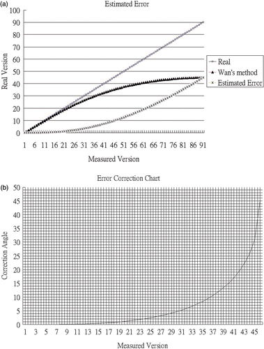
According to Equation 3, we can calculate the error in measuring radiographic version from the intra-ellipse angle, which is the method of Wan et al. The error and correction charts shown in were then calculated. The correction angle increased with increasing radiographic version angle, because the trigonometric function tan differs increasingly from sin with increasing angle. We have also incorporated Equation 3 into Microsoft Office Excel, so that the surgeon can easily use it to calculate the corrected acetabular radiographic version from Wan's angle. The Excel formula is “=ASIN(TAN((A2*PI()/180)))*180/PI()”.
Figure 2. (a) Theoretical error of Wan's method. (b) Error correction chart for Wan's method. After obtaining a measurement using Wan's method, the surgeon can add the correction angle from this chart.
To verify the theoretical error, we first used THR Simulator, a software program which has been used and reported previously Citation[17], to synthesize 48 radiographs with known radiographic version angle. These synthetic radiographs represented total hip arthroplasties but without the femoral head. The radiographic version ranged from 5° to 52° with random inclination of 30 to 60°. All these radiographs were randomly ordered. We also collected 10 clinical radiographs following THA procedures. All of these radiographs were printed on paper to facilitate measurement. One of the authors measured the radiographic version of these radiographs (both clinical and synthetic) using Wan's method with and without mathematical correction. The measurements were corrected mathematically using Equation 3. The same author again measured the clinical radiographs using Wan's method one week later. The errors with Wan's method, with or without mathematical correction, were calculated as measured radiographic version minus synthetic radiographic version.
We synthesized the radiographic version angle with a range of 5 to 52°, because this was the range reported in a recent study of acetabular radiographic version Citation[18]. Random inclination was used because the inclination does not influence the radiographic version Citation[7], Citation[16].
Statistics
The paired t-test was used for all continuous variables, with p < 0.05 as the significance level.
Ethics
This study was approved by the Institutional Review Board of Tao-Yuan General Hospital (TYGH098019).
Results
The results obtained with the simulated radiographs are shown in and . The angles measured with Wan's method with and without correction ranged from 5° to 40° (mean ± SD = 24.3° ± 10.3°) and from 5° to 57° (mean: 28.9° ± 14.8°), respectively. The errors with Wan's method without correction ranged from −1° to 15°, and the mean value was 4.3° ± 4.0°. Following mathematical correction, the errors ranged from −4.3° to 7°, and the mean value was 0.4° ± 2.0°. There was significant difference between the measured radiographic version with Wan's method and the synthetic radiographic version (p < 0.05), and the difference increased with increasing radiographic version angle. We also calculated whether smaller radiographic version caused less difference. If the synthetic radiographic version was less than 20° (less than 18° with Wan's method), there was no significant difference (p = 0.054). There was also no statistical difference between the synthetic radiographic version and the mathematically corrected radiographic version with Wan's method (p = 0.15). The absolute difference for Wan's method ranged from 0° to 15° with a mean value of 4.3° ± 4.0°. The range after mathematical correction was 0° to 7° with a mean value of 1.3° ± 1.6°.
Figure 3. Error with Wan's method before and after mathematical correction.
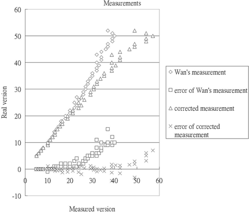
Table I. Results obtained with simulated radiographs.
For the inter-measurement study, the radiographic version measured twice with Wan's method ranged from 4° to 21° with a mean value of 12.0° ± 5.3°. The absolute difference between the two measurements ranged from 0° to 3° with a mean value of 1.1° ± 1.0°.
Discussion
Overview
Measuring radiographic version from a plain radiograph is a matter of measuring the ellipse formed by the outer shell of the acetabular component after total hip arthroplasty. Wan's method is relatively simple, and the surgeon can use any ordinary goniometer to make the measurement on plain radiographs (). Recently, digitalized radiograph systems have become increasingly popular. Vendors usually provide tools to measure the angle directly, which makes the digital measurement of Wan's angle even easier.
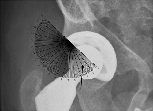
Figure 4. We use an ordinary goniometer to measure acetabular radiographic version directly. In this example, the goniometer is aligned with the long axis of the ellipse (the metal ring) and Wan's angle is then read on the middle point of the ellipse (arrow).
Possible causes of error
Most methods measure the ellipse at the middle point Citation[2], Citation[4], Citation[7–10]. However, this point is usually obscured by the femoral head and is thus a source of error. In our study, we synthesized the radiographs without the femoral head to eliminate such obscuration. The Trigonometric method Citation[2], Citation[4], Widmer's method Citation[9], our improved version of Widmer's method Citation[10], and the methods of Fabeck et al. Citation[8] and Lewinnek et al. Citation[2] all measure the ellipse horizontally. Therefore, mis-estimation of the long axis direction is eliminated. However, our own previously published method Citation[7] and that of Wan et al. Citation[15] measure it both horizontally and vertically, and mis-estimation in both the short and long axis directions may cause error. Our results show that the inter-measurement differences for the same observer ranged from 0° to 3° (mean: 1.1° ± 1.0°).
Recently, computerized measuring methods have become increasingly popular. The computer can generate a whole ellipse, in other words, approximating the ellipse with an ellipse, and we consider this approach to be more precise.
Every measuring method has its own advantages, disadvantages and precision. In any report, it is important to specify the measuring method used and its precision.
Acceptable range of Wan's method
In our study, Wan's method could be regarded as an appropriate radiographic measuring method only if the angle was smaller than 20°. However, after mathematical correction, the measured radiographic version was not statistically different from the synthetic radiographic version. It was also demonstrated that our mathematical correction is compatible with the results. We reviewed data from our previous study Citation[7] and found that 35.6% (16/45) of the acetabular radiographic version was over 20°, which makes our mathematical correction useful under such circumstances. When the radiographic version exceeds 20o (see , for example), in which case the range of motion will be affected Citation[1] and the dislocation rate will be increased Citation[2], along with the rate of wear Citation[3]. In these situations, measuring and documenting the correct radiographic version is more important than usual. Therefore, our mathematical correction is indispensable in such clinical settings.
For Wan's method, the mean absolute difference with synthetic version is 4.3° ± 4.0°, whereas after mathematical correction this becomes 1.3° ± 1.6°. Reviewing the literature, 1.3° is comparable to values reported previously Citation[2], Citation[4–13]. The correction is meaningful because the inter-measurement difference is smaller at 1.1° ± 1.0°.
Conclusions
Without correction, the directly measured Wan's angle is acceptable as the acetabular radiographic version when the version is less than 20°. However, acetabular radiographic version greater than 20° may require mathematical correction (Equation 3). A simple Microsoft Excel program or a correction chart () is provided for clinical use. Any report concerning acetabular radiographic version should specify the measuring method used and its precision.
Acknowledgments
We thank United Orthopedic Corporation, Hsinchu, Taiwan for providing us with the postoperative radiographs of U2 total hip arthroplasty.
Declaration of interest: This study was supported by a grant, NSC98-2314-B-087-001-MY2, from the National Science Council, Taiwan, ROC.
References
- Vendittoli PA, Ganapathi M, Nuno N, Plamondon D, Lavigne M. Factors affecting hip range of motion in surface replacement arthroplasty. Clin Biomech (Bristol, Avon) 2007; 22(9)1004–1012
- Lewinnek GE, Lewis JL, Tarr R, Compere CL, Zimmerman JR. Dislocations after total hip-replacement arthroplasties. J Bone Joint Surg Am 1978; 60(2)217–220
- Lusty PJ, Watson A, Tuke MA, Walter WL, Walter WK, Zicat B. Wear and acetabular component orientation in third generation alumina-on-alumina ceramic bearings: An analysis of 33 retrievals. J Bone Joint Surg Br 2007; 89(9)1158–1164, [erratum J Bone Joint Surg Br 2008;90(4):543]
- Ackland MK, Bourne WB, Uhthoff HK. Anteversion of the acetabular cup. Measurement of angle after total hip replacement. J Bone Joint Surg Br 1986; 68(3)409–413
- Pradhan R. Planar anteversion of the acetabular cup as determined from plain anteroposterior radiographs. J Bone Joint Surg Br 1999; 81(3)431–435
- Visser JD, Konings JG. A new method for measuring angles after total hip arthroplasty. A study of the acetabular cup and femoral component. J Bone Joint Surg Br 1981; 63B(4)556–559
- Liaw CK, Hou SM, Yang RS, Wu TY, Fuh CS. A new tool for measuring cup orientation in total hip arthroplasties from plain radiographs. Clin Orthop Relat Res 2006; 451: 134–139
- Fabeck L, Farrokh D, Tolley M, Descamps PY, Gebhart M, Delince P. A method to measure acetabular cup anteversion after total hip replacement. Acta Orthop Belg 1999; 65(4)485–491
- Widmer KH. A simplified method to determine acetabular cup anteversion from plain radiographs. J Arthroplasty 2004; 19(3)387–390
- Liaw CK, Yang RS, Hou SM, Wu TY, Fuh CS. Measurement of the acetabular cup anteversion on simulated radiographs. J Arthroplasty 2009; 24(3)468–474
- Blendea S, Troccaz J, Merloz P. [Accuracy measurements of acetabular cup positioning using CT less navigation] [in French]. Rev Chir Orthop Reparatrice Appar Mot 2007; 93(2)157–164
- Olivecrona H, Weidenhielm L, Olivecrona L, Beckman MO, Stark A, Noz ME, Maguire GQ, Jr, Zeleznik MP, Svensson L, Jonson T. A new CT method for measuring cup orientation after total hip arthroplasty: A study of 10 patients. Acta Orthop Scand 2004; 75(3)252–260
- Penney GP, Edwards PJ, Hipwell JH, Slomczykowski M, Revie I, Hawkes DJ. Postoperative calculation of acetabular cup position using 2-D-3-D registration. IEEE Trans Biomed Eng 2007; 54(7)1342–1348
- Hüfner T, Kendoff D, Citak M, Geerling J, Krettek C. [Precision in orthopaedic computer navigation] [in German]. Orthopade 2006; 35(10)1043–1045
- Wan Z, Malik A, Jaramaz B, Chao L, Dorr LD. Imaging and navigation measurement of acetabular component position in THA. Clin Orthop Relat Res 2009; 467(1)32–42
- Murray DW. The definition and measurement of acetabular orientation. J Bone Joint Surg Br 1993; 75(2)228–232
- Wu TY, Yang RS, Fuh CS, Hou SM, Liaw CK. THR Simulator – the software for generating radiographs of THR prosthesis. BMC Musculoskelet Disord 2009; 10: 8
- Minoda Y, Kadowaki T, Kim M. Acetabular component orientation in 834 total hip arthroplasties using a manual technique. Clin Orthop Relat Res 2006; 445: 186–191