Abstract
Objective: To develop and validate the efficacy and accuracy of a novel drill template for cervical pedicle instrumentation.
Materials and Methods: A CT scan of the cervical vertebrae was performed, and a 3D model of the vertebrae was reconstructed using MIMICS 10.01 software. The 3D vertebral model was then exported in STL format, and opened in a workstation running UGS Imageware 12.0 software to determine the optimal pedicle screw size and orientation. A virtual navigational template was established according to the laminar anatomic trait, and physical navigational templates were manufactured using rapid prototyping. The navigational templates were used intraoperatively to assist in the placement of cervical pedicle screws.
Results: In all, 84 pedicle screws were placed, and the accuracy of screw placement was confirmed with postoperative X-rays and CT scans. Eighty-two screws were rated as Grade 0, 2 as Grade 1, and no screws as Grade 2 or 3. Hence, safer screw positioning was accomplished with the drill template technique.
Conclusions: This study demonstrates a patient-specific template technique that is easy to use, can simplify the surgical act, and generates highly accurate cervical pedicle screw placement. The advantages of this technology over traditional techniques are that it enables planning of the screw trajectory to be completed prior to surgery, and that the screw can be sized to fit the patient's anatomy.
Introduction
The use of pedicle screw-based spinal instrumentation systems in the lumbar and thoracic spine has increased tremendously over the last decade because of their superior biomechanical properties and reduction possibilities Citation[1], Citation[2]. Nevertheless, despite the perception that cervical pedicle fixation would provide superior holding strength Citation[3–5], cervical pedicle screws have not been implemented routinely because of the large anatomic variations in the size and shape of the cervical pedicles from one individual to the next Citation[6–8]. Since the first technical description of this surgical technique in 1994 by Abumi and co-workers Citation[9–11], several attempts have been made to enhance the safety and accuracy of cervical pedicle screw placement Citation[12–14]. It has been suggested that Computer Assisted Surgery (CAS) systems offer the safest approach to cervical pedicle screw placement. However, in laboratory studies, pedicle perforation could not be completely prevented with any technique Citation[15–18]. The present study introduces an ingenious, custom-fit rapid prototype drill template for the placement of pedicle screws in the cervical spine Citation[19–21]. The trajectory of the cervical pedicle screws was first identified based on preoperative CT scans. Subsequently, patient-specific drill templates were designed that would maintain close contact with the posterior surface of the cervical vertebrae and thus provide the best stability for drilling. This approach was further validated in a cadaveric study.
Materials and methods
Specimens
Six preserved and randomly chosen human cervical spine specimens (from four males and two females) were obtained from the Department of Anatomy at Kunming Medical College. All the cervical spines were examined with standard radiographs and CT scans to exclude anomalies, tumors, or severe multi-segmental changes. All showed varying degrees of osteoporosis and degenerative changes as expected in humans with an average age of 65 years (range: 55–79 years). Subsequently, 3.5-mm titanium screws were placed in each cervical pedicle. These screws were chosen because titanium does not create significant artifacts in CT images and allows precise postoperative determination of the screw position.
Planning of the screw trajectory
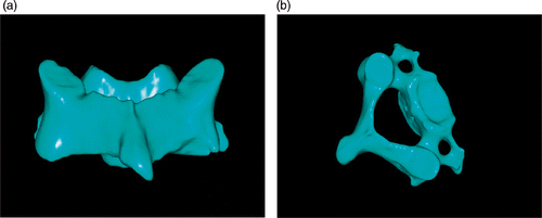
Based on the requirements of this technique, a spiral three-dimensional (3D) CT scan (LightSpeed VCT, GE Healthcare, Milwaukee WI) was performed preoperatively on the cervical spine specimen with a slice thickness of 0.625 mm and in-plane resolution of 0.35 mm. The images were stored in DICOM format and transferred to a workstation running MIMICS 10.01 software (Materialise, Leuven, Belgium) to generate a 3D reconstruction model of the desired cervical vertebrae ().
Figure 1. 3D model of the cervical vertebra. (a) Posterior view. (b) Superior view.
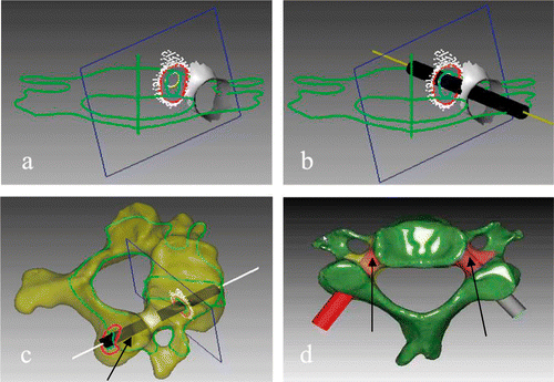
The 3D cervical vertebra model was then exported in STL format to a workstation running the reverse engineering software UGS Imageware 12.0 (EDS, Plano, TX). The cervical pedicle height (PH) and pedicle width (PW) were measured to determine the optimal screw size and orientation. Using UGS Imageware, the left and right pedicles were projected toward the vertebrae and lamina (). Since the thickness and cross-section of the pedicle vary along its length, the smaller diameter of the elliptical inner boundary of the pedicle's projection was used in determining the maximum allowable screw diameter (). This diameter was further used to draw a circle which was projected between the vertebrae and the lamina to obtain the optimal pedicle screw trajectory (). A 3D vertebral model was then reconstructed with a virtual screw placed on both sides ().
Figure 2. Analysis of cervical pedicle screw trajectory by the reverse engineering software. (a) Pedicle and its positive projection. (b) Best trajectory of pedicle screw projection. (c) Pedicle screw channel (arrow). (d) Planned screw trajectories (arrows).
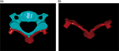
Following the determination of the optimal pedicle screw trajectory, a navigational template was constructed with a drill guide on either side. The template surface was created as the inverse of the posterior vertebral surface, thus potentially enabling a near-perfect fit. We also ensured that the template did not overlap with adjacent segments ().
Figure 3. Design of the navigational template.
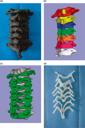
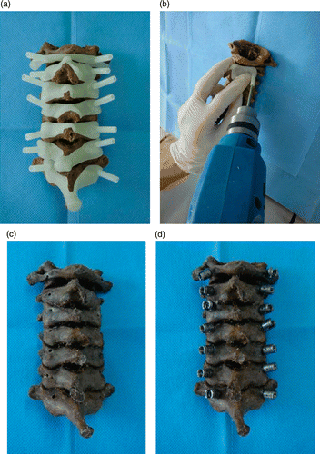
The biomodel of the navigational template was produced in acrylate resin (Somos 14120; DSM Desotech, Inc., Elgin, IL) by stereolithography, a rapid prototyping (RP) technique (Hen Tong Company, China). The biomodel was used as a cervical pedicle guide template ().
Figure 4. (a) Cervical spine specimen. (b) 3D reconstruction of cervical spine. (c) Drill template and corresponding cervical vertebrae. (d) Physical biomodel of the navigational template from C1–C7.
Surgery
Surgery was performed in a laboratory setting by the third author (G.P.C.) using the following procedures in each case. The bodies were placed in a prone position on a table. The spinous process, laminae, and lateral masses of the spine were then exposed as necessary. Spinous process and lamina of spine (), with a good fit being achieved between the template and spinous process. A high-speed drill was then used to drill the trajectory of the pedicle screw along the navigation channel (). Using a hand drill, the trajectory of the pedicle screw was carefully drilled to the desired depth in the preoperative plan (), then a 3.5-mm screw was carefully inserted along the same trajectory ().
Figure 5. (a) The navigational templates fitted perfectly onto the posterior surface of their corresponding vertebrae. (b) A high-speed drill was used along the navigational channel to drill the trajectory of the pedicle screw. (c) Pedicle channel of cervical spine specimen. (d) Pedicle screws inserted into the cervical spine specimens.
Evaluation of cervical pedicle screw
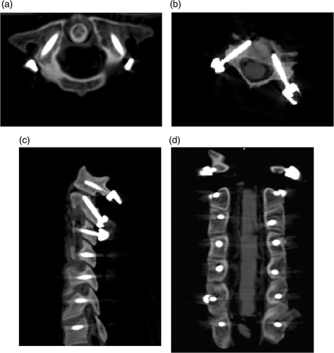
After surgery, the positions of the pedicle screws were evaluated using CT scans (). An axial image including the entire length of each screw was obtained, and the medial and lateral deviations of the screws were classified in 4 grades as follows: Grade 0 – no deviation, i.e., the screw was entirely contained within the pedicle; Grade 1 – deviation of less than 2 mm or less than half the screw diameter; Grade 2 – deviation of more than 2 mm and less than 4 mm, or half the screw diameter; Grade 3 – deviation of more than 4 mm or complete deviation. The screw position was independently evaluated by the sixth author (Y.B.C.) in terms of the extent of pedicle wall violation.
Figure 6. Postoperative CT scan showing good positioning of the cervical pedicle screw. (a) C2 pedicle screw – axial view. (b) C4 pedicle screw – axial view. (c) Sagittal view of cervical pedicle screw. (d) Coronal view of cervical pedicle screw.
Results
Using the virtual 3D model, the optimal entry point for the drill can be chosen, thus determining the entry point and direction for the cervical pedicle screw. The drill template was created to fit the posterior surface of the cervical vertebrae very well.
During the surgery, the best fit for positioning the template could easily be found manually because no significant free motion of the template occurred when it was pressed lightly against the spinous process and lamina. The navigational template can therefore be used as an in situ drill guide. The individual navigational template technique enabled the placement and size of each screw to be customized based on the unique morphology of each cervical vertebra, as well as permitting the surgical plan to be prepared preoperatively. To achieve this, exact preparation of the bone surface is essential, including thorough removal of the attached muscle and fat tissue without damage to the bony surface.
Using these novel custom-fit navigational templates, each vertebral pedicle screw insertion took approximately 50 seconds. Fluoroscopy was not required, which considerably reduced the radiation exposure for the surgical team. Currently, the production time for an RP model is approximately 1 day, and the cost of each template is approximately $20 per vertebral level.
A total of 84 pedicles were instrumented with cervical pedicle screws. displays the number and incidence of pedicle perforations in relation to the vertebral level (C1–C7). Of the 84 screws, 82 were rated as Grade 0, 2 as Grade 1, and no screws as either Grade 2 or 3. Hence, safer screw positioning was accomplished using the drill template technique.
Table I. Number and incidence of pedicle perforations.
Discussion
Biomechanical data confirm the outstanding stability of cervical pedicle screw fixation Citation[22], Citation[23]. However, this technique is technically demanding and carries the risk of severe complications Citation[24], Citation[25]. Successful placement of pedicle screws in the cervical spine requires a thorough 3D understanding of pedicle morphology to accurately identify the ideal screw axis Citation[3], Citation[26], Citation[27]. Several methods have been explored for precise cervical pedicle screw placement, including the use of anatomic studies, image guided techniques Citation[18], computer assisted surgery systems, and drill templates Citation[28–32]. These techniques can be broadly classified into five types: (1) techniques relying on anatomical landmarks and averaged angular dimensions; (2) techniques requiring direct exposure of the pedicle, e.g., by laminotomy; (3) CT-based computer assisted surgery (CAS); (4) fluoroscopy-based CAS techniques; and (5) drill template techniques.
The accuracy of computer assisted screw insertion has been demonstrated recently. The rate of pedicle perforation in 52 consecutive patients who received posterior cervical or cervico-thoracic instrumentations using pedicle screws was 8.6% in the conventional group and 3.0% in the computer assisted surgery group Citation[18]. Another group has reported similar results, in which the rate of pedicle wall perforation was significantly lower in the computer assisted group (1.2%) compared to the conventional group (6.7%) Citation[33]. However, despite advances in instrumentation techniques and intraoperative imaging, the successful implementation of posterior cervical instrumentation remains a challenge. As reported by Ludwig et al. Citation[34], 18% of the pedicles (C3–C7) of human cadaveric cervical spines instrumented using computer assisted image guided surgical systems and the Abumi technique had a critical breach. Thus, several caveats should be considered: (1) mastering these techniques requires a lot of time; (2) errors may occur when adjacent segments of the spine shift intraoperatively, or if the registration frame and optical array shift; (3) tracking of optical array devices can be obscured by the surgeons or surgical tools; (4) the cost of the technology is high; and (5) these techniques can lengthen the duration of surgical procedures.
The use of drill templates was initially demonstrated in hip and knee surgery Citation[32]. Several studies have also described their use in spinal procedures, including cervical spine surgery Citation[28–31]. Berry et al. Citation[28] used a three-V-shaped knife design to support the drill template, while Goffin et al. Citation[29] designed a template with clamps to interface with the posterior course of the cervical vertebra. More recently, Owen et al. Citation[30] introduced a drill template designed to match the posterior surface of the cervical vertebra around the entry point. The concept of using the drill template to assist in the placement of cervical pedicle screws was promoted, but its practical realization was not so well described. The approach of Owen et al. differs from our method in several aspects. First, in our method, the best cervical pedicle screw can be determined through the reverse engineering software analysis, thus the optimal pedicle channel can be derived from an irregular channel. Second, our template complements the spinal process, lamina, and lateral mass, thus providing more stability and improving accuracy. In the approach of Owen et al., the template is only in contact with the lamina, making it difficult to obtain adequate stability and potentially reducing the accuracy of the drill template. The design of the templates is also different, and our template is made using stereolithography, further improving its accuracy.
Based on comparisons with other template designs, the present study has successfully introduced a novel, custom-fit navigational template for the placement of pedicle screws in the cervical spine and has further validated the technique through a cadaveric study, which demonstrated the high accuracy of this approach. A preoperative CT scan was obtained to customize the placement of each screw based on the unique morphology of the patient's cervical vertebrae. The availability of a high-resolution CT scanner and advanced technologies facilitated high geometric accuracy of the drill template. The MIMICS software used to reconstruct the 3D model of the vertebrae from the CT scan data is capable of providing fast, easy, and powerful 3D image processing and editing. However, designing and producing the patient-specific template is a specialized task beyond the ability of surgeons, so the cervical templates should be made by engineering personnel. In addition, rapid prototyping (RP) technology, in which a model is built by stacking thin layers instead of milling, has been employed. RP can reproduce more complex designs because of its high accuracy and versatility. The resolution of the RP machine used is well above the 0.35-mm resolution of the template model, thus the accuracy of reproduction is not a limiting factor Citation[30]. As the template is produced in brittle acrylate resin, the thickness of the navigational template must be at least 2 mm to assure the rigidity of the template, and it should be carefully manipulated during the operation. A little debris may be produced during drilling.
Using the patient-specific drill template design has several advantages. First, the surgeon can decide the location, orientation and size of each screw based on the unique morphology of the cervical vertebrae even before going to the operating table. As observed in this study, the pedicle anatomy may vary widely from patient to patient. Therefore, preoperative CT evaluation is suggested as a mandatory step for precise planning of the surgical procedure. The appropriate screw diameter should be selected for each individual pedicle. During the course of this study, we encountered five cases of abnormal cervical pedicles; however, the use of patient-specific drill templates enabled us to handle even these abnormal cases successfully by helping us to choose the correct screws and decide on the best orientation of the screw insertion for each pedicle. Second, this technique is simple as it requires little expertise on the part of the surgeon and the preoperatively prepared drill template can be used intraoperatively to assist with surgical navigation and precise placement of instrumentation. Third, in contrast with image guided techniques, our method eliminates the need for complex equipment and time-consuming procedures in the operating theater, thereby reducing the duration of the surgical procedure. Fourth, screw placement is accurate, thus avoiding perforation of the spinal canal and blood vessels. In abnormal cases with very narrow pedicles, this technique also provides the flexibility to examine the accuracy of the drill templates by inserting screws into the biomodel of the vertebrae. Lastly, the need for fluoroscopy during screw insertion is eliminated, considerably reducing the radiation exposure for the surgical team.
Nevertheless, there are potential sources of error in this technique. It requires clean preparation of the bone surface, including thorough removal of the attached muscle and fat tissue without damaging the bony surface structures, to ensure the proper fit of the drill template on the lamina. Even with this limitation, our technique has proven better than other techniques reported in the literature. For example, the use of V-shaped knifes to support the drill template by Berry et al. Citation[28] did not require excessive soft tissue dissection from the vertebra. However, the surgeons were not confident of the screw positioning in the cervical spine. Another issue is that, in the clinical setting, a template should be capable of being used as an in situ drill guide, and any movement between the bone and template caused by vibration while drilling will greatly affect the accuracy of screw placement. Therefore, the template has to be firmly placed in position by the surgeon while drilling is underway. As mentioned above, thorough removal of the attached muscle and fat tissue without damage to the bony surface is very important for the application of the template. Based on the findings from the study of D’Urso et al. Citation[35], the use of this technique in the thoracic and lumbar regions of the spine is not recommended due to the presence of excessive soft tissue.
Conclusion
The positioning of pedicle screws in the cervical spine is a technically demanding surgical method burdened with potential risks. Although data obtained in clinical studies have revealed a relatively low risk of complications, results achieved under laboratory conditions have been less favorable: pedicle perforations occur while employing any pedicle screw placement technique, even in association with CAS navigation. This template design is unique because it was created based on the reverse engineering principle, and could therefore perfectly match the posterior surface of the cervical vertebra. After preparing the bone surface by thorough removal of attached muscle and fat tissue without damaging the bony surface structures, the templates can be securely held in place by the surgeon's free hand.
Acknowledgments
The authors would like to thank Dr. You H. Cheng for his assistance with the CT data collection.
Declaration of interest: This work was supported by the Yunnan Natural Science Foundation (2008CD210 and 2010ZC183).
References
- Metz LN, Burch S. Computer-assisted surgical planning and image-guided surgical navigation in refractory adult scoliosis surgery. Spine 2008; 33(9)E287–E292
- Ludwig SC, Kramer DL, Vaccaro AR, Albert TJ. Transpedicle screw fixation of the cervical spine. Clin Orthop Relat Res 1999; (359): 77–88
- Richter M, Amiot LP, Neller S, Kluger P, Puhl W. Computer assisted surgery in posterior instrumentation of the cervical spine – an in-vitro feasibility study. Eur Spine J 2000; 9(Suppl 1)65–70
- Schmidt R, Wilke HJ, Claes L, Puhl W, Richter M. Pedicle screws enhance primary stability in multilevel cervical corporectomies: Biomechanical in-vitro comparison of different implants including angle- and non-angle stable instrumentations. Spine 2003; 28(16)1821–1828
- Kotani Y, Cunningham BW, Abumi K, McAfee PC. Biomechanical analysis of cervical stabilization systems: An assessment of transpedicular screw fixation in the cervical spine. Spine 1994; 19(22)2529–2539
- Ebraheim NA, Xu R, Knight T, Yeasting RA. Morphometric evaluation of lower pedicle and its projection. Spine 1997; 22(1)1–6
- Karaikovic EE, Daubs MD, Madsen RW, Gaines RW, Jr. Morphologic characteristics of human cervical pedicles. Spine 1997; 22(5)493–500
- Xu R, Nadaud MC, Ebraheim NA, Yeasting RA. Morphology of the second cervical vertebra and the posterior projection of the C2 pedicle axis. Spine 1995; 20(3)259–263
- Abumi K, Ito M, Taneichi H, Kaneda K. Transpedicular screw fixation for traumatic lesions of the middle and lower cervical spine: Description of the techniques and preliminary report. J Spinal Disord 1994; 7(1)19–28
- Abumi K, Kaneda K, Shono Y, Fujiya M. One-stage posterior decompression and reconstruction of the cervical spine by using pedicle screw fixation systems. J Neurosurg 1999; 90(1 Suppl)19–26
- Abumi K, Shono Y, Ito M, Taneichi H, Kotani Y, Kaneda K. Complications of pedicle screw fixation in reconstructive surgery of the cervical spine. Spine 2000; 25(8)962–969
- Karaikovic EE, Yingsakmongkol W, Gaines RW, Jr. Accuracy of cervical pedicle screw placement using the funnel technique. Spine 2001; 26(22)2456–2462
- Reinhold M, Bach C, Audigé L, Bale R, Attal R, Blauth M, Magerl F. Comparison of two novel fluoroscopy-based stereotactic methods for cervical pedicle screw placement and review of the literature. Eur Spine J 2008; 17(4)564–575
- Kowalski JM, Ludwig SC, Hutton WC, Heller JG. Cervical spine pedicle screws: A biomechanical comparison of two insertion techniques. Spine 2000; 25(22)2865–2867
- Holly LT, Foley KT. Percutaneous placement of posterior cervical screws using three-dimensional fluoroscopy. Spine 2006; 31(5)536–540
- Ludwig SC, Kramer DL, Balderston RA, Vaccaro AR, Foley KF, Albert TJ. Placement of pedicle screws in the human cadaveric cervical spine: Comparative accuracy of three techniques. Spine 2000; 25(13)1655–1667
- Reinhold M, Magerl F, Rieger M, Blauth M. Cervical pedicle screw placement: Feasibility and accuracy of two new insertion techniques based on morphometric data. Eur Spine J 2007; 16(1)47–56
- Richter M, Cakir B, Schmidt R. Cervical pedicle screws: Conventional versus computer-assisted placement of cannulated screws. Spine 2005; 30(20)2280–2287
- Lu S, Xu YQ, Zhang YZ, Li YB, Xie L, Shi JH, Guo H, Chen GP, Chen YB. A novel computer-assisted drill guide template for lumbar pedicle screw placement: A cadaveric and clinical study. Int J Med Robot 2009; 5(2)184–191
- Lu S, Xu YQ, Zhang YZ, Xie L, Guo H, Li DP. A novel computer-assisted drill guide template for placement of C2 laminar screws. Eur Spine J 2009; 18(9)1379–1385
- Lu S, Xu YQ, Lu WW, Ni GX, Li YB, Shi JH, Li DP, Chen GP, Chen YB, Zhang YZ. A novel patient-specific navigational template for cervical pedicle screw placement. Spine 2009; 34(26)E959–E966
- Jones EL, Heller JG, Silcox DH, Hutton WC. Cervical pedicle screws versus lateral mass screws. Anatomic feasibility and biomechanical comparison. Spine 1997; 22(9)977–982
- Kotani Y, Abumi K, Ito M, Minami A. Improved accuracy of computer-assisted cervical pedicle screw insertion. J Neurosurg 2003; 99(3 Suppl)257–263
- Neo M, Sakamoto T, Fujibayashi S, Nakamura T. The clinical risk of vertebral artery injury from cervical pedicle screws inserted in degenerative vertebrae. Spine 2005; 30(24)2800–2805
- Kast E, Mohr K, Richter HP, Borm W. Complications of transpedicular screw fixation in the cervical spine. Eur Spine J 2006; 15(3)327–334
- Rezcallah AT, Xu R, Ebraheim NA, Jackson T. Axial computed tomography of the pedicle in the lower cervical spine. Am J Orthop 2001; 30(1)59–61
- Steinmann JC, Herkowitz HN, el-Kommos H, Wesolowski DP. Spinal pedicle fixation. Confirmation of an image-based technique for screw placement. Spine 1993; 18(13)1856–1861
- Berry E, Cuppone M, Porada S, Millner PA, Rao A, Chiverton N, Seedhom BB. Personalised image-based templates for intra-operative guidance. Proc Inst Mech Eng 2005; 219(2)111–118
- Goffin J, Van Brussel K, Martens K, Vander Sloten J, Van Audekercke R, Smet MH. Three-dimensional computed tomography-based, personalized drill guide for posterior cervical stabilization at C1-C2. Spine 2001; 26(12)1343–1347
- Owen BD, Christensen GE, Reinhardt JM, Ryken TC. Rapid prototype patient-specific drill template for cervical pedicle screw placement. Comput Aided Surg 2007; 12(5)303–308
- Mac-Thiong JM, Labelle H, Rooze M, Feipel V, Aubin CE. Evaluation of a transpedicular drill guide for pedicle screw placement in the thoracic spine. Eur Spine J 2003; 12(5)542–547
- Radermacher K, Portheine F, Anton M, Zimolong A, Kaspers G, Rau G, Staudte HW. Computer assisted orthopaedic surgery with image based individual templates. Clin Orthop Relat Res 1998; 354: 28–38
- Kothe R, Ruther W, Schneider E, Linke B. Biomechanical analysis of transpedicular screw fixation in the subaxial cervical spine. Spine 2004; 29(17)1869–1875
- Ludwig SC, Kowalski JM, Edwards CC, 2nd, Heller JG. Cervical pedicle screws: Comparative accuracy of two insertion techniques. Spine 2000; 25(13)2675–2681
- D’Urso PS, Williamson OD, Thompson RG. Biomodeling as an aid to spinal instrumentation. Spine 2005; 30(24)2841–2845