Abstract
Anterior cruciate ligament (ACL) injury may cause knee instability and may result in damage to the menisci and the articular cartilage. The pivot shift test is commonly used to identify rotational instability of the knee following injury to the ACL. The magnitude of lateral compartment translation correlates well with the grade of the pivot shift. However, commonly used navigation systems do not readily provide individualized compartmental translation. We aimed to develop software to (a) quantify individual medial and lateral compartmental translation in the knee during the pivot shift test, and (b) generate animated three-dimensional renderings of recorded pivot shift examinations. Twelve paired cadaveric knees were used to test the software. Three mechanized pivot shift tests were performed on each knee with the ACL intact and again after sectioning the ACL. Using the Pivot Shift Processor, we successfully analyzed the data recorded using the navigation system. After sectioning the ACL, there was a greater increase in tibiofemoral translation in the lateral compartment compared to the medial compartment. The Pivot Shift Visualizer successfully produced a 3D rendering of the knee joint and the recorded pivot shift maneuvers. This virtual representation of the pivot shift phenomenon from multiple points of view allows for efficient side-by-side comparison of tibiofemoral motion tracking across conditions, which is not possible in the in vivo / in vitro settings. This, in turn, could lead to a better understanding of the kinematics in play during the pivot shift phenomenon.
Introduction
Anterior cruciate ligament (ACL) injury may cause knee instability and may result in damage to the menisci and the articular cartilage Citation[1], Citation[2]. The pivot shift test is commonly used to identify rotational instability of the knee following injury to the ACL Citation[3]. It is a dynamic, multiplanar maneuver that attempts to reproduce the rotational and translational instability of the ACL deficient knee Citation[4], Citation[5].
The grade of the pivot shift test correlates well with functional stability of the knee and patient outcomes following ACL injury Citation[6], Citation[7]. Furthermore, in a cadaveric and clinical study using computer navigation, Bedi et al. Citation[8] showed that the magnitude of lateral compartment translation correlated well with the grade of the pivot shift. However, commonly used navigation systems do not readily provide individualized compartmental translation.
The aim of this study was to develop software to (a) quantify individual medial and lateral compartmental translation in the knee during the pivot shift test, and (b) generate animated three-dimensional (3D) renderings of recorded pivot shift examinations. To test the software, we designed a cadaveric study to record, quantify and visualize medial and lateral tibiofemoral translation in the ACL-intact and ACL-deficient knee.
Materials and methods
Six pelvis-to-toes specimens (12 paired knees) were used for this study. The specimens were fresh frozen post mortem and were then thawed for 48 hours at room temperature prior to testing. A standard medial parapatellar arthrotomy was performed prior to registration by the navigation system, and all specimens were inspected visually to rule out the presence of osteoarthritic changes. Manual examination was also performed to rule out abnormal ligamentous laxity or range of motion restrictions. The Praxim Surgetics surgical navigation system (Praxim, Grenoble, France), running dedicated ACL software with an accuracy of 1 mm and 1° Citation[9], Citation[10], was used for kinematic data acquisition. The use of surgical navigation systems during clinical laxity examinations has been shown to be reliable and repeatable, and the results correlate well with those of similar examinations performed with a robotic manipulator (intraclass correlation coefficient = 0.998) Citation[11]. This approach allows for kinematic measurements during the actual examination as opposed to “simulated pivot shifts,” which may be unable to capture the dynamic stability of the joint.
For each knee, rigid marker triads with passive reflectors were fixed to Steinman pins in the distal femur and proximal tibia and tracked using infrared cameras Citation[12], Citation[13]. Surface landmarks and intra-articular surface geometry were recorded with a digitizer fitted with a marker triad, and this data was used to generate 3D models of each knee. The knees were cycled from full extension to 90° of flexion with an axial force to maintain tibiofemoral contact throughout the motion. This passive flexion path provided the reference for comparisons among pivot shift examinations.
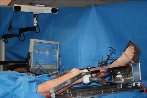
Three mechanized pivot shift tests were performed on each knee in both ACL-intact and ACL-deficient conditions, totaling 60 measurements Citation[15]. The mechanized pivot shifting device reproducibly induces a positive pivot shift test in ACL-deficient knees and has been shown to provide better repeatability than a manual examination Citation[14] (). During each mechanized examination, the navigation system recorded the 3D motion path of the center of the tibia, the center of the medial tibial plateau, and the center of the lateral tibial plateau relative to the femur. The navigation system stores all the registered anatomical geometric mesh model data and the appropriate coordinate data in MDL 3D model files.
Figure 1. The experimental set-up for mechanized pivot shift testing. The lower limb is mounted in full extension on the mechanized pivot shifter, abducted by at least 15° at the hip joint. The foot is affixed to the base plate and is externally rotated. The base plate can then be pushed by the examiner along two linear bearing surfaces, effectively cycling the knee from 0° to 90° of flexion. Using a load cell attached to the mechanized pivot shifter through a three-degrees-of-freedom arm, a valgus load of 4–5 kg is applied to the proximal third of the tibia to reproduce the pivot shift phenomenon. The computer navigation system tracks the tibiofemoral motion path using a camera that emits and then captures a signal reflected by the passive reflective markers attached to the femur and tibia. The arc of motion during the pivot shift test is compared to a flexion-extension path acquired during the registration process of the intact knee and is then visually represented on the screen.

The motion paths and anatomical models acquired from the navigation system and stored in MDL files were analyzed with a custom Pivot Shift Processor program developed in MATLAB (Mathworks, Natick, MA). Using the Grood and Suntay coordinate system, the Pivot Shift Processor calculates knee flexion for each acquired point in all 3D motion paths. At the flexion points provided by the reference path, the anterior tibial translation for each point (center, medial tibial plateau, lateral tibial plateau) is calculated. In cases where an exact flexion angle match was not found, a linear interpolation was performed. The magnitude of the maximum anterior tibial translation for each compartment of the knee was then obtained, along with the angle of flexion at which it occurs.
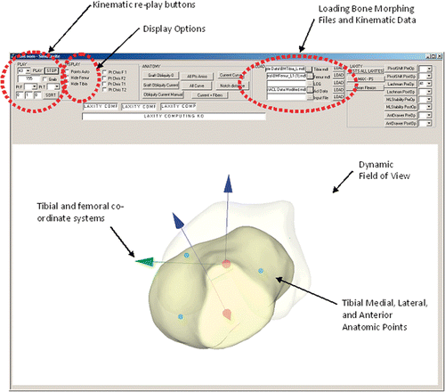
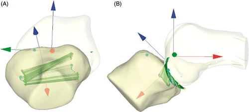
A Pivot Shift Visualizer program was also developed in the C++ programming environment. After loading the navigation system's ACL data files for the tibia, femur and acquired pivot shift points, the software is able to reproduce a 3D virtual model of the knee joint on the screen (). A drop-down menu allows the user to select any particular test, while a “play” button initiates a playback of the recorded pivot shift phenomenon. The graphic interface also permits manipulation of the virtual 3D image, allowing the user to change the point of view from which to visualize the tibiofemoral translation ().
Figure 2. Screenshot of the Pivot Shift Visualizer interface. Once the bone morphing and kinematic data files are loaded, the program displays a 3D rendering of the tibia and the femur, along with a coordinate system. The user may then replay the tibiofemoral motion recorded by the navigation system during the pivot shift test.

Figure 3. During kinematic replay, the tibiofemoral translation path is represented by yellow dots connected by green lines. This visual representation aids the examiner in tracking the motion of the tibia over the femur during each test. The bones may be rotated through 360° to allow visualization from any angle (A, B).

Paired Student's t-tests were used to compare the differences in tibiofemoral translation in the medial and lateral compartments in the ACL-intact and ACL-deficient states. Significance was set to α = 0.05. Results are presented as the mean measurement ± the standard deviation of the mean.
Results
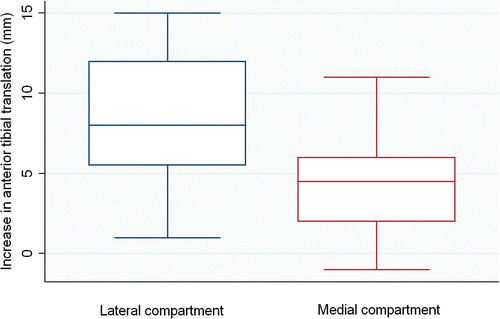
Mean tibiofemoral translation in the intact knee was −0.5 ± 3.4 mm for the lateral compartment and 5.1 ± 3.3 mm for the medial compartment. After sectioning the ACL, translation in the lateral compartment increased to 8 ± 2.7 mm (p < 0.05), while medial compartment translation increased to 9.3 ± 3.9 mm (p < 0.05). The increase in tibiofemoral translation of 8.5 ± 4.2 mm for the lateral compartment after sectioning the ACL was significantly greater than the 4.25 ± 3.3 mm increase observed in the medial compartment (p < 0.05) ().
Discussion
The grade of the pivot shift test is a good predictor of outcome following ACL injury and subsequent treatment Citation[6], Citation[7]. Lateral compartment translation during the pivot shift correlates well with the grade of the pivot shift Citation[8]. However, isolated compartmental translation is not readily available in current computer navigation systems for quantifying the pivot shift Citation[13].
In this study, we have presented two computer programs developed to obtain isolated compartmental tibiofemoral translation and to visualize navigated pivot shift tests a posteriori. The Pivot Shift Processor was able to analyze MDL files created by the navigation system to store anatomic landmarks and kinematic data and to calculate individual compartmental tibiofemoral translation compared to the reference flexion-extension path obtained prior to pivot shift testing. Consistent with previous research, our results showed that, after sectioning the ACL, there was a greater increase in translation in the lateral compartment compared to the medial compartment. This was explored using the 3D rendering Pivot Shift Visualizer program. Using the program, we were able to review the path created by the tibia over the femur during each pivot shift examination. This program may be useful for quality control of pivot shift maneuvers performed during in vitro or in vivo research. More importantly, it may be a valuable tool for exploring the kinematic properties of the pivot shift, which are still not fully understood despite recent technological advances Citation[15]. Improved understanding of the kinematics of the knee before and after ACL injury could lead to better surgical planning by providing the surgeon with a biomechanical rationale for choosing the most appropriate surgical technique given the characteristics and needs of each patient.
The main limitation of this study is the proprietary nature of the navigation files used to perform the analysis. While the software was designed for use with a particular navigation system, changes in the programming code could be made to analyze kinematic data acquired using other navigation systems that use similar data acquisition and storage formats.
Conclusion
Using the Pivot Shift Processor, we successfully analyzed the data recorded using the navigation system. After sectioning the ACL, there was a greater increase in tibiofemoral translation in the lateral compartment compared to the medial compartment. The Pivot Shift Visualizer successfully produced a 3D rendering of the knee joint and the recorded pivot shift maneuvers. This virtual representation of the pivot shift phenomenon from multiple points of view allows for efficient side-by-side comparison of tibiofemoral motion tracking across conditions, which is not possible in the in vivo/in vitro settings. This, in turn, could lead to a better understanding of the kinematics in play during the pivot shift phenomenon.
Acknowledgments
We thank Ms. Carinne Granchi for her contributions to the development and programming of the Pivot Shift Visualization software. We also thank Ms. Clara Hilario for her assistance during the cadaveric testing portion of this study.
Declaration of interest: Dr. Plaskos is an employee of Praxim, Inc. None of the other co-authors have any affiliations that may have biased this study.
References
- Keene GC, Bickerstaff D, Rae PJ, Paterson RS. The natural history of meniscal tears in anterior cruciate ligament insufficiency. Am J Sports Med 1993; 21(5)672–679
- Roos H, Adalberth T, Dahlberg L, Lohmander LS. Osteoarthritis of the knee after injury to the anterior cruciate ligament or meniscus: The influence of time and age. Osteoarthritis Cartilage 1995; 3(4)261–267
- Lane CG, Warren R, Pearle AD. The pivot shift. J Am Acad Orthop Surg 2008; 16(12)679–688
- Tamea CD, Jr, Henning CE. Pathomechanics of the pivot shift maneuver. An instant center analysis. Am J Sports Med 1981; 9(1)31–37
- Pearle AD, Kendoff D, Musahl V, Warren RF. The pivot-shift phenomenon during computer-assisted anterior cruciate ligament reconstruction. J Bone Joint Surg Am 2009; 91(Suppl 1)115–118
- Kaplan N, Wickiewicz TL, Warren RF. Primary surgical treatment of anterior cruciate ligament ruptures. A long-term follow-up study. Am J Sports Med 1990; 18(4)354–358
- Kocher MS, Steadman JR, Briggs KK, Sterett WI, Hawkins RJ. Relationships between objective assessment of ligament stability and subjective assessment of symptoms and function after anterior cruciate ligament reconstruction. Am J Sports Med 2004; 32(3)629–634
- Bedi A, Musahl V, Lane C, Citak M, Warren RF, Pearle AD. Lateral compartment translation predicts the grade of pivot shift: A cadaveric and clinical analysis. Knee Surg Sports Traumatol Arthrosc 2010; 18(9)1269–1276
- Hüfner T, Kendoff D, Citak M, Geerling J, Krettek C. [Precision in orthopaedic computer navigation] [in German]. Orthopade 2006; 35(10)1043–1055
- Khadem R, Yeh CC, Sadeghi-Tehrani M, Bax MR, Johnson JA, Welch JN, Wilkinson EP, Shahidi R. Comparative tracking error analysis of five different optical tracking systems. Comput Aided Surg 2000; 5(2)98–107
- Pearle AD, Solomon DJ, Wanich T, Moreau-Gaudry A, Granchi CC, Wickiewicz TL, Warren RF. Reliability of navigated knee stability examination: A cadaveric evaluation. Am J Sports Med 2007; 35(8)1315–1320
- Brophy RH, Voos JE, Shannon FJ, Granchi CC, Wickiewicz TL, Warren RF, Pearle AD. Changes in the length of virtual anterior cruciate ligament fibers during stability testing: A comparison of conventional single-bundle reconstruction and native anterior cruciate ligament. Am J Sports Med 2008; 36(11)2196–2203
- Colombet P, Robinson J, Christel P, Franceschi JP, Djian P. Using navigation to measure rotation kinematics during ACL reconstruction. Clin Orthop Relat Res 2007; 454: 59–65
- Citak M, Suero EM, Rozell JC, Bosscher MR, Kuestermeyer J, Pearle AD. A mechanized and standardized pivot shifter: Technical description and first evaluation. Knee Surg Sports Traumatol Arthrosc 2011; 19(5)707–711
- Lane CG, Warren RF, Stanford FC, Kendoff D, Pearle AD. In vivo analysis of the pivot shift phenomenon during computer navigated ACL reconstruction. Knee Surg Sports Traumatol Arthrosc 2008; 16(5)487–492
