Abstract
Objective: The widespread use of image guided surgery in the frontolateral skull base region has been limited by the need for a reliable and non-invasive registration procedure that provides sub-millimetric accuracy. We developed and validated preclinically a non-invasive, easy-to-use registration device based on a dental splint with a laterally mounted fiducial carrier.
Methods: Repeated accuracy measurements were performed on six titanium target fiducials which were screwed into the lateral skull base region of a cadaver head and could be unequivocally identified both on the CT image and in reality. The system accuracy was evaluated by determining the deviation of the real target position from the position indicated in the CT scan. The accuracy of the dental splint-based registration was compared to that of two standard registration procedures: contour-based laser surface registration and fixed marker registration.
Results: The mean accuracy of 0.55 ± 0.28 mm obtained when using the maxillary splint device was similar to that obtained with the “gold standard” registration using bone-implanted markers (0.33 ± 0.26 mm), while being clearly superior to that obtained with contour-based laser surface registration (1.91 ± 0.74 mm).
Conclusions: Registration using the non-invasively fixed maxillary fiducial platform can provide sub-millimetric accuracy in the lateral skull base region. In vivo validation may prove dental splint-based registration to be an accurate and non-invasive alternative option for image guided surgery of the lateral skull base, and may facilitate the application of navigation systems in this delicate region.
Introduction
In ENT surgery, particularly sinus surgery, surgical navigation systems have been successfully used for some years. They are widely accepted as useful tools for intraoperatively identifying important anatomical structures and assisting in the exact positioning of surgical instruments, especially when conditions are complex because of the loss of surgical landmarks as a result of previous surgeries or tumor destruction Citation[1-3].
A prerequisite for the reliable use of image guided surgery (IGS) is a successful and accurate registration process, i.e., the matching of the patient's preoperatively acquired image data set (e.g., a CT scan) with the intraoperative reality. The quality of the registration process directly affects the accuracy of the navigation system, which is defined as the deviation of the real position of a surgical target or instrument from its position as indicated in the CT scan Citation[4]. Accuracy is highly dependent on the chosen registration method, and tends to increase with the distance between the registration area and surgical target Citation[5-7]. In ENT surgery, non-invasive registration methods are commonly used, such as laser surface scanning Citation[8-11] or tracking of external fiducial markers that are self-adhesive or fixed on headbands Citation[10], Citation[11]. In most cases, these registration methods provide sufficient clinical accuracy for paranasal sinus surgery Citation[12], Citation[13].
Lateral skull base surgery, performed in close proximity to vital structures such as the brain, the middle and inner ear, the vestibular organ, the carotid artery, or the venous sinus, carries considerable risks and requires sub-millimetric precision. IGS can facilitate orientation, especially in complex surgeries such as those for recurrent cholesteatoma, paraganglioma, or CSF leaks. However, as the currently available registration methods are either highly invasive or demonstrate low reliability when applied to targets in the lateral skull base Citation[14], Citation[15], IGS has not yet been established as a standard surgical procedure in this region.
To facilitate and improve the application of IGS in frontolateral skull base surgery, a new registration method providing both high clinical accuracy and minimal invasiveness is desirable. For this purpose, we developed a new registration device consisting of a dental splint with a lightweight laterally mounted fiducial platform. The splint can be reversibly fixed to the maxillary dentition, and no invasively applied markers are needed. For intraoral interventions, such as the navigated placement of dental implants, the use of dental splint registration has already proven its applicability Citation[16], Citation[17]. However, standard dental registration splints are not suitable for IGS of the lateral skull base as the considerable distance between the registration area (the oral cavity) and surgical area results in a high target registration error Citation[4], Citation[5], Citation[18]. We hypothesized that the use of our modified dental splint with immovable, non-invasively fixed registration markers in close proximity to the region of interest would improve navigational accuracy when compared the registration methods commonly applied in ENT surgery. In the present study, we evaluated the navigational accuracy in the lateral skull base region when using this new registration device on a cadaver head, and compared it to two standard registration procedures: the widely used (particularly in ENT surgery) contour-based laser surface registration method and registration via implanted bone screws.
Methods
CT scan and navigation system
The measurements were performed on a cadaver head (obtained from the Institute of Anatomy at the LMU Munich). Axial spiral CT scans were obtained (SOMATOM Sensation 64® CT scanner; Siemens, Germany) (slice thickness 1 mm, continous scan with overlapping slices, 0.5 mm reconstruction increment) and transferred to a VectorVision Compact® passive optical navigation system with the ENT 7.8 software update (BrainLAB, Feldkirchen, Germany).
Methods of registration
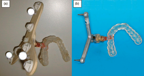
Dental splint registration: We developed a new registration device by modifying a common dental registration splint. The key feature was to apply the fiducial carrier unilaterally, in contrast to the large bilateral extraoral reference frames commonly used. The fiducial carrier was attached to an individually adjusted dental splint to bring the registration area as close as possible to the target area, i.e., the lateral skull base. After taking impressions of the upper jaw and determining the maximal intercuspidation, an individually formed dental splint was created from polymethylmethacrylate (PMMA, Ortocryl®; Dentaurum J. P. Winkelstroeter KG, Ispringen, Germany) in the Department of Prosthodontics at the LMU Munich, and adjusted to the maxilla of the cadaver head. The precise fit with the contours of the maxillary teeth held the splint in place and ensured its stable position; adhesive or metal fastening was not required, nor was it necessary for the upper and lower teeth to be closed on the splint. The three-dimensional polyethylene reference body, carrying six fiducial markers for localization in the registration process and three passive reflecting balls for tracking during surgery, was attached with a screw thread to the individually formed PMMA pedicule on the left side of the splint (), as the measurements were to be performed on the left lateral skull base. The reference body can be easily removed and is fully sterilizable, while the individual dental splint is discarded after use. CT scanning was performed with the registration splint fitted onto the dentition of the maxilla and the fiducial platform fixed in place. For registration, each fiducial screw was selected with the referenced pointer and detected by the cameras of the navigation system as a defined registration landmark (). This was then matched exactly with the previously saved image data set.
Figure 1. Dental splint (a) attached with a screw thread to a 3D polyethylene body carrying six fiducial markers for registration close to the lateral skull base, and (b) attached, again with a screw-thread, to the reference array intended to carry passive reflecting balls for referencing during navigation.
Fixed marker registration: The highest accuracy values are generally obtained with registration methods that use implanted bone screw fiducials Citation[19], Citation[20]. This minimizes the risk of registration markers being displaced between the CT imaging session and the registration process Citation[21], which can easily occur with soft tissue-mounted adhesive fiducials Citation[22]. At present, fixed marker registration is still considered the “gold standard” for IGS at the lateral skull base Citation[19].
Four screws were drilled into the bone in the area of the left lateral skull base prior to performing the CT scan. Following scanning, the locations of all these registration fiducials were obtained as described above to register the physical space to the image data set.
Contour-based laser surface registration: Contour-based laser surface scanning is commonly chosen in (endoscopically controlled) sinus surgery as a convenient, non-invasive and touchless registration method. The individual contours of the patient's face are aligned with the image data set, thus making the application of external markers unnecessary Citation[8], Citation[10].
Laser scanning was performed with a handheld laser scanner device (z-touch®; BrainLAB, Heimstetten, Germany). The laser reflections detected by the infrared cameras of the navigation system provide the localization data. The system automatically correlates the position of this data to the reference star and calculates each coordinate link with the corresponding point from the CT data set. For effective matching, an adequate number of points (approximately 100) must be detected Citation[8]. The choice of the area to be scanned is crucial to the accuracy of the registration procedure. The scanned regions should be hairless, and the skin should cover the bones tightly so that tissue shift during the interval between CT scanning and registration is negligible Citation[5]. To keep the registration area as close as possible to our measurement points in the frontolateral skull base region, we chose points for scanning that were distributed along the aurical. As the suitable surfaces in the lateral area did not yield a sufficient number of points, it was necessary to expand the registration area along the forehead and orbital rim.
Determination of navigational accuracy
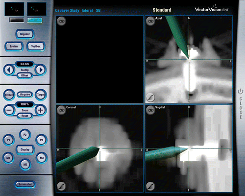
The navigational accuracy was determined as previously described Citation[8], Citation[10], Citation[11], Citation[23], Citation[24]. Six titanium screws fixed in the lateral skull base () served as precisely defined target points. Each was pointed to by an infrared pointer tracked by the navigation system. The distance between the target point and the tip of the pointer was measured separately for the three planes (axial, coronal, sagittal) using the scale on the navigation screen (). The highest value among these three measurements, i.e., the maximum deviation, represents a reasonable approximation to the accuracy of the navigation system. Twenty replicate measurements were performed per registration method. Each measurement process consisted of shutting down and restarting the system, registration using the respective method, and localization of the target fiducials. When using the dental splint for registration, the splint was removed and newly adjusted every time. To avoid inter-operator variation, all measurements were performed by the same person.
Figure 2. The locations of the six precisely defined bone-implanted target fiducials demonstrated on an anatomical model.
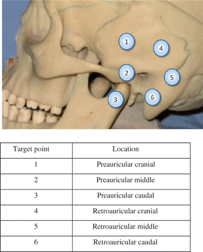
Figure 3. Navigational screenshot. Titanium screws served as target points and were aimed at by the referenced infrared pointer of the navigation system. The distance between the target point and the tip of the pointer was measured separately for each of the three planes (axial, coronal, sagittal) using the scale on the navigation screen.
Statistics
Data are expressed as mean values (in mm) ± standard deviation. Differences between groups were tested for statistical significance using the Kruskal-Wallis test and Tukey's test. A P-value of < 0.05 was considered statistically significant.
Results
Measurements were performed on the left lateral skull base at six target points, three located preauricularly and three postauricularly (). Mean accuracy values (n = 20) are shown in . The highest accuracy was obtained following fixed marker registration (overall accuracy: 0.33 ± 0.26 mm; range: 0.00–1.50 mm). Accuracy measured following dental splint registration was highly reproducible and was only slightly higher than after fixed marker registration (0.55 ± 0.28 mm; range: 0.00–1.20 mm). Following laser surface registration the accuracy was significantly lower at every measuring point compared to that with fixed marker or dental splint registration (p < 0.05; overall accuracy 1.91 ± 0.74 mm; range: 0.40–3.30 mm) ().
Figure 4. Target registration error for fixed marker registration, laser surface registration and dental splint registration at the six measuring points (MP) located pre- or postauricularly, as indicated in . Results are expressed as box-plots of n = 20 independent measurements. *p < 0.05 versus fixed marker registration.
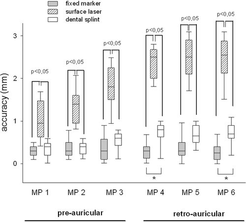
Table I. Navigational accuracy (NA) for different registration methods (target points as outlined in ). Data represent the mean of n = 20 measurements.
Following fixed marker registration, no differences in accuracy between the single target points could be detected. In contrast, when registration was performed by laser surface scanning or with the use of the dental splint device, the increasing distance between the registration area and target points corresponded with a significant decline in accuracy (pre- versus retroauricular; p < 0.001) which was particularly pronounced for the postauricularly located target points after laser surface registration.
Discussion
The reliability of image guided surgery is highly dependent on accurate matching of the patient's preoperative image data to the intraoperative anatomy. In IGS of the lateral skull base, rigid fixed marker registration with bone screws is still considered the gold-standard registration method Citation[19]. In our study, we confirmed the high accuracy (0.33 ± 0.26 mm) of this registration method; however, its high invasiveness has prevented IGS from becoming a routine procedure in skull base surgery.
In oral and cranio-maxillofacial surgery, registration is often performed using intraoral fiducial markers mounted on an intraoral dental splint device Citation[16], Citation[21]. However, the great distance from the registered region (the oral cavity) prevents the use of such intraoral templates for operations in the lateral skull base area Citation[25]. In fact, in a recent in vitro study published by Luebbers and coworkers, adequate precision in regions beyond the mid-face could only be achieved by combining the occlusal splint with two bone-implanted markers on the lateral orbital rim Citation[18]. In the present study, we developed a modified dental splint device with a unilateral reference frame carrying the registration fiducials. Thus, a close spatial relationship between the registration area and the target fiducials was assured. The splint integrates easily into common navigation systems such as the BrainLAB VectorVision, and does not require any additional software. The high accuracy and reliability of this novel registration device was proven by the measurement of values in the submillimetric range for target fiducials located in the lateral skull base region, which almost matched the accuracy of fixed bone-implanted marker registration. Although the more distant targets yielded slightly larger values, confirming the significant impact of the distance between the registration and target points on the accuracy Citation[7], Citation[8], Citation[21], even the maximum deviation of 1.2 mm is still within the range (1 to 2 mm) that is often considered “clinically acceptable” for navigation system accuracy Citation[1], Citation[21], Citation[25], Citation[26].
Maxillary splint-based systems with extraoral extensions for reference markers have been used previously with sufficient navigational accuracy in neurosurgery (0.29-0.86 mm Citation[17], 0.0-2.0 mm Citation[27]), in sinus surgery (1.56 ± 0.76 mm Citation[28]), and in the temporal region (0.73 ± 0.25 mm Citation[17], Citation[29]); however, only a few of these systems have been tested for targets in the lateral skull base region Citation[15], Citation[29], Citation[30], Citation[31]. Bale et al. used a mouthpiece-based registration template held in place by a vacuum system to successfully cannulate the foramen ovale Citation[30], but did not test its applicability for other locations in the lateral skull base. In contrast, insufficient accuracy was reported in an in vitro model of mastoidectomy when using a navigation bow with integrated registration markers fixed to the upper jaw Citation[15]. A major problem with the existing devices is the large bilateral extraoral reference frame Citation[17], Citation[29], Citation[31], which does not allow the patient's head to be positioned on one side for surgery – a prerequisite for surgery at the lateral skull base. Furthermore, depending on the size and weight of the extensions, the extraoral parts might act as a lever, leading to dislocation of the maxillary splint and poor registration accuracy. Additionally, extraoral support of the reference frame by the nasion or external auditory canal Citation[28] is not advisable, as the attachment of reference structures to soft tissue tends to increase inaccuracy because of soft tissue deformity, a problem also known from non-rigidly mounted registration devices like headbands or headsets Citation[32]. In an attempt to solve this problem, we took special care to make our dental splint device as lightweight and as small as possible. This smaller splint-mounted reference system should not interfere with surgical access in common skull base procedures, such as surgery of the middle ear or paraganglioma.
As the reference array is held in place solely by the close fit with the teeth, a proper dental status is a prerequisite for a rigid attachment of the splint to the maxilla to guarantee high navigational accuracy.
As a non-invasive alternative registration method, laser surface contour scanning has found widespread use in paranasal sinus surgery, and there have been preliminary studies to assess its usefulness in lateral skull base surgery as well Citation[11], Citation[33]. However, common laser-based registration systems could not match the accuracy of bone-implanted fiducial marker registration Citation[11]. The lack of an appropriate registration surface in close proximity to the lateral skull base most likely accounts for the rather poor navigational accuracy reported. The hairy skin, the soft auricle, and the lack of bony anatomical landmarks prevent a reliable registration. On the other hand, scanning of the facial skull, while providing sufficient accuracy in the frontal or paranasal sinuses Citation[8], Citation[12], has been found to be an unsuitable method of registration for targets located in the lateral skull base due to the considerable distance between the reference points and the surgical area Citation[11]. Accordingly, the deviation measured in our study of accuracy after laser surface scanning was more than three times that measured after fixed marker or dental splint registration. With maximum values of 3.3 mm, laser surface registration did not meet clinical requirements for interventions in the lateral skull base, even though the measurements were performed under ideal experimental conditions on a cadaver head, thereby excluding factors that might compromise accuracy in vivo, such as soft tissue shifting or altered skin reflection following disinfection. Still, it is possible that future laser scanning devices will overcome the difficulties of marker-less registration in the skull base area. Promising in this respect are preliminary results by Marmulla et al., who used a special laser scanner system that was able to record far more surface points than are recorded by common laser scanners Citation[33]. To minimize the distance between the target and the registration area, they used the auricle as a spatial reference area and assumed an acceptable navigational accuracy in the lateral skull base. One problem with this approach is the possibility of auricular deformation during CT acquisition.
Prior to testing a new registration method in vivo, it is important to know the system-immanent (in)accuracy. For an exact evaluation of the system accuracy, it is necessary to exclude any unrelated factors which might additionally influence accuracy in a patient. We therefore conducted the evaluation of the respective registration methods under optimized experimental conditions. According to our measurements, the easy-to-use and non-invasive dental splint registration device provided sufficient accuracy, comparable to that obtained with fixed marker registration and much better than that obtained with ordinary contour-based laser registration. Although it is possible that the accuracy will decrease slightly when tested in vivo, the initial results give reason to expect clinical applicability of the device.
Conclusion
The results show that submillimetric accuracy is achievable in the lateral skull base region with a modified, non-invasive maxillary fiducial carrier. This new dental splint device has been proven to provide navigational accuracy similar to that obtained with standard registration methods based on bone-implanted markers, while being clearly superior to standard laser surface registration in this area. The study emphasizes the importance of a close spatial relationship between the registration and target areas. Ongoing clinical studies to evaluate the applicability of this approach in vivo may reveal the dental splint-based registration method to be an accurate and non-invasive alternative option for image guided surgery of the lateral skull base.
Conflict of interest: The authors declare that they have no conflict of interest.
References
- Cartellieri M, Vorbeck F. Endoscopic sinus surgery using intraoperative computed tomography imaging for updating a three-dimensional navigation system. Laryngoscope 2000; 110: 292–296
- Reardon EJ. Navigational risks associated with sinus surgery and the clinical effects of implementing a navigational system for sinus surgery. Laryngoscope 2002; 112: 1–19
- Strauss G, Limpert E, Strauss M, Hofer M, Dittrich E, Nowatschin S, Lüth T. [Evaluation of a daily used navigation system for FESS] [In German]. Laryngorhinootologie 2009; 88: 776–781
- Labadie RF, Davis BM, Fitzpatrick JM. Image-guided surgery: What is the accuracy?. Curr Opin Otolaryngol Head Neck Surg 2005; 13: 27–31
- Gunkel AR, Thumfart WF, Freysinger W. [Computer-aided 3D-navigation systems. Survey and location determination] [In German]. HNO 2000; 48: 75–90
- Knott PD, Maurer CR, Gallivan R, Roh HJ, Citardi MJ. The impact of fiducial distribution on headset-based registration in image-guided sinus surgery. Otolaryngol Head Neck Surg 2004; 131: 666–672
- West JB, Fitzpatrick JM, Toms SA, Maurer CR, Jr, Maciunas RJ. Fiducial point placement and the accuracy of point-based, rigid body registration. Neurosurgery 2001; 48: 810–816, discussion 816-817
- Ledderose GJ, Stelter K, Leunig A, Hagedorn H. Surface laser registration in ENT-surgery: Accuracy in the paranasal sinuses – a cadaveric study. Rhinology 2007; 45: 281–285
- Marmulla R, Mühling J, Wirtz CR, Hassfeld S. High-resolution laser surface scanning for patient registration in cranial computer-assisted surgery. Minim Invasive Neurosurg 2004; 47: 72–78
- Raabe A, Krishnan R, Wolff R, Hermann E, Zimmermann M, Seifert V. Laser surface scanning for patient registration in intracranial image-guided surgery. Neurosurgery 2002; 50: 797–801, discussion 802-793
- Schlaier J, Warnat J, Brawanski A. Registration accuracy and practicability of laser-directed surface matching. Comput Aided Surg 2002; 7: 284–290
- Stelter K, Andratschke M, Leunig A, Hagedorn H. Computer-assisted surgery of the paranasal sinuses: Technical and clinical experience with 368 patients, using the Vector Vision Compact system. J Laryngol Otol 2006; 120: 1026–1032
- Strauss G, Spitzer C, Dittrich E, Hofer M, Strauss M, Lüth T. [Modified procedure for patient registration for navigation control instruments in ENT surgery] [In German]. HNO 2009; 57: 153–159
- Hofer M, Grunert R, Dittrich E, Müller E, Möckel M, Koulechov K, Strauss M, Korb W, Schulz T, Dietz A, et al. Surgery on the lateral skull base with the navigated controlled drill employed for a mastoidectomy (pre clinical evaluation). Stud Health Technol Inform 2007; 125: 179–184
- Strauss G, Dittrich E, Baumberger C, Hofer M, Strauss M, Stopp S, Koulechov K, Dietz A, Lüth T. [Improvement of registration accuracy for navigated-control drill in mastoidectomy (autopilot)] [In German]. Laryngorhinootologie 2008; 87: 560–564
- Brief J, Edinger D, Hassfeld S, Eggers G. Accuracy of image-guided implantology. Clin Oral Implants Res 2005; 16: 495–501
- Howard MA, 3rd, Dobbs MB, Simonson TM, LaVelle WE, Granner MA. A noninvasive, reattachable skull fiducial marker system. Technical note. J Neurosurg 1995; 83: 372–376
- Luebbers HT, Messmer P, Obwegeser JA, Zwahlen RA, Kikinis R, Gratez KW, Matthews F. Comparison of different registration methods for surgical navigation in cranio-maxillofacial surgery. J Craniomaxillofac Surg 2008; 36: 109–116
- Pfisterer WK, Papadopoulos S, Drumm DA, Smith K, Preul MC. Fiducial versus nonfiducial neuronavigation registration assessment and considerations of accuracy. Neurosurgery 2008; 62: 201–207, discussion 207-208
- Schramm A, Gellrich NC, Gutwald R, Schipper J, Bloss H, Hustedt H, Schmelzeisen R, Otten JE. Indications for computer-assisted treatment of cranio-maxillofacial tumors. Comput Aided Surg 2000; 5: 343–352
- Eggers G, Mühling J, Marmulla R. Template-based registration for image-guided maxillofacial surgery. J Oral Maxillofac Surg 2005; 63: 1330–1336
- Eggers G, Mühling J, Marmulla R. Image-to-patient registration techniques in head surgery. Int J Oral Maxillofac Surg 2006; 35: 1081–1095
- Khan M, Ecke U, Mann WJ. [The application of an optical navigation system in endonasal sinus surgery] [In German]. HNO 2003; 51: 209–215
- Schmerber S, Chassat F. Accuracy evaluation of a CAS system: Laboratory protocol and results with 6D localizers, and clinical experiences in otorhinolaryngology. Comput Aided Surg 2001; 6: 1–13
- Eggers G, Mühling J. Template-based registration for image-guided skull base surgery. Otolaryngol Head Neck Surg 2007; 136: 907–913
- Caversaccio M, Bächler R, Lädrach K, Schroth G, Nolte LP, Häusler R. Frameless computer-aided surgery system for revision endoscopic sinus surgery. Otolaryngol Head Neck Surg 2000; 122: 808–813
- Bale RJ, Burtscher J, Eisner W, Obwegeser AA, Rieger M, Sweeney RA, Dessl A, Giacomuzzi SM, Twerdy K, Jaschke W. Computer-assisted neurosurgery by using a noninvasive vacuum-affixed dental cast that acts as a reference base: Another step toward a unified approach in the treatment of brain tumors. J Neurosurg 2000; 93: 208–213
- Hauser R, Westermann B, Probst R. Noninvasive tracking of patient's head movements during computer-assisted intranasal microscopic surgery. Laryngoscope 1997; 107: 491–499
- Labadie RF, Shah RJ, Harris SS, Cetinkaya E, Haynes DS, Fenlon MR, Juscyzk AS, Galloway RL, Fitzpatrick JM. In vitro assessment of image-guided otologic surgery: Submillimeter accuracy within the region of the temporal bone. Otolaryngol Head Neck Surg 2005; 132: 435–442
- Bale RJ, Laimer I, Martin A, Schlager A, Mayr C, Rieger M, Czermak BV, Kovacs P, Widmann G. Frameless stereotactic cannulation of the foramen ovale for ablative treatment of trigeminal neuralgia. Neurosurgery 2006; 59(4 Suppl 2)ONS394–ONS401, discussion ONS402
- Balachandran R, Fitzpatrick JM, Labadie RF. Accuracy of image-guided surgical systems at the lateral skull base as clinically assessed using bone-anchored hearing aid posts as surgical targets. Otol Neurotol 2008; 29: 1050–1055
- Bumm K, Federspil PA, Klenzner T, Majdani O, Raczkowsky J, Strauss G, Schipper J. [Update on computer- and mechatronic-assisted head and neck surgery in Germany] [In German]. HNO 2008; 56: 908–915
- Marmulla R, Eggers G, Mühling J. Laser surface registration for lateral skull base surgery. Minim Invasive Neurosurg 2005; 48: 181–185