Abstract
Femoral intramedullary guides have been shown to be insufficiently accurate in creating a distal femoral resection perpendicular to the mechanical axis in total knee arthroplasty (TKA), as they make assumptions regarding the difference between the patient's femoral mechanical and anatomical angles. The aim of this cadaveric study was to validate the accuracy of a portable accelerometer-based navigation device for alignment of the distal femoral cutting block in TKA. Twenty-nine trials were performed on five cadaveric specimens (hip-to-ankle), in which the distal femoral cutting block was placed using the KneeAlign 2™ navigation device. For each specimen, a preoperative “target” was assigned for varus/valgus and flexion/extension alignment of the cutting block. The actual alignment of each cutting block was then measured using the ORTHOsoft Computer Assisted Surgery (CAS) system. The mean absolute difference between the preoperative target and the alignment of the cutting block was 0.83 ± 0.60° for varus/valgus, and 0.83 ± 0.83° for flexion/extension. The KneeAlign 2™ navigation device can set and align the distal femoral resection guide with the same accuracy as a large-console CAS system, thus demonstrating that portable accelerometer-based navigation can be used reliably in total knee arthroplasty.
Introduction
Although total knee arthroplasty (TKA) is a successful procedure for the treatment of degenerative joint disease, implant malalignment has been shown to decrease component survival and increase the rate of revision surgery Citation[1–5]. In a review of 6070 TKA cases, a mechanical tibiofemoral alignment angle between 2.4° and 7.2° of valgus (0.5%) resulted in significantly lower rates of component revision compared to knees implanted in varus (1.8%) Citation[6]. In addition, a review of 115 TKA cases noted that the incidence of component loosening at a median period of 8 years postoperatively was 24% in those TKAs aligned >3° outside of a neutral mechanical axis, versus 3% in those aligned within 3° of the axis Citation[7].
Currently, an intramedullary (IM) distal femoral alignment guide is the most commonly used means of aligning the distal femoral cutting block in TKA. This approach involves the insertion of a rod into the femoral canal through the intercondylar notch, to which a cutting block is then attached. The cutting block is fixed to the distal femur at a specific angle relative to the alignment rod prior to performing the distal resection. Standard practice for most orthopaedic surgeons is to set the distal femoral resection angle to the same degree of valgus for all TKAs with a preoperative varus alignment (e.g., 6°), and to a different degree for all TKAs with a preoperative valgus alignment (e.g., 3°), with the goal of creating a distal femoral resection perpendicular to the femoral mechanical axis Citation[8]. This assumes there is no variation in the femoral mechanical-anatomical angle between patients, and relies on the assumption that, in most patients, the difference between the femoral mechanical and anatomic axes is approximately 6°. Unfortunately, numerous studies have demonstrated this assumption to be inaccurate, especially in osteoarthritic patients, and that using a fixed resection angle for performing the distal femoral resection commonly fails to achieve a neutral resection Citation[8–11]. A radiographic analysis of 174 TKA cases demonstrated that both the femoral offset and neck-shaft angle can strongly affect the distal femoral valgus cut angle required to obtain a neutral mechanical axis, with up to 51% of patients requiring an angle of less than 5° or greater than 6°. In addition, the appropriate starting point for insertion of the intramedullary rod varies based on the patient's distal femoral mechanical angle, and substantial malalignment has been shown to occur with minor variations in this starting point Citation[12].
Concerns regarding the accuracy of conventional alignment systems and the goal of more accurate component positioning have led to the development of computer-assisted surgery (CAS) techniques. Several comparative studies have demonstrated statistically significant improvements in both the overall mechanical alignment and the femoral component positioning, yet CAS techniques have not been widely embraced Citation[2], Citation[13], Citation[14]. Concerns over increased operative times, capital costs, and the learning curve associated with such techniques have limited their widespread acceptance: less than 3% of TKA procedures are performed using CAS in the United States Citation[13].
The KneeAlign 2™ (OrthAlign, Inc., Aliso Viejo, CA) is a portable accelerometer-based navigation device for use in performing the distal femoral resection in TKA. This device works in a similar manner to computer-assisted surgical systems, but does not require the use of a large console for registration and alignment feedback. In addition, it relies on accelerometer-based navigation, as opposed to the CT-guided, image-based, or imageless navigation technologies used in most CAS systems. The aim of this cadaveric study was to validate the accuracy of a portable accelerometer-based device in navigating alignment of the distal femoral cutting block in total knee arthroplasty. Our hypothesis was that accelerometer-based navigation can be used to set and align the distal femoral resection guide with the same accuracy as a predicate device, the ORTHOsoft Sesamoid Computer Aided Surgery TKA Navigation System (Zimmer, Inc., Warsaw, IN).
Materials and methods
Prior to initiation of the study, approval for the appropriate use of cadaveric specimens was obtained from the Institutional Review Board of the Hospital for Special Surgery. The cadaveric data reported in this paper formed part of the accuracy data evaluated to obtain FDA 510(k) clearance, to ensure that the device was able to determine precisely the mechanical axis of the femur, and to enable the surgeon to align the distal femoral cutting block precisely at the desired varus/valgus and flexion/extension angles. Five fresh-frozen cadaveric specimens (hip-to-toe lower extremities; 3 right, 2 left) were included in this controlled laboratory study. Prior to formal testing of the specimens, a pilot study was performed using a single fresh-frozen specimen to test the experimental protocol. All specimens were thawed at room temperature for at least 24 hours prior to testing.
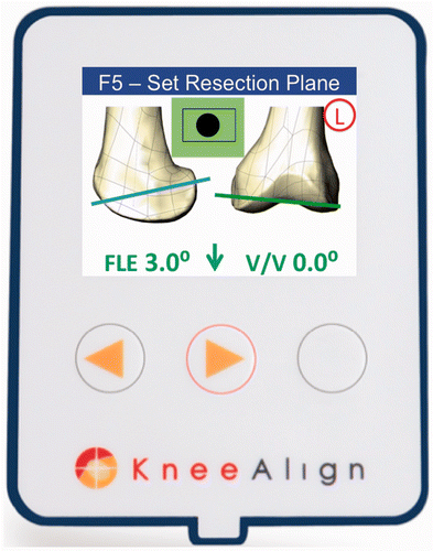
The KneeAlign 2™ (OrthAlign, Inc., Aliso Viejo, CA) is a portable accelerometer-based surgical navigation system for use in performing the distal femoral resection in TKA. It consists of a KneeAlign 2™ display console (), a reference sensor, and a femoral jig. The original KneeAlign™ device was created for use in performing the proximal tibia resection in TKA, and has demonstrated encouraging clinical results Citation[15], Citation[16]. The KneeAlign 2™ device is an adaptation of the same technology, but required the additional design of a femoral jig and cutting block. Of note, the same display console is used for both the proximal tibial and distal femoral resections.
To perform femoral navigation, a femoral jig is pinned to the distal femur. The jig has a component that is fixed to the femur, and an adjustable component that guides the distal femoral cutting block. The display unit, which contains a triaxial accelerometer, is connected to the fixed component of the jig, and the reference sensor is connected to the adjustable component. The reference sensor contains a combination 3-axis accelerometer and 3-axis angular rate sensor, which measures angular velocity. The jig is mechanically located to reference the distal mechanical axis of the exposed knee, and is placed at the midpoint of the most distal point of the sulcus of the trochlea, or the deepest point of the intercondylar notch. During hip center registration, the knee is briskly maneuvered through a range of motion then returned to its starting point. During this movement, the outputs of the accelerometers and angular rate sensors are logged.
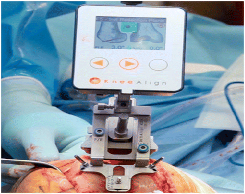
An algorithm is then applied which integrates the angular rate sensor data to propagate the directional cosine matrix, which is used to subtract gravity from the acceleration data. This leaves measured acceleration values that are due to the circular motion experienced by the sensor. This is integrated to establish the measured velocity of the sensor data at each point. Numerical techniques are applied to the data set to establish an optimal vector R. This is the estimated hip point. Once this vector is known, 3D trigonometry is used to calculate the orientation of the sensor relative to the femoral mechanical axis and present this information to the surgeon as the varus/valgus angle and flexion/extension angle via the graphical user interface of the display console. The resection plane of the cutting block is set to the desired varus/valgus and flexion/extension angles (using a 2.5-mm ball driver to adjust screws on the cutting block) as the display console provides real-time feedback on its orientation relative to the hip center of rotation (). During this phase, the accelerometers in the display console and sensor module are used as tiltmeters to measure the fine changes in the angle of the cutting block. In addition, the depth of resection is set by sliding the cutting block relative to the distal femoral condyles. Once the surgeon is satisfied with the cutting block's position, it is pinned to the anterior femur with two headless pins, after which the distal femoral resection can be performed.
Figure 2. Intraoperative photograph of the KneeAlign 2™ device. The display console attaches to the front of the femoral jig, and provides real-time feedback on both the distal cutting block's mechanical axis varus/valgus alignment and flexion/extension alignment.

For each cadaveric specimen, the femoral head was secured to a fixture that stabilized the lower extremity but allowed internal/external rotation and flexion/extension of the femur for registration of the hip center of rotation. For each specimen, a specific target for the varus/valgus and flexion alignment of the distal femoral cutting block, relative to the mechanical axis of the femur, was assigned (). An anterior midline incision and standard medial parapatellar arthrotomy was performed for exposure of the knee joint. The knee was then brought into flexion, and navigation with the KneeAlign 2™ device was used to set the femoral cutting block to the target alignment for each specimen as described above. The femoral cutting block was then secured with two headless pins, but the distal femoral cut was not made. The remainder of the femoral jig was then removed, leaving the distal femoral cutting block in place.
Table I. The specific targets for varus/valgus and flexion alignment of the distal femoral cutting block for each specimen. The targets are relative to the mechanical axis of the femur. The experimental protocol was repeated a minimum of five times for each specimen. For convention, valgus alignment was assigned a negative value.
The ORTHOsoft Sesamoid Computer Aided Surgery TKA Navigation System (Zimmer, Inc., Warsaw, IN) is an imageless navigation system that uses an optical camera, a computer display screen, femoral and tibial reference sensors, and a registration pointer to provide guidance for appropriate bone resections, implant sizing, and correct component positioning in TKA procedures. Imageless navigation systems have demonstrated accuracy to within 1–2° for component alignment in TKA, and numerous clinical studies have demonstrated their superior accuracy when compared to conventional methods Citation[2], Citation[17], Citation[18].
The next step in our study protocol was to implant the femoral and tibial references for use with the ORTHOsoft CAS system. The femoral reference was inserted medially through the vastus medialis, using two pins inserted into the distal femur, while the tibial reference was inserted into the medial surface of the tibia, again using two pins. The center of the femoral head was registered by moving the leg in a conical pattern, as distinct positions of the femur are registered by the optical camera and computer system. The femoral mechanical entry point, defined as the deepest point of the intercondylar notch, was then digitized using the registration pointer. The posterior condyles of the distal femur were also digitized. Once appropriate registration of the ORTHOsoft CAS system was performed, the ORTHOsoft CAS paddle and reference sensor were used to record the varus/valgus and flexion/extension alignment of the cutting block previously set by the KneeAlign 2™ device by placing the ORTHOsoft paddle in the distal femoral cutting slot of the KneeAlign 2™. The alignment of the cutting block, as determined by the CAS system, was then recorded.
For each cadaveric specimen, with the cutting block still in place, the KneeAlign 2™ device was re-applied to again register the hip center of rotation and measure the cutting block's alignment. Then, the above steps using the ORTHOsoft CAS system were repeated to again determine the cutting block's alignment. These steps were performed a minimum of five times for each cadaveric specimen. All trials were performed by the same attending surgeon (D.J.M).
Statistical analysis
For each trial, the alignment of the distal femoral cutting block placed by the KneeAlign 2™ device was compared to the alignment measured by the ORTHOsoft CAS system. For each cadaveric specimen, the mean absolute difference and standard deviation (±s.d.) between the preoperative “target” alignment and the alignment reported by the CAS system was calculated. In addition, 95% confidence intervals for deviation from the target alignment were calculated. A one-way ANOVA analysis was performed to test the null hypothesis that the mean absolute differences when comparing each specimen would not be significantly different. A p-value of <0.05 was significant. All values for varus alignment were recorded as positive values, while valgus alignments were recorded as negative values. Similarly, flexion alignment was assigned a positive value, while extension alignment was assigned a negative value. All data were collected and analyzed using Microsoft Excel software (Microsoft Corporation, Redmond, WA).
Results
A total of 29 trials were performed on the five cadaveric specimens included in this study protocol. With regard to varus/valgus alignment of the distal femoral cutting block, as navigated by the KneeAlign 2™ device and measured using the ORTHOsoft CAS system, the mean absolute difference between the preoperative target and the cutting block's alignment was 0.83 ± 0.60°. The maximum deviation from the preoperative target was 1.5°, as 100% of the trials were measured to have a varus/valgus alignment within 2° of the preoperative target. The 95% confidence interval of the mean absolute difference was between 0.61° and 1.04°.
With regard to flexion/extension alignment of the distal femoral cutting block, the mean absolute difference between the preoperative target and the cutting block's alignment was 0.83 ± 0.83°. The maximum deviation from the preoperative target was 2.5°, with 86% of the trials having a flexion/extension alignment within 2° of the preoperative target, and 100% having an alignment within 3°. The 95% confidence interval of the mean absolute difference was between 0.53° and 1.13°. No significant difference was observed with respect to the accuracy of positioning of the cutting block among the cadaveric specimens (and each preoperative target) for either varus/valgus (p = 0.36) or flexion/extension (p = 0.61).
Discussion
The aim of this study was to validate the performance and accuracy of portable accelerometer-based navigation in aligning the distal femoral cutting block in total knee arthroplasty. Our study protocol has demonstrated that the KneeAlign 2™ can set and align the distal femoral cutting block with a similar degree of accuracy to that of the navigated resection plane measured using the ORTHOsoft Sesamoid CAS Navigation system. Over the course of 29 navigated trials, the mean absolute difference between the preoperative target and the cutting block's alignment was 0.83 ± 0.60° for varus/valgus alignment, and 0.83 ± 0.83° for flexion/extension alignment.
A limitation of this study is the relatively small number of cadaveric specimens used. However, multiple trials of navigation and alignment of the cutting block were performed for each specimen to purposely demonstrate that accelerometer-based navigation is reproducible. Actual femoral resections were not performed or measured in this study, as the goal was to demonstrate the accuracy of the navigated alignment of the distal femoral cutting block. The most important factor in creating a well-aligned distal femoral resection is accurate placement of the distal femoral cutting block, and thus the alignment of the cutting block itself was the emphasis of this study. The results demonstrate that accelerometer-based navigation is accurate when compared to large-console CAS navigation systems for determination of the mechanical axis of the femur, the hip center of rotation, and the alignment of the distal femoral cutting block in TKA.
Currently, an intramedullary (IM) distal femoral alignment guide is most commonly used when performing the distal femoral resection in TKA, having shown improved accuracy relative to extramedullary (EM) alignment guides. Cates et al. Citation[9] compared the accuracy of the distal femoral resection in 125 TKA procedures in which an IM femoral alignment system was used to that in 75 TKA procedures in which an EM femoral alignment system was used. They noted that the percentage of distal-femoral resections outside the accepted range (within 3° of varus/valgus) was higher in the EM group (28%) than in the IM group (14.4%, p = 0.019). However, based on this study, it is evident that, although superior to EM femoral guides, IM guides are still highly inaccurate in achieving a distal femoral resection perpendicular to the mechanical axis of the femur. Using a fixed resection angle based on assumptions regarding the relationship of the mechanical and anatomic axes of the femur, along with inaccuracies in the starting point of the intramedullary rod, decreases the ability of IM alignment systems to consistently achieve a neutral resection Citation[8–11]. Another reported drawback of using IM alignment guides is the potentially increased risk of elevated pulmonary pressures intraoperatively and of pulmonary embolism following TKA Citation[19–21].
Numerous comparative studies have demonstrated CAS techniques to be superior to conventional alignment systems for achieving a neutral overall mechanical alignment and improved femoral component positioning in TKA Citation[3], Citation[14], Citation[22–26]. In a comparative study of 60 TKA procedures performed with conventional techniques and 60 performed using computer navigation, Ensini et al. demonstrated more accurate femoral component alignment in the CAS group in the frontal, sagittal and transverse planes. Overall, the use of computer-assisted navigation reduced the number of cases with a final mechanical axis alignment greater than 3° from neutral from 20.0% to 1.7% Citation[24]. However, CAS techniques have not been widely adopted due to concerns over increased operative times, capital costs, and the learning curve required to use these systems effectively Citation[13].
The KneeAlign 2™ (OrthAlign, Inc., Aliso Viejo, CA) is a portable accelerometer-based navigation device for performing the distal femoral resection in TKA. It offers several advantages compared to conventional IM alignment systems. First, assumptions regarding the difference between the mechanical and anatomic axes of the femur are not necessary, as the hip center of rotation is registered using this device. In addition, the intramedullary canal of the femur is not violated. The advantage of portable accelerometer-based navigation when compared to the majority of CAS systems is that it is able to provide immediate intraoperative feedback on the distal femoral cutting block alignment without requiring additional reference sensors or large-console platforms.
Although the use of accelerometer-based navigation for performing the tibial resection in TKA has previously been shown to be highly accurate Citation[15], the validity of accelerometer-based navigation for determining the hip center of rotation and achieving an accurate alignment of the distal femoral cutting block has not previously been demonstrated. Based on this cadaveric study, the authors believe that portable accelerometer-based navigation can be used to align the distal femoral cutting block with the same accuracy as large-console computer-assisted surgical techniques, and this technology can thus be used reliably in total knee arthroplasty.
Declaration of interest: The senior author has stock options in OrthAlign, Inc. None of the other authors have any conflicts of interest to declare.
References
- Berend ME, Ritter MA, Meding JB, Faris PM, Keating EM, Redelman R, Faris GW, Davis KE. Tibial component failure mechanisms in total knee arthroplasty. Clin Orthop Relat Res 2004, 428: 26–34
- Mason JB, Fehring TK, Estok R, Banel D, Fahrbach K. Meta-analysis of alignment outcomes in computer-assisted total knee arthroplasty surgery. J Arthroplasty 2007; 22(8)1097–1106
- Mulhall KJ, Ghomrawi HM, Scully S, Callaghan JJ, Saleh KJ. Current etiologies and modes of failure in total knee arthroplasty revision. Clin Orthop Relat Res 2006; 446: 45–50
- Sharkey PF, Hozack WJ, Rothman RH, Shastri S, Jacoby SM. Insall Award paper. Why are total knee arthroplasties failing today?. Clin Orthop Relat Res 2002, 404: 7–13
- Bargren JH, Blaha JD, Freeman MA. Alignment in total knee arthroplasty. Correlated biomechanical and clinical observations. Clin Orthop Relat Res 1983, 173: 178–183
- Fang DM, Ritter MA, Davis KE. Coronal alignment in total knee arthroplasty: Just how important is it?. J Arthroplasty 2009; 24(6 Suppl)39–43
- Jeffery RS, Morris RW, Denham RA. Coronal alignment after total knee replacement. J Bone Joint Surg Br 1991; 73(5)709–714
- Deakin AH, Basanagoudar PL, Nunag P, Johnston AT, Sarungi M. Natural distribution of the femoral mechanical-anatomical angle in an osteoarthritic population and its relevance to total knee arthroplasty. Knee 2012; 19(2)120–123
- Cates HE, Ritter MA, Keating EM, Faris PM. Intramedullary versus extramedullary femoral alignment systems in total knee replacement. Clin Orthop Relat Res 1993, 286: 32–39
- Kinzel V, Scaddan M, Bradley B, Shakespeare D. Varus/valgus alignment of the femur in total knee arthroplasty. Can accuracy be improved by pre-operative CT scanning?. Knee 2004; 11(3)197–201
- Bardakos N, Cil A, Thompson B, Stocks G. Mechanical axis cannot be restored in total knee arthroplasty with a fixed valgus resection angle: A radiographic study. J Arthroplasty 2007; 22(6 Suppl 2)85–89
- Reed MR, Bliss W, Sher JL, Emmerson KP, Jones SM, Partington PF. Extramedullary or intramedullary tibial alignment guides: A randomised, prospective trial of radiological alignment. J Bone Joint Surg Br 2002; 84(6)858–860
- Canale ST, Beaty JH. Campbell's Operative Orthopaedics. Eleventh Edition. Elsevier, Inc., Philadelphia, PA 2008
- Mullaji A, Kanna R, Marawar S, Kohli A, Sharma A. Comparison of limb and component alignment using computer-assisted navigation versus image intensifier-guided conventional total knee arthroplasty: A prospective, randomized, single-surgeon study of 467 knees. J Arthroplasty 2007; 22(7)953–959
- Nam D, Jerabek SA, Haughom B, Cross MB, Reinhardt KR, Mayman DJ. Radiographic analysis of a hand-held surgical navigation system for tibial resection in total knee arthroplasty. J Arthroplasty 2011; 26(8)1527–1533
- Nam D, Cross M, Deshmane P, Jerabek S, Kang M, Mayman DJ. Radiographic results of an accelerometer-based, handheld surgical navigation system for the tibial resection in total knee arthroplasty. Orthopedics 2011; 34(10)e615–e621
- Krackow KA, Phillips MJ, Bayers-Thering M, Serpe L, Mihalko WM. Computer-assisted total knee arthroplasty: Navigation in TKA. Orthopedics 2003; 26(10)1017–1023
- Zorman D, Etuin P, Jennart H, Scipioni D, Devos S. Computer-assisted total knee arthroplasty: Comparative results in a preliminary series of 72 cases. Acta Orthop Belg 2005; 71(6)696–702
- Dorr LD, Merkel C, Mellman MF, Klein I. Fat emboli in bilateral total knee arthroplasty. Predictive factors for neurologic manifestations. Clin Orthop Relat Res 1989, 248: 112–118, discussion 118–119
- Fahmy NR, Chandler HP, Danylchuk K, Matta EB, Sunder N, Siliski JM. Blood-gas and circulatory changes during total knee replacement. Role of the intramedullary alignment rod. J Bone Joint Surg Am 1990; 72(1)19–26
- Stern SH, Sharrock N, Kahn R, Insall JN. Hematologic and circulatory changes associated with total knee arthroplasty surgical instrumentation. Clin Orthop Relat Res 1994, 299: 179–189
- Anderson KC, Buehler KC, Markel DC. Computer assisted navigation in total knee arthroplasty: Comparison with conventional methods. J Arthroplasty 2005; 20(7 Suppl 3)132–138
- Confalonieri N, Manzotti A, Pullen C, Ragone V. Computer-assisted technique versus intramedullary and extramedullary alignment systems in total knee replacement: A radiological comparison. Acta Orthop Belg 2005; 71(6)703–709
- Ensini A, Catani F, Leardini A, Romagnoli M, Giannini S. Alignments and clinical results in conventional and navigated total knee arthroplasty. Clin Orthop Relat Res 2007; 457: 156–162
- Pang CH, Chan WL, Yen CH, Cheng SC, Woo SB, Choi ST, Hui WK, Mak KH. Comparison of total knee arthroplasty using computer-assisted navigation versus conventional guiding systems: A prospective study. J Orthop Surg (Hong Kong) 2009; 17(2)170–173
- Mihalko WM, Krackow KA. Differences between extramedullary, intramedullary, and computer-aided surgery tibial alignment techniques for total knee arthroplasty. J Knee Surg 2006; 19(1)33–36
