Abstract
Objective: Two key issues in image guided surgery are accurate patient-to-image registration and ongoing tracking of the patient's motion. To address these concerns, a novel device for preoperative registration and automatic tracking was designed, and the accuracy attainable with the device was evaluated in experiments with a skull and in a clinical study.
Methods: The device consists of a system of four titanium screws and four fluorescent spheres fixed to carbon bars which can be easily mounted on the maxillary dentition splint. Before surgery, CT image data of a skull with the device in place was acquired and registered in a navigation system. The rigidity and reproducibility of positioning of the device were measured in 15 repeated CT acquisitions of the skull with the device in place. The registration accuracy was compared to that obtained using micro-screw markers fixed to the maxillary alveolus. To determine the potential of the device in aiding image guided cranio-maxillofacial surgery, registration accuracy and surgical outcome were assessed.
Results: Fifteen tests were performed for CT scanning with no loosening of the splint and device. The arithmetic mean of the standard deviation (SD) ranged from 0.47 mm to 0.70 mm. When the device was used for registration, the mean deviations for the eight anatomical structures investigated ranged from 0.56 mm at the left infra-orbital foramen to 0.96 mm at the right temple. Compared with the method in which titanium screws are fixed to the maxillary alveolus, the target registration error (TRE) obtained using the new device was much less. Using this device, clinical reduction of a zygomatic-orbital-maxillary complex fracture was successfully completed with a registration discrepancy of less than 0.5 mm.
Conclusions: By successfully addressing the two key issues of image guided surgery, the device could be considered accurate and potentially useful for assisting in cranio-maxillofacial surgery.
Introduction
In standard image guided surgery, the surgeon is informed in real time about the position of an instrument relative to the patient's anatomy. This requires two steps to be performed prior to the use of the navigation system. The first step is registration, a computational procedure that maps preoperative images or planning information onto the position of the patient. The second step is tracking of the patient's movement by the navigation system; the treatment plan can then be guided using the patient position with respect to the dynamic reference frame.
Registration is usually performed by manually identifying points in the space of the patient and mapping them onto corresponding points in the image data Citation[1]. Fiducial-based registration is regarded as the “gold standard” when performing retrospective analyses of registration accuracy. To track the patient's motion, a rigid and invasive dynamic reference frame (DRF) has often been adopted for conventional cranio-maxillofacial image guided surgical procedures. However, despite being an established and reliable navigation method, this approach is invasive, tedious, time-consuming and susceptible to errors by the surgeon, which may ultimately result in inaccurate navigation.
To address these problems, a new device has been designed and a novel preoperative registration method developed which enables precise initial patient set-up and frequent detection and correction for patient motion during surgery. In this paper we describe the mechanical design of the device, assess its registration accuracy in an experimental set-up, and evaluate its effect on surgical outcome in a clinical pilot study.
Materials and methods
System description
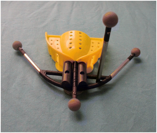
The device consists of four support bars held together with a plastic connector. At the free end of these bars are fixed four titanium micro-screws (Stryker Leibinger Micro Implants, Freiburg, Germany) and four fluorescent spheres (Northern Digital Inc., Waterloo, Ontario, Canada) (). The support configuration was designed to allow the centroid of the four micro-screw markers to be positioned near the central part of the maxillofacial region, thus allowing a more realistic registration parameter determination. The support was held securely in place on the acrylic-polymer splint, which can be attached to the skull's dentition. Once the splint was inserted and fixed to the dentition, the device was tightened against the splint as well as being fixed to the skull.
Figure 1. Structure of the device.
The skull was then scanned using a 64-slice CT unit (GE LightSpeed VCT 64-slice scanner) with the following parameters: helical scan; 0.625-mm slice thickness; 0° gantry tilt; and high-resolution filter. The basic projections were transferred as raw image data to the AccuNavi navigation workstation (Universal Enterprises Group Co., Shanghai, China). The primary reconstruction was performed using filtered back-projection techniques to build a 3D volume. The positions of the micro-screws were determined using three multi-planar reconstruction views and a 3D volume-rendering view. Multi-planar views or just a single plane in the full-screen window could be selected to facilitate micro-screw location. This enabled a reliable and clear localization of the head centers of the micro-screws.
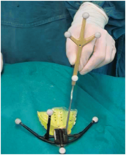
The registration algorithm used for this application was a rigid and global least square method. The position of each micro-screw marker was described by the x, y and z coordinates of its head center. Each head center was evaluated using a semi-automatic procedure beginning with a mouse-click in proximity to the marker under consideration. The corresponding micro-screws were located on the device using the tracked pointer of the navigation system. Thus, the device was registered before the operation (). At the beginning of the operation, the splint with the device was inserted into the skull again to detect the skull's motion during image guided surgery (IGS). Based on the position of this device, the position of the skull was also known; hence, the patient coordinates and image data coordinates were easily transformed into a single coordinate system. Since the position of the patient was detected using the infrared tracking system of the navigation system, the four fluorescent spheres mounted on the device could be used as a dynamic reference frame.
Figure 2. Preregistration of the device.
Device evaluation experiments
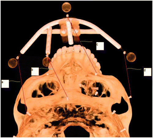
Primary requirements of the device are the ability to be repositioned reproducibility and long-term stability. To test the reproducibility and stability of the device when attached to the dentition, the spatial distances between the four micro-screw markers on the device and those on the skull were evaluated by repeated CT acquisitions of the skull. The skull, with the device re-fitted each time, was scanned using a 64-slice CT scanner daily for 15 days. The image files were then transferred to the navigation workstation for 3D reconstruction. The four markers on the device (a1, b1, c1, d1) and four markers on the skull (a2, b2, c2, d2) were defined, and the distances between them (a, b, c, d) measured in a multi-planar view ().
Figure 3. Measurement of 3D distances.
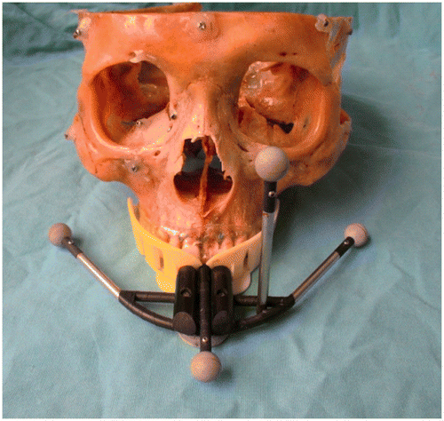
A skull was then used to obtain an estimate of the registration accuracy of the device. For this purpose, the device was attached to the maxillary dentition (). Eight micro-screws, fixed on the external surface of the skull (at the infraorbital foramens, inner canthus, orbit floor, supra-orbital foramen, forehead, temple and zygomatic process point that represent a common situation in cranio-maxillofacial surgery) were defined as target fiducials for measurement of target registration error (TRE). A further seven micro-screws, attached to the maxillary alveolus where they can be easily touched in real registration, were defined as registration fiducials for image-to-world registration. Each group of registration fiducials and target fiducials composed a fiducial marker configuration. Frameless stereotactic localizations were performed using a standard calibrated probe. The registration was performed by a senior surgeon according to the rigid-body point-based alignment of the coordinate systems described by Bale et al. Citation[2]. After each registration procedure, the target fiducials were digitized and used to determine the TRE. The TRE, defined as the Euclidean distance between the position of a target and the probe point in the image space (is) when the probe reaches the target in the world space (ws), was displayed on the navigation workstation after registration. Although the TRE measured at each fiducial was a vector quantity, the TRE was generally reported as a scalar value, – the length of the vector, i.e., the root mean square (RMS) of the vector components. The RMS was calculated as follows:Fifty separate registrations were performed for each configuration, and the average was regarded as the TRE.
Figure 4. The skull fitted with the device.
Clinical trial
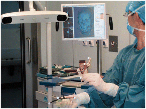
One patient with zygomatic-orbital-maxillary complex fractures, caused by a vehicular accident, was enrolled for image guided reduction with the device. The patient had facial asymmetry (flattened malar eminence and increased facial width on the right side), enophthalmos, diplopia, ocular motility restriction, and infraorbital hypoesthesia. A splint was manufactured and the device was fixed to the patient's dentition. A helical CT scan (GE LightSpeed RT16) was obtained. The CT data were transferred to the navigation system workstation and a 3D volume was reconstructed. The median sagittal plane was used as the reference plane. The normal anatomic structures of the target area were mirrored from the unaffected side, such that the desired contour of the affected zygomatic-orbital-maxillary complex could be visualized. Using the side-to-side comparison, the position of the displaced segments to be reduced was determined and displayed on a 3D reconstruction image using different colors (). Virtual osteotomy and reduction were performed on the 3D model. Pre-registration of the device was completed before the operation (), and intraoperative navigation was performed using frameless stereotaxy. Instrument orientation was determined by means of reference markers (light-reflecting spheres) attached to the handle of the surgical instrument. Tracking information was then processed by the navigation system and merged with the 3D cranio-maxillofacial volume model, providing the surgeons with continuous 3D positional information for the instruments. After image registration was accomplished, the registration accuracy was checked by pointing to the anatomic landmarks of the skull.
Figure 5. Comparison between the mirror of the left side (yellow) and the affected right side (blue).
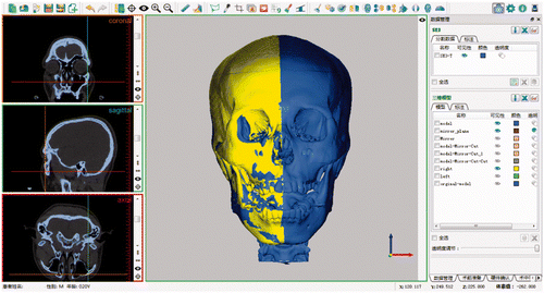
Figure 6. Pre-registration of the device before surgery.
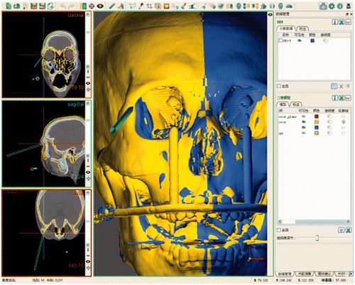
The patient underwent anatomic reduction of the fracture fragments and orbital floor reconstruction with the guidance of the navigation system. The zygomatic-orbital-maxillary complex was exposed by minimal supraorbital, subconjunctival, and intra-oral incisions. The fractured segment was released for reduction and precisely confirmed or modified with the navigation probe (). Once the actual reduction was accurately approximated to the preoperative plan, internal fixation was applied. The reduction of the fracture segments was checked by CT scanning one week postoperatively. The symmetry of the suturae zygomaticofrontalis, suturae zygomaticotemporalis, and zygomatic process was measured
Figure 7. Reduction confirmation during the operation.
Statistical analysis
The statistical analysis was carried out using the SPSS software (SPSS v.14, SPSS, Chicago, IL). Student's t-test was used, and a P value of ≤0.05 was assigned statistical significance.
Results
Reproducibility of device position
No loosening of the splint and device was observed during the 15 CT acquisitions. The results of the distance measurements based on the CT data volume are summarized in . The arithmetic mean of the standard deviation (SD) ranged from 0.47 mm to 0.70 mm. The coefficient of variance (CV) ranged from 0.54% to 0.97%. The maximum difference (MD) was less than 1.23 mm.
Table I. 3D distance measurements on the same skull.
Registration accuracy
The TREs for two different fiducial configurations are summarized in and shown with color-coded visualization in . Seven registration fiducials were defined on the maxillary alveolus to simulate the fiducial arrangements often used by maxillofacial surgeons. The mean deviations at the eight anatomical structures investigated ranged from 0.93 mm at the left infraorbital foramen to 3.19 mm at the right supraorbital foramen, and the target fiducial color-coded visualization values were red or yellow (). When the device was used as the registration fiducials, the mean deviations at the eight anatomical structures investigated ranged from 0.56 mm at the left infraorbital foramen to 0.96 mm at the right temple, and the target fiducials’ color-coded visualization values were green or sky blue (). The TRE obtained using the device was thus less than that obtained using fiducial markers fixed to the maxillary alveolus.
Figure 8. Target fiducial color-coded visualization value (colorwash dots) of the TRE for each registration fiducial (black dots) configuration. (A) Using registration fiducials on the maxillary alveolus. (B) Using registration fiducials on the device.
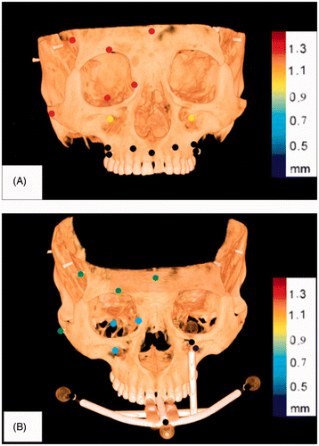
Table II. TRE of the investigated anatomical points for the fiducial configuration in which registration fiducials are defined on the maxillary alveolus and on the device.
Clinical trial
With the guidance of the navigation system, the positions of the probe and surgical instruments were displayed on a screen in real time. Both preoperative registration and intraoperative navigation were successfully achieved in this patient. The registration discrepancy was less than 0.5 mm, as verified by anatomic landmarks during surgery. The probe of the AccuNavi navigation system was used not only to navigate the deformed contour of the affected side, but also to check the desired position to which the displaced bone was to be reduced. The patient underwent uneventful healing with no serious complications. The ophthalmologic examination performed at one week after surgery verified the elimination of diplopia, enophthalmos, and ocular motility restriction. The previous facial asymmetry caused by the zygomatic-orbital-maxillary complex fracture was corrected satisfactorily upon clinically examination. Postoperative CT showed the symmetric deviation to be 0.73 at the suturae zygomaticofrontalis, 0.82 at the suturae zygomaticotemporalis, and 0.98 at the zygomatic process.
Discussion
Image guided surgery is based on the synchronization of the intraoperative position of the patient with the image of the patient's anatomy previously obtained by CT or MRI Citation[3]. This synchronization is realized through image registration and motion tracking. Firstly, the coordinate space of the actual patient is registered with that of the preoperative planning CT images, and then the orientation and position of the patient and instrument are tracked. The real-time relationship between the preoperative images and actual surgical anatomy is displayed on-screen. With the help of the device evaluated in this study, the registration could be performed prior to surgery, and the motion of the patient was detected automatically, reducing the operation time and increasing safety.
The fundamental prerequisite for this method is accurate and reproducible repositioning of the device on the dentition, as well as on the skull. Any deviation in the device's position, even tiny deformations or loosening, e.g., at the connective splint on the dentition, would result in tremendous error. Such deformations were minimized in this study by careful fabrication and short storage time of the device before surgery (i.e., avoiding any mechanical stress or temperature changes). The reproducibility and stability of the device were evaluated based on the distances between points in the device data sets and corresponding points in the skull data sets. The deviations were the lengths of the vectors between corresponding points according to the measurement coordinate system. For the rigidity test, the arithmetic mean of the standard deviation (SD) ranged from 0.47 mm to 0.70 mm. These results, together with the maximum difference found over all the experiments (1.23 mm), demonstrated that the device behaves like a rigid body attached to the dentition and has good positioning reproducibility.
The overall accuracy of IGS is influenced by numerous factors, such as head motion during the CT scan, accurate and reproducible repositioning of the device on the dentition, the environment in the operating theater (e.g., the optical tracking system, handling with surgical instruments, or the infrared component of the radiation from the operating lamp) and registration. These factors can hardly be separated for a systematic investigation, but from the clinical point of view the most relevant question concerns the overall accuracy. Therefore, an evaluation of the target registration error (TRE) is a fundamental prerequisite for any routine clinical use of this device. An implanted fiducial system has been considered the “golden standard” for image guided cranio-maxillofacial surgery, but this is invasive to the patient. Alternative non-invasive systems based on the external contours of the face and head or a custom bite-block have been developed. Ashamalla et al. showed relocation accuracy of 1.5 mm ± 0.8 mm using the Gildenberg-Laitinen adapter device Citation[4]. Das et al. reported variations of 1.0 mm, 0.8 mm, and 1.7 mm in the x, y and z directions respectively, using a relocatable Gill-Thomas-Cosman frame Citation[5]. Generally, the accuracy of a non-invasive system is less than that of an invasive system. However, the TRE of the navigation process with the device was better than that with the micro-screws implanted in the alveolar bone. One possible factor in error reduction might be the optimal distribution of the registration points. Our previous study has shown that optimal distribution of the fiducial positions improved registration accuracy Citation[6]. The fiducial markers in this device were designed to be scattered with the centroid of the markers being located near the target region. The markers were fixed far from the patient's head and around it, maximizing the probability of detecting even slight differences and permitting accurate registration. Another important factor in error reduction was that the device was fixed well to the dentition and was in the same position during CT scanning and the operation.
Using preregistration and automatic tracking, interactive, intraoperative application of 3D image data has been realized. By matching the contours of the mobile segment with the preoperative plan, computer-assisted navigation could be used to guide reduction of the zygomatic-orbital-maxillary complex fracture Citation[7]. The protocols in the present report are for unilateral maxillofacial fractures. Because most traumatic or tumor-related facial deformities are unilateral, the mirror tool (normal side to affected side) is an ideal adjunct to precise rehabilitation of the facial symmetry. It is based on the assumption that the facial bony contours of any given individual are symmetrical. In fact, studies have shown that measurable differences exist in the orbital volumes for any given individual, but the differences are small and have no significant effect on facial appearance and function Citation[8]. The technical system accuracy has been widely reported to be less than 1 mm, and the intraoperative precision for a patient has been reported as 1 to 2 mm [9, 10]. The intraoperative precision in this study was less than 1 mm, which shows the device to be promising for achieving precise and predictable results in the treatment of zygomatic-orbital-maxillary complex fracture reduction.
Conclusions
In summary, this article has introduced a pre-registration and automated tracking device for cranio-maxillofacial image guided surgery. The device's ability to be fixed to the patient's head in a reproducible fashion was confirmed through tests on a skull. The registration accuracy met the clinical requirement well. Operation and set-up of the device is convenient, and general clinical application should be possible. Future plans for the device include tests on patients undergoing other cranio-maxillofacial IGS procedures.
Declaration of interest: This study was supported by grants from the National Natural Science Foundation of China (30872906), the Hi-Tech Research and Development Program of China (SQ2009AA04ZX1485930), and the Key Project of Shanghai Scientific and Technological Commission (074119511).
References
- Fitzpatrick JM. The role of registration in accurate surgical guidance. Proc Inst Mech Eng H 2010; 224(5)607–622
- Bale RJ, Laimer I, Martin A, Schlager A, Mayr C, Rieger M, Czermak BV, Kovacs P, Widmann G. Frameless stereotactic cannulation of the foramen ovale for ablative treatment of trigeminal neuralgia. Neurosurgery 2006; 59(4 Suppl 2)ONS394–ONS402
- Nardy C, Alon W, Ron E. Computerized navigation for the lower jaw: comparison of 2 navigation systems. J Oral Maxillofac Surg 2008; 7: 1467–1475
- Ashamalla H, Addeo D, Ikoro NC, Ross P, Cosma M, Nasr N. Commissioning and clinical results utilizing the Gildenberg-Laitinen adapter device for X-ray in fractionated stereotactic radiotherapy. Int J Radiat Oncol Biol Phys 2003; 56: 592–598
- Das S, Isiah R, Rajesh B, Ravindran BP, Singh RR, Backianathan S, Subhashini J. Accuracy of relocation, evaluation of geometric uncertainties and clinical target volume (CTV) to planning target volume (PTV) margin in fractionated stereotactic radiotherapy for intracranial tumors using relocatable Gill-Thomas-Cosman (GTC) frame. J Applied Clin Med Phys 2010; 12: 3260
- Zhang WB, Wang CH, Yu HB, Liu YC, Shen GF. Effect of fiducial configuration on target registration error in navigation cranio-maxillofacial surgery. J Cranio-maxillofac Surg 2011; 39: 407–411
- Yu HB, Shen GF, Wang XD, Zhang SL. Navigation-guided reduction and orbital floor reconstruction in the treatment of zygomatic-orbital-maxillary complex fractures. J Oral Maxillofac Surg 2010; 68: 28–34
- Hassfeld S, Zöller J, Albert FK, Albert FK, Wirtz CR, Knauth M, Mühling J. Preoperative planning and intraoperative navigation in skull base surgery. J Cranio-maxillofac Surg 1998; 26: 220–225
- Metzger MC, Hohlweg-Majert B, Schön R, Tescher M, Gellrich NC, Schmelzeisen R, Gutwald R. Verification of clinical precision after computer-aided reconstruction in craniomaxillofacial surgery. Oral Surg Oral Med Oral Pathol Oral Radiol Endod 2007; 104: e1–e10
- Collyer J. Stereotactic navigation in oral and maxillofacial surgery. Br J Oral Maxillofac Surg 2010; 48: 79–83