Abstract
Knowledge of consistent anatomical relationships is an important criterion for establishing registration procedures for orthopedic navigation systems. Based on an analysis of 420 CT data sets, we investigated whether a robust registration of the pelvis in a lateral decubitus position could be achieved based on anatomical relationships. For this purpose, we assessed basic statistics and variation in anatomical parameters. It was found that inter-teardrop and inter-fossa distances exhibit a high degree of consistency in pelvises of the same gender. Additionally, stable relationships were found between the anterior pelvic plane (APP) and other reference planes that rely on acetabular points instead of pubic points. Based on these results, a registration procedure for the pelvis was developed which uses only landmarks that are accessible intra-operatively from the ipsilateral side. The deviation between a standard APP registration and this new registration method was assessed. For a standard cup position (40° inclination, 15° anteversion), the resulting deviations were found to be 0.15 ± 2.86° for inclination and 0.27 ± 3.46° for anteversion. Of the registrations, 99% had cup positions within the Lewinnek safe zone. This shows that accurate lateral pelvis registration based on anatomical relationships is achievable.
Introduction
Anatomical relationships are an important aspect of the evaluation of individual anatomical structures, e.g., deformities, developmental dysplasia, or the anatomical constitution. This applies particularly to bony anatomy. In adults, the bone is basically considered to be a rigid object, and thus the relationships are constant for an individual person, though they may vary between individuals. Knowledge of such relationships is important for many surgical procedures and tasks, such as the planning of joint replacement procedures. With regard to total hip arthroplasties (THAs), for example, the placement of the implants relies on a detailed analysis of the individual anatomy. It is known that many parameters, such as femoral antetorsion Citation[1], Citation[2], acetabular position Citation[3], acetabular inclination and anteversion Citation[4–7], or pelvic tilt Citation[8–10] are highly variable. Only a few parameters have been found to be consistent between individuals, such as transverse diameters Citation[5], Citation[11] and the deviations between the anterior pelvic plane (APP) and other reference planes Citation[12], Citation[13].
Such relationships are not only important for diagnostic purposes and in the planning of surgeries, but also for developing techniques for computer-assisted surgery (CAS). CAS techniques rely on robust anatomical knowledge to provide a frame of reference for positioning the implants. For example, a reference coordinate system must be provided for the orientation of the cup, which is closely related to a neutral (i.e., upright) position of the pelvis. Even though there has been discussion concerning deviations between the APP and the frontal plane of the patient Citation[8–10], the APP is still used as a standard reference for cup positioning in computer-assisted THAs. The APP is defined by the two anterior superior iliac spine (ASIS) points (on the ipsi- and contralateral sides) and a marginal point on the pubic symphysis. This leads to more consistent results, as it is more easily reproducible than the frontal plane and is fairly independent of inter- and intra-individual differences. The frontal plane, by comparison, is highly dependent on patient posture (e.g., standing versus sitting) or the clinical condition of the patient (e.g., soft tissue tension or pathologies of the lumbar spine).
Intra-operatively, the acquisition of the APP for use with navigation systems is problematic when the surgery is performed in a lateral decubitus position. With this patient set-up, it is not possible to acquire (i.e., to palpate and record) the contralateral ASIS point in a standard sterile environment. For this reason, other anatomical relationships play a crucial role in providing a frame of reference for the pelvis. Such relationships have to focus on landmarks that are accessible in a sterile environment from the ipsilateral side during a THA surgery. For these landmarks, relationships to standard frames of reference are of major importance in enabling comparisons between the results of new registration techniques and those of established procedures.
In this paper, we analyzed anatomical relationships which can be used for registration procedures with reference to those landmarks alone. In particular, the inter-teardrop distance (ITD) and inter-fossa distance (IFD) were studied. The teardrop figure is a structure that is well known to surgeons. It is identified in anterior-posterior (a-p) X-ray images and is used for various diagnostic procedures such as measuring leg-length differences Citation[14], Citation[15] or shifts of a previously inserted cup implant Citation[16], Citation[17]. The ITD is measured as a distance between the teardrop figures on the left and right sides in a left-right direction. The teardrop figure is a projection of the structures around the acetabular fossa Citation[18]. Thus, IFD and ITD are closely related. Additionally, the analyzed relationships included deviations between the APP and other reference planes having the same alignment as the APP in a coronal projection, but a different one in a sagittal projection. The center of rotation, the deepest fossa, and a point on the anterior rim of the acetabulum are used to define these planes instead of points on the pubic tubercles.
A thorough statistical analysis is required in order to be able to use these relationships in navigation systems. For this purpose, a comprehensive analysis based on an ensemble of 420 computed tomography (CT) data sets was performed in this study. Overall statistics were assessed, along with variations between genders, different geographical regions (populations), and conditions of the pelvis (distinguishing between normal and abnormal pelvic anatomy). All this information was then combined in a way that enabled a complete registration of the pelvis in a lateral decubitus position, i.e., a reconstruction of the APP based on the determined anatomical relationships. The main focus of this study was to establish the statistics of anatomical relationships for the pelvis and to assess the accuracy of the proposed lateral registration method based on CT data sets. The main question we posed was whether, when using this new technique, at least 95% of the targeted cup positions from this CT-based simulation study lie within the Lewinnek safe zone Citation[19], i.e., 40 ± 10° radiographic inclination and 15 ± 10°anteversion.
Material and methods
In the study, 420 CT data sets (174 male, 246 female) from previous studies were analyzed retrospectively. These comprised pre- and post-operative data sets from THA surgeries, as well as 60 data sets from cadaver studies. All available data sets representing a valid pre-operative situation for at least one joint were included in the study; data sets from patients with bilateral hip replacements were excluded. Additionally, in the overall ensemble of available data sets, there were four cases with extreme deformities of the femoral head/acetabular cavity, which prevented an appropriate definition of the center of rotation. These data sets were also excluded.
The data sets were subdivided according to their origin, i.e., whether they were from Asian countries, the United States (US), or the rest of the world (RoW), where RoW mainly reflected a European origin. This grouping was used to discriminate between different populations. The condition of all pelvises was rated as either normal or abnormal by a single observer (Michael Wörner). If the hip joint showed significant deformity compared to normal human hip joint anatomy, such as deformities caused by acetabular fracture, distortion of the bony pelvis resulting from a previous surgery, the effects of previous hip surgery that affected the pelvic anatomy (such as THA, surface replacement, or peri-acetabular osteotomy), or hip joint modifications caused by dysplasia, coxarthrosis, or necrosis, the hip joint was rated as Abnormal. A rating of Normal was given to hip joints exhibiting no or only minor abnormalities. See for details of the separation of the data sets into subgroups.
Table I. Number of observations and separation into subgroups.
All 3D pelvic landmarks were defined using iPlan 3.0 software (Brainlab AG, Feldkirchen, Germany). A threshold-based segmentation was applied to the CT data set to identify the bony structures of the pelvis. The landmarks were first defined roughly on the resulting 3D model, then the locations of the landmark points were fine-tuned in the axial, sagittal, and coronal slices to accurately determine the landmarks on the bone contour. Both ASIS points and a point on the anterior margin of the pubis were acquired to define the APP. The APP was used as the frontal plane, and the line between both ASIS points as the medial-lateral direction.
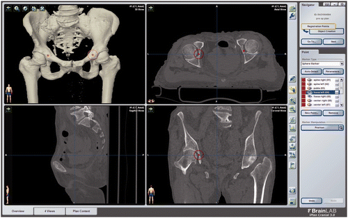
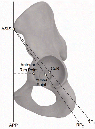
Further landmarks on the acetabulum were defined in order to perform a simulated lateral registration of the pelvis. A sphere was manually fitted around the acetabulum in the CT data set. The center of this sphere was used as the center of rotation (CoR). One point (the anterior rim point) was acquired on the anterior side of the acetabular rim. Additionally, the deepest point in the acetabular fossa was determined on each side of the pelvis. shows the planning of these landmarks. The acetabular landmarks were used to specify alternative reference planes, which were defined by both ASIS points and one acetabular point. For the remainder of this paper, these reference planes will be referred to as RP1 (ASIS-ASIS-CoR), RP2 (ASIS-ASIS-deepest fossa point), and RP3 (ASIS-ASIS-anterior rim point). shows the relationship between the APP and RP1, and between the APP and RP2. RP3 was constructed in a slightly different way by assuming a fixed distance between the anterior rim point and the APP. A fourth reference plane, RP4, was calculated as a combination of the other reference planes, RP1, RP2 and RP3, by averaging the information concerning those other planes. The spina distance was defined as the distance between both ASIS points. In addition, one point on the middle of the spinous process of the L5 vertebra was determined. This point lies approximately on the midsagittal plane, and is referred to as the midplane point in this paper.
Figure 1. Planning of the pelvic landmarks, including the deepest point in each fossa, performed with iPlan 3.0 software (Brainlab AG).

Figure 2. Alternative reference planes RP1 (ASIS-ASIS-CoR) and RP2 (ASIS-ASIS-Fossa) to define the sagittal orientation of the pelvis. The distance between the anterior rim point and the APP is also shown: This distance was used to define RP3.

Descriptive statistics were calculated for the following measurements: The IFD was measured as the sum of the distances between both fossa points and the midsagittal plane. Angular deviations between the APP and the alternative reference planes, i.e., rotational differences according to a rotation around the ASIS-ASIS line, were also obtained.
Subsequently, a true lateral registration of the pelvis was performed based on statistics of the IFD, as well as the deviations between the APP and RP4. That is, the APP was reconstructed based on the anatomical relationships using only those landmarks which are accessible in a lateral decubitus position (the ipsilateral ASIS, deepest fossa point, CoR, anterior rim, and midplane point). The statistics of the IFD, along with an estimation of the spina distance, were used to basically determine the medial-lateral direction. The statistics of the deviation between the APP and the other reference planes were used to fix the remaining degree of freedom, i.e., rotation in the medial-lateral direction. The differences between the gold standard and the described lateral registration were calculated for each data set. They were assessed in terms of the deviation of inclination/anteversion for a standard cup position, i.e., the cup was virtually placed in a position of 40° inclination and 15° anteversion (as recommended by Lewinnek et al. Citation[19]). In this paper, all values for inclination/anteversion are specified in the radiographic definition (according to Murray Citation[20]). This position was set in the gold-standard coordinate system, then the deviations of the cup position were obtained when switching between the gold standard and the described lateral registration. Additionally, it was determined how many virtual cup positions lay in the Lewinnek safe zone Citation[19].
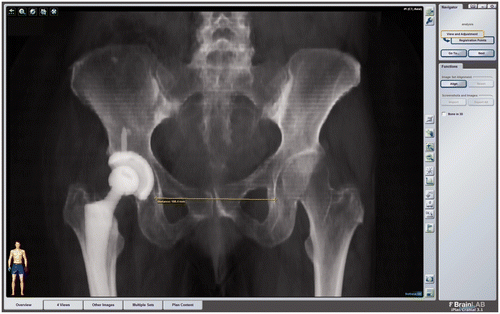
To measure the inter-teardrop distance (ITD), an iPlan 3.1 prototype (Brainlab AG, Feldkirchen, Germany) was used. Virtual antero-posterior X-ray images (digitally reconstructed radiographs – DRRs) were generated with this software using a volume rendering technique, i.e., a virtual X-ray projection image was calculated based on the Hounsfield values given by the CT data sets. The teardrop figure was identified in the DRRs, and the inter-teardrop distance was determined as the distance between the most caudal parts of the teardrop figure in a left-right direction (see ).
Figure 3. Measurement of the inter-teardrop distance (ITD). The measurement was performed virtually based on digitally reconstructed radiographs (DRRs) with iPlan 3.1 prototype software (Brainlab AG).

The point planning procedures and measurements were performed by five different observers (M.W., V.S., S.S., L.P., and S.R. – see Acknowledgments), who were not involved in the subsequent analysis of the data. Statistical analyses were performed for geographical regions and different conditions of the pelvis. These groups were subdivided according to gender. Mean values and standard deviations, as well as range values, were calculated for each group. Student's two-sample t-tests assuming equal variances (p-value = 0.05) were applied to determine statistical differences between populations from different geographical regions, genders, and conditions of the pelvis. Additionally, differences between ITD and IFD were analyzed by Student's two-sample t-tests assuming equal variances (p-value = 0.05). The tests were performed with and without a shift in the values according to the estimated differences between ITD and IFD. The correlation between ITD and IFD was calculated using the coefficient of determination R2.
Results
Regarding IFD, the statistical results for the subgroups are summarized in . No significant differences were found for IFD in normal and abnormal conditions of the pelvis in any analyzed subgroup. The differences between the male and female groups were statistically significant, instead. This proved to be true not only for the entire group, but also within the subgroups (e.g., those representing normal and abnormal conditions of the pelvis). After separating the test population by gender, no statistically significant differences for IFD were found for the different geographical regions, except for a slight deviation between RoW and Asia in the female group.
The mean value of the differences between IFD and ITD was 3.6 ± 3.7 mm (95% CI: [−3.7 mm; 10.9 mm], min: −6.0 mm, max: 15.3 mm) for the female group, and 4.5 ± 3.1 mm (95% CI: [−1.7 mm; 10.7 mm], min: −3.6 mm, max: 12.2 mm) for the male group. The coefficient of determination between IFD and ITD was R2 = 0.7975 (female) and R2 = 0.7744 (male). There were statistically significant differences between IFD and ITD as long as no correction of the base value was applied to one of the two measurements. After shifting the ITD values according to the observed mean differences, the differences between the IFD and ITD were no longer found to be statistically significant.
The angular differences between the APP and the new reference planes RP1 to RP4 according to the performed registrations are shown in . No statistically significant differences were found between the genders, but significant differences were found between some of the subgroups (e.g., those representing ethnicities and conditions of the pelvis).
Table II. Summary of statistics (mean ± standard deviation) for anatomical relationships used in the proposed pelvis registration method.
The final deviations between the gold-standard APP and the new lateral registration for a cup orientation of 40° inclination and 15° anteversion were 0.15 ± 2.86° (95% CI: [−5.25°; 6.34°], min: −10.65°, max: 9.91°) for inclination and 0.27 ± 3.46° (95% CI: [−6.10°; 7.48°], min: −8.7°, max: 10.93°). This means that statistically 99.9% of the cases were in the Lewinnek safe zone for inclination (40 ± 10°) and 99.6% were in the safe zone for anteversion (15 ± 10°). Out of the entire test ensemble, only three cases (0.71%) were outside the combined Lewinnek safe zone (40 ± 10° inclination, 15 ± 10° anteversion). Again, there were no statistically significant differences based on gender, but minor differences were found for ethnicity and condition of the pelvis. See for details of these results.
Table III. Summary of statistics (mean ± standard deviation) for the deviation between the gold-standard APP and lateral registration, assuming a standard cup position (40° inclination, 15° anteversion).
Discussion
When considering the varying shapes of human pelvises, only a few anatomical relationships have been found to be consistent Citation[5], Citation[11], Citation[21], Citation[22]. Some measurements with a low level of variation have been found, e.g., in obstetrics when analyzing the pelvic cavity. In particular, the diameters of the pelvic cavity were found to be consistent Citation[11]: The mean value of the transverse diameter of the pelvic inlet was 130 ± 8 mm for white males and 134 ± 8 mm for white females. The biacetabular distance was 118 ± 7 mm (males) and 126 ± 7 mm (females). Maruyama et al. Citation[5] measured the floor-to-floor distance between both acetabula for a set of 50 male and 50 female pelvises from human skeletons. In comparison to the IFD values in our present study, they found slightly different values for the floor-to-floor distance, i.e., 115 ± 9 mm for the entire group, 111 ± 7 mm for the male group, and 118 ± 9 mm for the female group. The different measurement methods used may explain these differences. They also recognized a significant difference between male and female pelvises.
The consistency of the transverse diameters has a strong relationship to ITD and IFD, since these values also represent transverse measurements which are basically determined by the pelvic cavity. Additionally, points on the acetabular fossa (ipislateral side) can easily be acquired intra-operatively after luxation of the joint. Thus, the IFD is a good candidate for use in registration techniques. Direct access to the ITD is not possible in navigation applications, but it could be used as an indirect measurement, since it has been shown that IFD and ITD have high correlations. Thus, individual IFD values can be estimated when the ITD has, for example, already been obtained by measurements on anterior-posterior X-rays.
Information about the IFD and ITD can be used to basically determine the left-right direction of the pelvis when points in the acetabular fossa and on the midsagittal plane (e.g., at the sacrum/lumbar spine) are digitized by the user of a navigation system. Deviations between the APP and other reference planes can be used to estimate the angular orientation of the pelvis according to a rotation around the medial-lateral rotation Citation[12], Citation[13]. For example, the center of rotation, the deepest fossa point, or an anterior rim point may be used to define an appropriate reference plane. Thornberry et al. Citation[13] determined that deviations between the APP and a reference plane spanned by the two ASIS points and CoR are low. They found deviations in the range of 36.5 ± 3.5° in a CT-based study of 16 male and 16 female pelvises. No statistically significant differences were found between genders. A similar approach was used in our lateral registration study by combining the values for the deviations between the APP and three other reference planes. One of these (RP1) refers directly to the definitions used by Thornberry et al. Citation[13]. The other reference planes (RP2 and RP3) use a fossa and an anterior rim point instead of the CoR. These points are considered to be more robust, especially in the presence of deformed acetabula. Using combinations of the alternative reference planes can help to improve the robustness of the registration techniques which rely on these statistics.
In this study, it has been shown that anatomical relationships can be used to implement a lateral registration technique. In comparison to standard APP registration, this approach allows for the pubis points to be replaced by points around the acetabulum. The contralateral ASIS point is replaced by a midplane point. In addition, the distance between the ASIS points has to be determined. This can be done by pre-operative measurements on the patient, using appropriate hardware tools, or measurements based on X-ray images. Using this approach, the deviations between a gold-standard APP registration and the new lateral registration approach were found to be 0.15 ± 2.86° in radiographic inclination and 0.27 ± 3.46° in anteversion. Individual deviations of the IFD, ITD, or other statistical relationships did not directly affect the registration accuracy but only had a limited influence on the overall registration accuracy. In total, more than 99% of the cases had virtual cup positions within the Lewinnek safe zone. There were some differences between the analysed subgroups, which likewise only had a minor impact on the final registration accuracy.
Of course, these results must be confirmed in a clinical set-up. Regarding the analysis of regional differences, another weakness of this study was the low number of CT data sets available for some subgroups. In particular, only 12 Asian males and 19 US males were included. Additionally, there was no clear distinction between ethnic groups, but rather between the geographical regions from which patients came. However, we showed that the regional differences had only limited influence on the results. Furthermore, it is not clear whether the APP or the new reference planes show less variation when compared to the frontal plane of the patient, and if either one better accounts for pelvic tilt. No information concerning the pelvic tilt of the patients was available for our study. Currently, the APP is still considered an established reference for navigated hip surgeries and was thus used as a gold standard.
In summary, it has been demonstrated that a true lateral registration of the pelvis can be achieved based on the anatomical relationships analyzed. The results from this extensive CT data analysis are very promising. The procedure only requires a few landmarks which are accessible from the ipsilateral side in a lateral decubitus position. This is a huge benefit for navigation systems, as nowadays they usually rely on direct acquisition of the anterior pelvic plane. Currently, registration of the contralateral ASIS and additional points on the pubis is required, but this is not possible in a standard sterile environment when surgery is performed with the patient in a lateral decubitus position. Thus, utilization of these methods requires considerable additional OR time. In contrast, the proposed method only requires the user to palpate points that are accessible from the ipsilateral side. Thus, it qualifies as a true lateral registration of the pelvis. Such a registration is a key element in improving the integration of navigation systems into the clinical workflow for hip surgeries.
Acknowledgments
The authors thank Dr. Michael Wörner, Verena Schmalzl (V.S.), Luise Poitzsch (L.P.), Silke Schubert (S.S.), Stefan Richter (S.R.), and Nicole Ehret (N.E.) for their contributions to this study.
Declaration of interest: All the authors are employees of Brainlab AG.
References
- Pierchon F, Pasquier G, Cotten A, Fontaine C, Clarisse J, Duquennoy A. Causes of dislocation of total hip arthroplasty. CT study of component alignment. J Bone Joint Surg Br 1994; 76: 45–48
- Wines AP, McNicol D. Computed tomography measurement of the accuracy of component version in total hip arthroplasty. J Arthroplasty 2006; 21: 696–701
- Dandachli W, Nakhla A, Iranpour F, Kannan V, Cobb JP. Can the acetabular position be derived from a pelvic frame of reference?. Clin Orthop Relat Res 2009; 467(4)886–893
- Bargar WL, Jamali AA, Nejad AH. Femoral anteversion in THA and its lack of correlation with native acetabular anteversion. Clin Orthop Relat Res 2010; 468(2)527–532
- Maruyama M, Feinberg JR, Capello WN, D’Antonio JA. The Frank Stinchfield Award: Morphologic features of the acetabulum and femur: anteversion angle and implant positioning. Clin Orthop Relat Res 2001; 393: 52–65
- Murtha PE, Hafez MA, Jaramaz B, DiGioia AM. Variations in acetabular anatomy with reference to total hip replacement. J Bone Joint Surg Br 2008; 90(3)308–313
- Tohtz SW, Sassy D, Matziolis G, Preininger B, Perka C, Hasart O. CT evaluation of native acetabular orientation and localization: Sex-specific data comparison on 336 hip joints. Technol Health Care 2010; 18(2)129–136
- Babisch JW, Layher F, Amiot LP. The rationale for tilt-adjusted acetabular cup navigation. J Bone Joint Surg Am 2008; 90(2)357–365
- Parratte S, Pagnano MW, Coleman-Wood K, Kaufman KR, Berry DJ. The 2008 Frank Stinchfield Award: Variation in postoperative pelvic tilt may confound the accuracy of hip navigation systems. Clin Orthop Relat Res 2009; 467(1)43–49
- Zhu J, Wan Z, Dorr LD. Quantification of pelvic tilt in total hip arthroplasty. Clin Orthop Relat Res 2010; 468(2)571–575
- LaVelle M. Natural selection and developmental sexual variation in the human pelvis. Am J Phys Anthropol 1995; 98(1)59–72
- Hakki S. Acetabular center axis: A novel alternative registration point to anterior pelvic plane in navigated THA [abstract]. Proceedings of the 10th Annual Meeting of the International Society for Computer Assisted Orthopaedic Surgery (CAOS-International), Paris, France, June 2010. pp 255–256.
- Thornberry RL, Martin JD, Toole GC, Barbu A. Statistical variability of the anterior pelvic plane to the hip center ASIS pelvic plane [abstract]. Proceedings of the 10th Annual Meeting of the International Society for Computer Assisted Orthopaedic Surgery (CAOS-International), Paris, France, June 2010. pp 328–330.
- Murphy SB, Ecker TM. Evaluation of a new leg length measurement algorithm in hip arthroplasty. Clin Orthop Relat Res 2007; 463: 85–89
- Woolson ST, Hartfort JM, Sawyer A. Results of a method of leg-length equalization for patients undergoing primary total hip replacement. J Arthroplasty 1999; 14(2)159–164
- Massin P, Schmidt L, Engh CA. Evaluation of cementless acetabular component migration. An experimental study. J Arthroplasty 1989; 4(3)245–251
- Nunn D, Freeman MA, Hill PF, Evans SJ. The measurement of migration of the acetabular component of hip prostheses. J Bone Joint Surg Br 1989; 71-B(4)629–631
- Bowerman JW, Sena JM, Chang R. The teardrop shadow of the pelvis; anatomy and clinical significance. Radiology 1982; 143(3)659–662
- Lewinnek GE, Lewis JL, Tarr R, Compere CL, Zimmerman JR. Dislocations after total hip-replacement arthroplasties. J Bone Joint Surg Am A 1978; 60(2)217–220
- Murray DW. The definition and measurement of acetabular orientation. J Bone Joint Surg Br 1993; 75(2)228–232
- Motzny S. Bestimmung des Rotationszentrums über die Landmarken des Beckens. PhD thesis. Tübingen, 2011.
- Tague RG. Variation in pelvic size between males and females in nonhuman anthropoids. Am J Phys Anthropol 1995; 97(3)213–233