Abstract
Intramedullary nail removal can be demanding, especially in cases of implant breakage or bony overgrowth at the end-cap, if the exact insertion depth of the nail is neglected in the index surgery. In the presented case, two challenging nail removals were necessary. The first was performed in a re-nailing procedure due to a pseudarthrosis with implant breakage, and the second was performed during hardware removal after fracture healing in a situation where there was deep intramedullary placement of the exchange nail. For the second implant removal a minimally invasive approach based on instrument placements over a navigated guide-wire was used to reduce the iatrogenic morbidity associated with an extensive open approach to the nail and to decrease the radiation exposure for the patient and the operating team.
Introduction
Intramedullary nails have to be removed in cases of re-operation due to non-union, peri-implant fracture, and infection, or in preparation for arthroplasty surgery, when the two implants would interfere with one another.
In patients with asymptomatic nails in the lower extremities, the indication for hardware removal is relative. For these elective procedures, good preoperative planning of the surgery is mandatory to prevent complications and medico-legal problems. Already in the index surgery, control of the exact insertion depth of the nail, as well as the use of an end-cap, are of similar importance as the use of a dynamometric screwdriver for locking screws in plate fixation procedures.
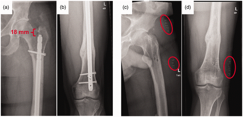
In the presented case, a young patient requested a nail removal which was challenging due to the deep intramedullary placement of the nail with bony overgrowth at the end-cap ().
According to Gösling et al. Citation[1], nail intrusion is a common pattern, which was observed in 27% of antegrade inserted femoral nails (40 of 148 nails), and results in a prolonged operating time, as well as in increased radiation exposure for the patient and operating team.
To reduce the iatrogenic morbidity associated with an extensive open approach to the proximal end of the nail, a minimally invasive approach based on instrument placements over a navigated guide-wire was used. Reductions in fluoroscopy exposure and operating time were postulated as additional advantages due to the first-pass accuracy of the navigated guide-wire placement.
Patient and methods
Patient
A 43-year-old man presented at our department with persistent pain in the left leg three years after nailing of a femoral fracture (AO classification: 32-B3.3) resulting from a motorcycle accident. In the radiographs a hypertrophic pseudarthrosis with a broken nail (Grosse-Kempf System, Howmedica Osteonics) on the same level was diagnosed. Hardware removal was performed using a modification of the technique of Magu et al. Citation[2] () and a larger reamed nail with internal compression (T2 femoral nail, Stryker) was used for the fixation (). Postoperatively, the patient was free of pain under full weight-bearing and fracture healing was observed on the follow-up radiographs. Two years after the last surgery, the patient requested hardware removal. Due to the two previous operations and the deep intrusion of the nail (18 mm according to the measurement procedure of Gösling et al. Citation[1]) with bony overgrowth of the end-cap (), we decided to use a minimally invasive navigated procedure for nail removal.
Figure 1. Radiographs of the first broken-nail removal by a modification of the technique of Magu et al. Citation[2]. (a) Lateral view of the femoral pseudarthrosis with the broken nail. (b) Intraoperative fluoroscopic image showing an antegrade inserted guide-wire, which was bent outside the knee after distal perforation. (c) Retraction of the bent guide-wire removes the distal part of the broken nail.
![Figure 1. Radiographs of the first broken-nail removal by a modification of the technique of Magu et al. Citation[2]. (a) Lateral view of the femoral pseudarthrosis with the broken nail. (b) Intraoperative fluoroscopic image showing an antegrade inserted guide-wire, which was bent outside the knee after distal perforation. (c) Retraction of the bent guide-wire removes the distal part of the broken nail.](/cms/asset/81c8c034-5877-48ae-8af4-d74dbd440d4e/icsu_a_741623_f0001_b.gif)
Figure 2. Radiographs of the navigated nail removal. (a, b) Healed femoral pseudarthrosis after re-nailing; deep placement of the nail in the medullar cavity (18 mm) made the implant removal demanding. (c, d) Postoperative X-ray control showing complete nail removal via less invasive incisions (red circles).

Navigation system
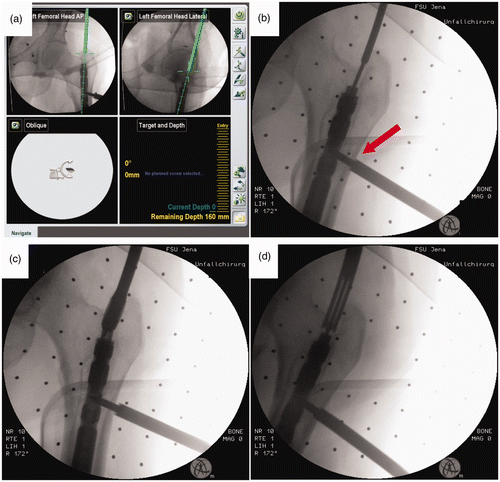
For the navigation procedure an optoelectronic navigation system based on 2D fluoroscopic images (VectorVision, Trauma Software Version 3.0, BrainLAB AG, Feldkirchen, Germany) was used (). The system facilitates real-time tracking of instruments and visualization of their movements in relation to the patient's anatomy as displayed on a sterile covered touch screen (). Therefore, image acquisition was done with a standard image intensifier (Ziehm Imaging, Inc.). For the registration of the two acquired 2D images (antero-posterior and axial projection) a plexiglass plate with reference markers had to be attached to the C-arm detector and a X-Spot® device had to be placed in the cone beam during each image acquisition Citation[3].
Figure 3. Navigated technique for minimally invasive removal of buried nails. (a) Screenshot of the navigation system display. Based on two orthogonal 2D fluoroscopic images (antero-posterior and axial) a navigated guide-wire can be placed precisely in the hole of the end-cap. (b) Intraoperative fluoroscopic control image showing the precise guide-wire placement via a navigated drill sleeve. The dynamic reference base for the navigation procedure was placed in an existing drill hole after removal the most proximal locking screw (red arrow). (c) Preparation of the osseous tunnel to the nail with a cannulated driller. (d) Removal of bone overgrowth around the end cap using a cannulated crown drill.

In addition, reflective passive reference markers were attached to the drill sleeve to localize its position and the drill trajectory in relation to the femur, which was marked with a rigidly fixed dynamic reference base. After calibration of a 3-mm drill sleeve, the navigated procedure was begun.
Operation procedure
The patient was placed on a standard operating table in the supine position with a freely moveable left leg. After removal of all locking screws via the old skin incisions using the standard technique, the proximal dynamic screw hole was used for the fixation of a 5 mm Schanz screw to attach a reference base rigidly to the femur for the navigated guide-wire placement ().
An optimized skin incision was determined by the alignment of the drill trajectory in the center of the end-cap of the nail in both acquired images. After skin incision the drill sleeve was placed at the bone, the visualized trajectory again adjusted (), and a 3-mm k-wire inserted.
After verification of the exact position in two orthogonal fluoroscopic images, a cannulated 10-mm reamer (No. 1806-2010, Stryker Corporation) was used to core out an osseous tunnel to the nail, after which a crown drill (No. 1806-2020, Stryker Corporation) was used to remove the bone around the end-cap (). The end-cap was removed with a self-holding screwdriver (No. 1806-6005, Stryker Corporation) to avoid disengagement and loss of the end-cap in the soft tissues during retraction. The nail extraction rod was connected and, after Schanz screw removal, the nail was pulled out with a slotted hammer. A suction drain was placed and the skin stapled. The time from skin incision to wound closure was 80 minutes (including the navigation-specific set-up), with a fluoroscopy time of 58 seconds and radiation dosage of 270 cGy/cm2, mainly for verification and documentation of the procedure. In the postoperative X-rays the minimally invasive skin incision and the small osseous tunnel without excessive bone removal are documented (). The patient was discharged on full weight-bearing without crutches on the second postoperative day, but was prohibited from high-impact activities (sports and carrying anything heavy) for six weeks, due to the interlocking holes. At the six-month follow-up the patient had no complaints.
Discussion
A well-published cannulated technique for minimally invasive femoral nail removal Citation[4–7] was combined with the use of a 2D fluoroscopic navigation procedure to reduce the radiation exposure and operating time, as well as to facilitate nail removal in the supine position on a standard operating table.
The decision-making for operative removal of femoral nails is based on general aspects, such as the nail design Citation[8], composition and surface modifications (stainless steel versus titanium alloy, polished versus unpolished surfaces) Citation[9], as well as on patient- and surgeon-specific aspects, such as the age and co-morbidity of the patient Citation[10], a history of open fractures or infection Citation[10], a request by the patient Citation[11], intrusion of the femoral nail Citation[1], the experience of the surgeon Citation[1], Citation[12] and the equipment available at the hospital.
The best time for surgery should also be evaluated. Whereas early implant removal increases the risk of re-fracture, delayed removal may result in more difficult and extensive operating procedures due to a stronger bony integration and overgrowth of implants. In conclusion, each decision must be made on an individual basis and the procedure is sometimes not an “operation for beginners”, as is often thought. In the case reported here, the hardware removal was performed at the request of the patient in spite of there being no complaints, due to his young age and the absence of co-morbidities.
Removal of a buried nail (18 mm of intrusion with bony overgrowth) is a common problem, observed in 27% of 148 antegrade femoral nails, which is correlated with prolonged operating time Citation[1], Citation[11] and frequent fluoroscopic imaging to determine and visualize the correct path to the nail. Compared with the index surgery, extensive soft tissue dissections are usually necessary, abrogating the initial efforts to perform minimally invasive surgery. Furthermore, postoperative morbidity due to iatrogenic damage to the m. glutei and heterotopic ossifications have to be considered Citation[13].
Using a cannulated technique for minimally invasive nail removal is not a new approach and has been described by several groups Citation[4–7]. Whereas Ackermann et al. Citation[4] in most cases used the technique for gamma nail removal, in which deep intrusion is not a common problem due to the insertion depth being predefined by the femoral neck screw, Georgiadis Citation[6] stressed the difficulty of correct guide-wire placement in buried nails and exact determination of the skin incision, which often differed from the stab wound of the index surgery.
Combining the cannulated technique with navigated guide-wire placement in these demanding cases is a promising approach for several reasons:
Supine position of the patient on the standard operating table. For the conventional guide-wire placement and control of the osseous path preparation, repetitive fluoroscopic images acquired by C-arm movements in different projections (antero-posterior and axial) are mandatory. Therefore, a rigid placement of the leg on a traction table or a lateral position of the patient is preferred in order to maintain exact instrument alignment in the first antero-posterior plane when controlling for the second orthogonal axial plane.
Alternatively, the patient can be placed supine on a standard operating table with the leg in a neutral position to acquire the antero-posterior fluoroscopic projection, and in the FABER position (hip flexion, abduction and external rotation) to acquire the second orthogonal, axial fluoroscopic projection. However, for these maneuvers the primarily adjusted guide-wire alignment in the first plane will be lost and a “trial and error” placement of the guide-wire is necessary, as described by Kahler Citation[14].
With a navigation system, both projection images can be acquired by first using a dynamic reference base rigidly fixed to the femur, followed by a navigated instrument alignment on the leg in the neutral position without further leg movements, due to simultaneous control of the procedure on both virtual images.
Increased precision. An optimized skin entry point and path to the nail can be determined precisely before skin incision, due to the simultaneous virtual visualization of the drill sleeve with the planned trajectory on both acquired fluoroscopic images in two orthogonal projections, resulting in a first-pass accuracy of the navigated guide-wire placement.
Reduced radiation exposure and decreased operating time. The well-known “first-pass accuracy” of navigated guide-wire placement seems to reduce the radiation exposure and the operating time, as shown for different applications, such as the retrograde drilling of osteochondral lesions Citation[15]. Although a direct comparison with the published literature is difficult, due to the poor data records and different conditions (e.g., type of nail, number of locking screws, insertion point and depth of the nail, BMI and muscle status of the patient, experience of the surgeon, etc.), Husain et al. reported a mean operating time of 119 minutes for femoral nail removal Citation[11], while Georgiadis reported an average operating time of 98 minutes and a fluoroscopy time of 66 seconds Citation[6].
Preserved sterility. The sterility of the operating field is less jeopardized if the leg is moved in the FABER position to acquire the second axial view instead of performing a C-arm rotation under the operating table.
To our knowledge, the use of navigation for hardware removal, in order to reduce soft tissue damage and radiation exposure, has previously been reported only for pelvic screw and projectile removal Citation[16–18].
Common navigation-related complications are associated with the rigid fixation of the dynamic reference base in the bone via an additional skin incision. Postoperative heterotopic ossifications and soft tissue irritations Citation[19], Citation[20], as well as iatrogenic fractures of a bone that is weakened (by approximately 50% for torsion stiffness, with reduced capacity for axial energy absorption) caused by the osseous drill holes have been described Citation[21], Citation[22]. To prevent these severe complications, we used the existing stab wound and screw hole after removal of the most proximal locking screw for the rigid fixation of the dynamic reference base. This position was used to reduce the inaccuracy of the navigation system as far as possible by placing the dynamic reference base in the closest proximity to the target point of navigation.
Limitations of navigated hardware removal are the costs of a navigation system, as well as the need for specialized education and experience of the surgeon in computer assisted surgery, making navigation technologies predominantly available only in specialized major trauma centers at present.
Conclusion
This paper describes an alternative technique for removal of buried intramedullary nails and highlights the potential benefits of computer assisted navigation. The resulting reduction in radiation exposure, operative time and soft tissue damage, together with the minimization of bone loss and mechanical weakening of the bone, makes hardware removal more viable.
Declaration of interest: The authors report no declarations of interest.
References
- Gösling T, Hufner T, Hankemeier S, Zelle BA, Muller-Heine A, Krettek C. Femoral nail removal should be restricted in asymptomatic patients. Clin Orthop Relat Res 2004; 6(423)222–226
- Magu NK, Sharma AK, Singh R. Extraction of the broken intramedullary femoral nail - an innovative technique. Injury 2004; 35(12)1322–1323
- Gras F, Marintschev I, Klos K, Mückley T, Hofmann GO, Kahler DM. Screw placement for acetabular fractures. Which navigation modality (2D vs. 3D) should be used? - An experimental study. J Orthop Trauma 2012; 26(8)466–473
- Ackermann O, Maier K, Ruelander C, Vogel T, Schofer M. [Modfied technique for intramedullary femur nail removal] [In German]. Z Orthop Unfall 2011; 149(3)296–300
- Ciampolini J, Eyres KS. An aid to femoral nail removal. Injury 2003; 34(3)229–231
- Georgiadis GM. Percutaneous removal of buried antegrade femoral nails. J Orthop Trauma 2008; 22(1)52–55
- van Eerten PV, Reemst PH, Repelaer van Driel OJ. Minimally invasive femoral nail extraction. Arch Orthop Trauma Surg 2005; 125(3)197–200
- Seligson D, Howard PA, Martin R. Difficulty in removal of certain intramedullary nails. Clin Orthop Relat Res 1997; 7(340)202–206
- Hayes JS, Vos DI, Hahn J, Pearce SG, Richards RG. An in vivo evaluation of surface polishing of TAN intermedullary nails for ease of removal. Eur Cell Mater 2009; 18: 15–26
- Hora K, Vorderwinkler KP, Vecsei V, Gabler C. [Intramedullary nail removal in the upper and lower limbs: Should we recommend this operation?] [In German]. Unfallchirurg 2008; 111(8)599–606
- Husain A, Pollak AN, Moehring HD, Olson SA, Chapman MW. Removal of intramedullary nails from the femur: A review of 45 cases. J Orthop Trauma 1996; 10(8)560–562
- Boerger TO, Patel G, Murphy JP. Is routine removal of intramedullary nails justified. Injury 1999; 30(2)79–81
- Dodenhoff RM, Dainton JN, Hutchins PM. Proximal thigh pain after femoral nailing. Causes and treatment. J Bone Joint Surg Br 1997; 79(5)738–741
- Kahler D. Percutaneous screw insertion for acetabular and sacral fractures. Techniques in Orthopaedics 2003; 18(2)174–183
- Gras F, Marintschev I, Kahler DM, Klos K, Mückley T, Hofmann GO. Fluoro-Free navigated retrograde drilling of osteochondral lesions. Knee Surg Sports Traumatol Arthrosc 2011; 19(1)55–59
- Perlick L, Tingart M, Lerch K, Bäthis H. Navigation-assisted, minimally invasive implant removal following a triple pelvic osteotomy. Arch Orthop Trauma Surg 2004; 124(1)64–66
- Mosheiff R, Weil Y, Khoury A, Liebergall M. The use of computerized navigation in the treatment of gunshot and shrapnel injury. Comput Aided Surg 2004; 9(1-2)39–43
- Weil Y, Liebergall M, Khoury A, Mosheiff R. Using computerized fluoroscopic navigation to remove pelvic screws. Am J Orthop (Belle Mead NJ) 2004; 33(8)384–385
- Bonutti P, Dethmers D, Stiehl JB. Case report: Femoral shaft fracture resulting from femoral tracker placement in navigated TKA. Clin Orthop Relat Res 2008; 466(6)1499–1502
- Citak M, Kendoff D, O’Loughlin PF, Pearle AD. Heterotopic ossification post navigated high tibial osteotomy. Knee Surg Sports Traumatol Arthrosc 2009; 17(4)352–355
- Rosson J, Egan J, Shearer J, Monro P. Bone weakness after the removal of plates and screws. Cortical atrophy or screw holes?. J Bone Joint Surg Br 1991; 73(2)283–286
- Brooks DB, Burstein AH, Frankel VH. The biomechanics of torsional fractures. The stress concentration effect of a drill hole. J Bone Joint Surg Am 1970; 52(3)507–514