Abstract
Objective: Transfixation of the acromioclavicular (AC) joint is a well-established technique for treating Rockwood IV to VI lesions. However, several complications, including pin breakage or pin migration due to incorrect placement, have been reported in the literature. A cadaveric study was performed to investigate whether the use of 3D navigation might improve the accuracy of AC joint transfixation.
Methods: Seventeen transfixations of the AC joint (8 non-navigated, 9 navigated) were performed minimally invasively in cadaveric shoulders. For the navigated procedures, a 3D C-arm (Ziehm Vision FD Vario 3D) and a navigation system (BrainLab VectorVision) were used. Reference markers were attached to the spina scapulae, then a 3D scan was performed and the data transferred to the navigation system. Two Kirschner wires (K-wires) were placed either freehand under fluoroscopic control (in the non-navigated group) or with the use of a navigated drill guide. Radiological analysis was performed with OsiriX software, measuring the distance of the K-wires from the center of the AC joint. For statistical analysis, Student's t-test was performed, with the significance level being set to p < 0.05.
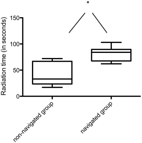
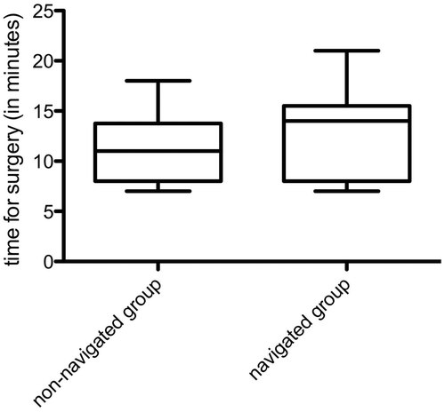
Results: The maximum distance of the K-wires from the center of the AC joint was 5.4 ± 1.1 mm for the freehand non-navigated group and 3.1 ± 1.6 mm for the navigated group (p = 0.0054). The minimum distance of the K-wires from the AC joint center was 3.0 ± 0.6 mm for the freehand group and 1.6 ± 0.6 mm for the navigated group (p = 0.0002). The radiation time was significant lower for the freehand group (41.25 ± 20.4 seconds versus 79.5 ± 13.3 seconds for the navigated group, p = 0.004). There was no statistical difference between the groups with respect to the time required for surgery (11.25 ± 3.6 min for the freehand group and 12.6 ± 4.6 min for the navigated group; p = 0.475). In the freehand group, the AC joint was penetrated by both K-wires in 87.5% of the procedures, compared to 100% in the navigated group. Both K-wires were placed completely intraosseously in the clavicula in 50% of the procedures in the freehand group, compared to 88% in the navigated group.
Conclusion: Three-dimensional navigation may improve the accuracy of AC joint transfixation techniques. However, the radiation time is increased when using the navigated procedure, while the overall operation time remains comparable. Nevertheless, a 3D C-arm with a variable isocentric design is recommended for the acquisition of the shoulder scans.
Introduction
Injuries of the acromioclavicular (AC) joint represent approximately 4% of all joint dislocations Citation[1], Citation[2]. Such injuries were classified by Tossy et al. in 1963 Citation[3], with the classification being updated by Rockwood and Young in 1991 Citation[4].
Until now, there have been no consistent treatment concepts reported in the literature for AC joint injuries Citation[2]. There is a consensus that conservative treatment is appropriate for Rockwood I and II injuries Citation[5–9], but the appropriate treatment for Rockwood III injuries remains controversial Citation[5], Citation[6], Citation[10]. While in the United States most Rockwood III injuries are treated conservatively Citation[11–15], in Germany the majority of patients with such injuries are subjected to surgery Citation[5], Citation[6]. Nevertheless, many authors agree that Rockwood IV–VI injuries should be treated operatively Citation[5], Citation[6].
A survey by Bäthis et al. Citation[5] of the preferred treatment options for AC joint injuries in Germany showed that 39% of such injuries were treated by AC joint transfixation, 22% with the use of hook plates Citation[16], and 9% with Bosworth screws Citation[17]. Another survey by Powers and Bach Citation[18] also found that 60% of the surgeons preferred transarticular stabilization of the AC joint as the main treatment option.
The most common complications after surgery are wound infections Citation[19], Citation[20], failure of the implant Citation[19], and iatrogeneous fractures of the clavicula or acromion Citation[21]. Particularly for transfixations of the AC joint, life-threatening dislocations and breakage of the implant have also been described Citation[22–24].
Over the past few years, three-dimensional (3D) fluoroscopy has been shown to improve the precision of surgical tool placement in a variety of surgical procedures, often in combination with computer navigation Citation[25–35]. Several studies have shown that the accuracy of procedures may be improved, and operative time, costs and the need for prolonged set-up reduced compared to conventional techniques or those using CT-based navigation Citation[25], Citation[32], Citation[36–39]. Additionally, CT-based navigation offers no information concerning the patient's position on the operating table.
Nevertheless, 3D scans of the shoulder region using C-arms with an isocentric design have been reported to be difficult, if not impossible, to perform due to the isocentric design Citation[31], Citation[40]. Therefore, a flat panel radiation detector (FD) 3D C-arm has been developed (Ziehm Vision FD Vario 3D, Ziehm Imaging Gmbh, Nuremberg, Germany) in order to overcome these known limitations. In comparison to conventional C-arm systems (for example, the Iso-C3D from Siemens Medical, Erlangen, Germany), the FD C-arm uses a variable isocentric design and the detector panel is much smaller. Image-quality output is 1024 × 1024 pixels for 2D and 512 × 512 pixels for the 3D model, compared to 256 × 256 pixels for the Siemens Iso-C3D. The FD C-arm employs pulsed fluoroscopy with an output power of 2000 W; the X-ray tube voltage is variable from 40 to 110 kV; and the X-ray current intensity ranges from 0.2 to 20 mA Citation[32].
So far, however, no information about the use of this technology for AC joint transfixations has been published. We hypothesized that 3D C-arm navigation would provide greater accuracy in K-wire positioning than the freehand technique, and conducted a human cadaveric study to evaluate the accuracy and feasibility of using a navigation system for minimally invasive procedures of this type.
Materials and methods
Five human cadavers (3 male, 2 female; median age 67 years) with no known deformities or previous injuries to the shoulder girdle were used for the present study. The cadaveric bodies were positioned in a beach-chair position on a radiolucent table. The shoulders were randomized for either the conventional technique with Kirschner-wire (K-wire) transfixation or the navigated procedure.
For the navigated procedure, the standard reference array (BrainLab VectorVision, Heimstetten, Germany) was positioned on the spina scapulae with two 3.0-mm Schanz screws pointed caudally, so as not to interfere with the K-wires for the transfixation ( shows the set-up for the navigated procedure). An optoelectronic camera was then positioned so as to be able to track both the C-arm and the reference array Citation[32]. A 3D C-arm (Ziehm Vision FD Vario 3D, Ziehm Imaging Gmbh, Nuremberg, Germany) was used initially to obtain 2D images to verify the correct positioning of the 3D C-arm.
Figure 1. Laboratory set-up. (a) Positioning on the radiolucent table with the flat detector 3D C-arm. The Kirschner wire is navigated via a navigated drill guide. The reference marker is attached to the spina scapulae. (b) Screenshot of a navigated AC joint transfixation. The upper left panel shows the aiming trajectory; the other panels show the planned trajectory.
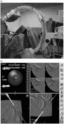
Once the set-up and registration process were completed, image acquisition was initiated by performing a 3D scan. The radiation source of the C-arm then produced pulsed fluoroscopy while the C-arm was moved automatically through a scan angle of 136°. The 3D reconstructed images were created on the screen and then transferred to the navigation system for further intervention and navigation with the use of a navigated drill guide (BrainLab VectorVision, Heimstetten, Germany) (see ).
For the freehand technique, the procedure was performed with the aid of intraoperative 2D fluoroscopy. In this procedure, two 2.0-mm K-wires should be placed centrally in the AC joint without perforation of the outer cortex of the clavicle Citation[2]. For the navigated technique, the K-wires were inserted through a navigated drill guide.
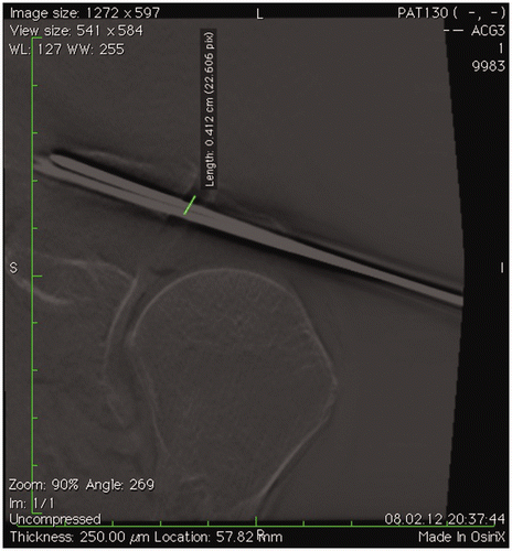
After implantation of the K-wires, another 3D scan was performed for documentation purposes and to permit further analysis. The radiologic analysis was performed using the OsiriX DICOM shareware viewer. Evidence of K-wire misplacement outside the AC joint was recorded, and the distance of the wire from the center of the AC joint determined, as well as any penetration of the cortex of the clavicle (see ).
Figure 2. Radiological analysis with the use of the OsiriX software. Multiplanar reformations are created, and the distance of the K-wires from the center of the AC joint is measured.
Statistical analysis
Student's t-tests were performed using GraphPad Prism (GraphPad Software, San Diego, CA). The significance level was set to 0.05.
Results
A total of 17 transfixation procedures (8 conventional, 9 navigated) were performed by an experienced orthopaedic surgeon (T.S.).
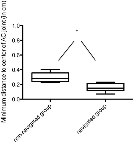
The maximum distance of the K-wire from the center of the AC joint was 5.4 ± 1.1 mm for the freehand group and 3.1 ± 1.6 mm for the navigated group (p = 0.0054) (). The minimum distance from the AC joint center was 3.0 ± 0.6 mm for the freehand group and 1.6 ± 0.6 mm for the navigated group (p = 0.0002) ().
Figure 3. Maximum distance of the K-wires from the center of the AC joint. Statistical analysis revealed significantly lower values in the navigated group.
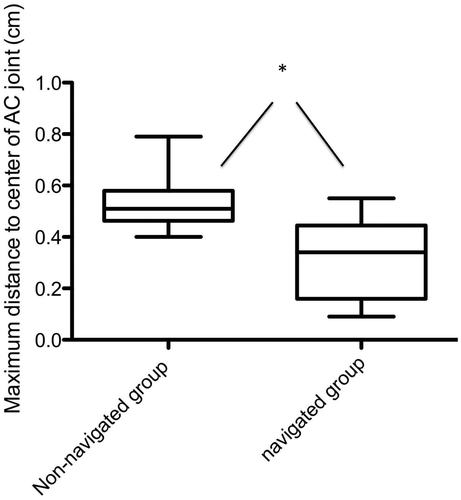
Figure 4. Minimum distance of the K-wires from the center of the AC joint. Statistical analysis revealed significantly lower values in the navigated group.
Total radiation time was significantly lower for the freehand group (41.25 ± 20.4 seconds versus 79.5 ± 13.3 seconds for the navigated group; p = 0.004) ().
Figure 5. Radiation time in the non-navigated and navigated groups. For the freehand group the radiation time covers only the intraoperative fluoroscopic imaging, while the significantly higher radiation time for the navigated group represents 2D fluoroscopic imaging along with the 3D scan.
There was no statistical difference between the two groups with respect to the overall time required for surgery (11.25 ± 3.6 min for the freehand group and 12.6 ± 4.6 min for the navigated group (p = 0.475) (). In the freehand group, both K-wires penetrated the AC joint in 87.5% of the procedures, compared to 100% in the navigated group. In the freehand group, both K-wires were placed completely intraosseously in the clavicula in 50% of the procedures, compared to 88% in the navigated group.
Figure 6. Surgery time in the non-navigated and navigated groups. The comparison revealed no statistical differences. The time for the navigated group included 60 seconds for the 3D scan and approximately 1 minute for the transfer of the 3D data from the 3D C-arm to the navigation system.
Discussion
In our cadaveric study, 3D C-arm navigation with a flat detector panel significantly improved the overall accuracy of AC joint transfixation compared to a freehand technique. The advantages of multiplanar visualization throughout transfixation, as displayed by the navigation system, made the procedure more manageable. Nevertheless, higher accuracy was achieved at the expense of longer radiation time due to the 3D scan, though the operation time remained comparable.
AC joint transfixation for Rockwood IV to VI injuries is an established technique Citation[7], Citation[41], Citation[42] and may be performed using a minimally invasive approach. Operative treatment for Rockwood III injuries should be reserved for young adults with a high demand for shoulder function Citation[43], Citation[44]. Such a procedure depends on the accuracy of the implant placement to avoid complications such as infections Citation[44], K-wire migration into the lung, heart or great vessels Citation[23], Citation[27], Citation[34], Citation[45–48], or breakage Citation[49], Citation[50] and secondary loss of reduction Citation[10], Citation[50].
Another possible complication is early acromioclavicular degeneration, which may not be caused by articular perforation of the K-wires as it has also been reported after conservative treatment and coracoclavicular fixation Citation[43], Citation[51].
Performing the transfixation procedure in a freehand manner may be easy, but the surgeon has to rely on tactile information and 2D fluoroscopic X-rays. In some cases, several attempts may be required to obtain satisfactory positioning of the K-wire; and in the clinical set-up, the clavicle must be maintained in the anatomical position by clamps or manual retention. Our results show that intraoperative 2D fluoroscopic visualization may not always be adequate, as both K-wires penetrated the AC joint in only 88% of the freehand group. Obtaining the lateral axillary view may be particularly demanding in the intraoperative set-up with a beach-chair table. The multiplanar reformations obtained by 3D scanning and presented by the navigation system in real time may be helpful for this procedure, as we noticed no failures to penetrate the AC joint in the navigated group. However, only 3D C-arm systems with a variable isocentric design can provide scans of the shoulder region Citation[40].
K-wire penetrations of the clavicular shaft were detected in both groups, but the incidence was significantly lower in the navigated group. K-wire penetration may be explained by the limited field of view of 3D C-arm systems in comparison to computer tomographs Citation[21], Citation[40]. In the current model, the 3D data cube is also limited to 13 × 13 × 13 cm, thus providing 1 cm more detail in each plane than a standard 3D C-arm system, with the peripheral K-wire position being sometimes not visible due to the limited data volume Citation[25].
As shown in our study, the accuracy of AC joint transfixation can be improved using 3D C-arm flat detector navigation. Accurate positioning of the K-wires, together with anatomical repositioning of the AC joint, is supposed to improve the biomechanical behavior of the repaired area. However, clinical data of this kind is not available in the literature and was not part of this study.
The present study had several limitations. First, the group size was rather small and inconsistent, with only 8 cadavers being used for the conventional approach and 9 for the navigated procedure. Second, the AC joint was not dissected prior to the procedure, leaving the ligaments intact for optimal accuracy analysis. Third, as already described above, the size of the data cube, which is provided by the flat detector 3D C-arm, is limited in comparison to a CT scan with variable data volume, and this may lead to misjudgment of implant position in a clinical setting.
Conclusion
Flat detector-based navigation seems to be promising for application in navigated AC joint transfixation. The accuracy of the procedure is improved, and while radiation time is increased, the overall time required for surgery remains comparable to that for the conventional technique. Misplacement of K-wires with respect to AC joint perforation occurred only in the freehand group, and not in the navigated group.
Declaration of interest: The authors report no declarations of interest.
References
- Riedl J, Genelin A. [Treatment of acromioclavicular dislocations by a pin and tension band fixation] [In German]. Unfallchirurgie 1991; 17(3)140–145
- Mayr E, Braun W, Eber E, Rüter A. [Treatment of acromioclavicular joint separations. Central Kirschner-wire and PDS-augmentation] [In German]. Unfallchirurg 1999; 102(4)278–286
- Tossy JD, Mead NC, Sigmond HM. Acromioclavicular separations: Useful and practical classification for treatment. Clin Orthop Relat Res 1963; 28: 111–119
- Rockwood CA, Young DC. Acromioclavicular injuries. Fractures in Adults3rd edn, CA Rockwood, Jr, DP Green, RW Bucholz. JB Lippincott, Philadelphia, PA 1991; 1: 1181–1239
- Bäthis H, Tingart M, Bouillon B, Tiling T. [The status of therapy of acromioclavicular joint injury. Results of a survey of trauma surgery clinics in Germany] [In German]. Unfallchirurg 2001; 104(10)955–960
- Prokop A, Helling HJ, Andermahr J, Mönig S, Rehm KE. [Tossy III injuries of the acromioclavicular joint. In what circumstances is surgery still justified? Personal results and literature review] [In German]. Orthopade 2003; 32(5)432–436
- Leidel BA, Braunstein V, Kirchhoff C, Pilotto S, Mutschler W, Biberthaler P. Consistency of long-term outcome of acute Rockwood grade III acromioclavicular joint separations after K-wire transfixation. J Trauma 2009; 66(6)1666–1671
- Guy DK, Wirth MA, Griffin JL, Rockwood CA, Jr. Reconstruction of chronic and complete dislocations of the acromioclavicular joint. Clin Orthop Relat Res 1998; 347: 138–149
- Lemos MJ. The evaluation and treatment of the injured acromioclavicular joint in athletes. Am J Sports Med 1998; 26(1)137–144
- Larsen E, Bjerg-Nielsen A, Christensen P. Conservative or surgical treatment of acromioclavicular dislocation. A prospective, controlled, randomized study. J Bone Joint Surg Am 1986; 68(4)552–555
- Bannister GC, Wallace WA, Stableforth PG, Hutson MA. The management of acute acromioclavicular dislocation. A randomised prospective controlled trial. J Bone Joint Surg Br 1989; 71(5)848–850
- Cox JS. Current method of treatment of acromioclavicular joint dislocations. Orthopedics 1992; 15(9)1041–1044
- Calvo E, Lopez-Franco M, Arribas IM. Clinical and radiologic outcomes of surgical and conservative treatment of type III acromioclavicular joint injury. J Shoulder Elbow Surg 2006; 15(3)300–305
- Fraser-Moodie JA, Shortt NL, Robinson CM. Injuries to the acromioclavicular joint. J Bone Joint Surg Br 2008; 90(6)697–707
- Nissen CW, Chatterjee A. Type III acromioclavicular separation: Results of a recent survey on its management. Am J Orthop (Belle Mead NJ) 2007; 36(2)89–93
- Wolter D, Eggers C. [Reposition and fixation of acromioclavicular luxation using a hooked plate] [In German]. Hefte Unfallheilkd 1984; 170: 80–86
- Bosworth BM. Acromioclavicular separation: New method of repair. Surg Gynecol Obstet 1941; 73: 866–887
- Powers JA, Bach PJ. Acromioclavicular separations. Closed or open treatment?. Clin Orthop Relat Res 1974; 104: 213–223
- Jerosch J. [The acromioclavicular joint] [In German]. Orthopade 2000; 29(10)895–908
- Mlasowsky B, Brenner P, Duben W, Heymann H. Repair of complete acromioclavicular dislocation (Tossy stage III) using Balser's hook plate combined with ligament sutures. Injury 1988; 19(4)227–232
- Wendl K, von Recum J, Wentzensen A, Grützner PA. [Iso-C(3D0-assisted) navigated implantation of pedicle screws in thoracic lumbar vertebrae]. Unfallchirurg 2003; 106(11)907–913
- Daud DF, de Campos MM. Migration of a Kirschner wire into the thoracic ascendent aorta artery. Rev Bras Cir Cardiovasc 2011; 26(3)508–510
- Sergides NN, Nikolopoulos DD, Yfadopoulos DK, Novi EA, Kanata MP. Intrathoracic migration of a Steinman wire: A case report and review of the literature. Cases J 2009; 2: 8321
- Nakayama M, Gika M, Fukuda H, Yamahata T, Aoki K, Shiba S, Eguchi K. Migration of a Kirschner wire from the clavicle into the intrathoracic trachea. Ann Thorac Surg 2009; 88(2)653–654
- Takao M, Yabuta K, Nishii T, Sakai T, Sugano N. Accuracy of a 3D fluoroscopic navigation system using a flat-panel detector-equipped C-arm. Comput Aided Surg 2011; 16(5)234–239
- Liu YJ, Tian W, Liu B, Li Q, Hu L, Li ZY, Yuan Q, Lu YW, Sun YZ. Comparison of the clinical accuracy of cervical (C2-C7) pedicle screw insertion assisted by fluoroscopy, computed tomography-based navigation, and intraoperative three-dimensional C-arm navigation. Chin Med J (Engl) 2010; 123(21)2995–2998
- Rajasekaran S, Karthik K, Chandra VR, Rajkumar N, Dheenadhayalan J. Role of intraoperative 3D C-arm-based navigation in percutaneous excision of osteoid osteoma of long bones in children. J Pediatr Orthop B 2010; 19(2)195–200
- Hott JS, Papadopoulos SM, Theodore N, Dickman CA, Sonntag VK. Intraoperative Iso-C C-arm navigation in cervical spinal surgery: Review of the first 52 cases. Spine (Phila Pa 1976) 2004; 29(24)2856–2860
- Grützner PA, Zheng G, Langlotz U, von Recum J, Nolte LP, Wentzensen A, Widmer KH, Wendl K. C-arm based navigation in total hip arthroplasty – background and clinical experience. Injury 2004; 35(Suppl 1)S-A90-S-A95
- Euler E, Heining S, Riquarts C, Mutschler W. C-arm-based three-dimensional navigation: A preliminary feasibility study. Comput Aided Surg 2003; 8(1)35–41
- Briem D, Ruecker AH, Neumann J, Gebauer M, Kendoff D, Gehrke T, Lehmann W, Schumacher U, Rueger JM, Grossterlinden LG. 3D fluoroscopic navigated reaming of the glenoid for total shoulder arthroplasty (TSA). Comput Aided Surg 2011; 16(2)93–99
- Citak M, Stübig T, Kendoff D, O’Loughlin PF, Hüfner T, Krettek C. Navigated minimally invasive thoracolumbar pedicle screw placement with flat panel 3-D imaging. A feasibility study. Technol Health Care 2010; 18(2)101–110
- Citak M, Kendoff D, Stübig T, Krettek C, Hüfner T. [Drilling with 3D fluoroscopic navigation in osteonecrosis of the femoral condyle] [In German]. Unfallchirurg 2008; 111(5)344–349
- Kendoff D, Citak M, Gaulke R, Gardner MJ, Geerling J, Krettek C, Hüfner T. [Navigation for placement of scaphoid screws: A new indication for intraoperative 3D navigation – a cadaver study] [In German]. Unfallchirurg 2007; 110(9)745–750
- Hüfner T, Geerling J, Gänsslen A, Kendoff D, Citak C, Grützner P, Krettek C. [Computer-assisted surgery for pelvic injuries] [In German]. Chirurg 2004; 75(10)961–966
- Schleicher I, Haselbacher M, Mayr E, Kaiser PM, Lenze FW, Keiler A, Nogler M. Accuracy of navigation in hip resurfacing with different surgeons and varying anatomy. Comput Aided Surg 2012; 17(2)77–85
- Zhang HL, Zhou DS, Jiang ZS. Analysis of accuracy of computer-assisted navigation in cervical pedicle screw installation. Orthop Surg 2011; 3(1)52–56
- Verborgt O, De Smedt T, Vanhees M, Clockaerts S, Parizel PM, Van Glabbeek F. Accuracy of placement of the glenoid component in reversed shoulder arthroplasty with and without navigation. J Shoulder Elbow Surg 2011; 20(1)21–26
- Arand M, Teller S, Gebhard F, Schultheiss M, Keppler P. [Clinical accuracy of fluoroscopic navigation at the thoracic and lumbar spine] [In German]. Zentralbl Chir 2008; 133(6)597–601
- Stübig T, Kendoff D, Citak M, Geerling J, Khalafi A, Krettek C, Hüfner T. Comparative study of different intraoperative 3-D image intensifiers in orthopedic trauma care. J Trauma 2009; 66(3)821–830
- Geaney LE, Miller MD, Ticker JB, Romeo AA, Guerra JJ, Bollier M, Arciero RA, DeBerardino TM, Mazzocca A. Management of the failed AC joint reconstruction: Causation and treatment. Sports Med Arthrosc 2010; 18(3)167–172
- Dumrongwanich P, Piyapittayanum P. Outcomes of percutaneous K-wire fixation for AC joint separation type III. J Med Assoc Thai 2009; 92( Suppl 6)S211–S216
- Lizaur A, Marco L, Cebrian R. Acute dislocation of the acromioclavicular joint. Traumatic anatomy and the importance of deltoid and trapezius. J Bone Joint Surg Br 1994; 76(4)602–606
- Leidel BA, Braunstein V, Pilotto S, Mutschler W, Kirchhoff C. Mid-term outcome comparing temporary K-wire fixation versus PDS augmentation of Rockwood grade III acromioclavicular joint separations. BMC Res Notes 2009; 2: 84
- Jerosch J, Filler T, Peuker E, Greig M, Siewering U. Which stabilization technique corrects anatomy best in patients with AC-separation? An experimental study. Knee Surg Sports Traumatol Arthrosc 1999; 7(6)365–372
- Sethi GK, Scott SM. Subclavian artery laceration due to migration of a Hagie pin. Surgery 1976; 80(5)644–646
- Lindsey RW, Gutowski WT. The migration of a broken pin following fixation of the acromioclavicular joint. A case report and review of the literature. Orthopedics 1986; 9(3)413–416
- Norrell H, Jr, Llewellyn RC. Migration of a threaded Steinmann pin from an acromioclavicular joint into the spinal canal. A case report. J Bone Joint Surg Am 1965; 47: 1024–1026
- Walz L, Salzmann GM, Fabbro T, Eichhorn S, Imhoff AB. The anatomic reconstruction of acromioclavicular joint dislocations using 2 TightRope devices: A biomechanical study. Am J Sports Med 2008; 36(12)2398–2406
- Kienast B, Thietje R, Queitsch C, Gille J, Schulz AP, Meiners J. Mid-term results after operative treatment of rockwood grade III–V acromioclavicular joint dislocations with an AC-hook-plate. Eur J Med Res 2011; 16(2)52–56
- Kawabe N, Watanabe R, Sato M. Treatment of complete acromioclavicular separation by coracoacromial ligament transfer. Clin Orthop Relat Res 1984; 185: 222–227