Abstract
Background: Previous work by our group to address the problem of acetabular positioning based on 2D methods resulted in the development of a measurement method with better precision – Liaw's version. This method may help the early diagnosis of acetabular loosening. In the present study, we hypothesized that our computerized ellipse method could improve the precision of measuring acetabular version.
Methods: We developed our Elliversion software to measure acetabular version. Using total hip replacement (THR) Simulator, 96 radiographs were synthesized with random femoral inclination and 5° to 52° version, half with the femoral head included and half without. These synthetic radiographs and 28 real radiographs were measured with both Elliversion and the trigonometric method twice by one of the authors with a one-week interval between measurements. We then calculated the difference in the repeated measurements. Student's t-test was used for statistical analysis of the measuring error and inter-measurement difference.
Results: In the precision study, for synthetic radiographs including the femoral head, the ellipse method was significantly better than the trigonometric method (p < 0.01). For synthetic radiographs without the femoral head, there was no significant difference between the ellipse method and the trigonometric method (p = 0.19). As for the repeated measurements, for synthetic radiographs including the femoral head, the ellipse method was significantly better than the trigonometric method (p = 0.001), whereas for synthetic radiographs without the femoral head, there was no significant difference between the two methods (p = 0.17). For real radiographs, there was no significant difference between the two measuring methods (p = 0.12). However, if we excluded the four poor-quality radiographs, there was a significant difference between the two measuring methods (p = 0.04).
Discussion: We developed a computerized ellipse method for measuring acetabular version on synthetic radiographs and good-quality real radiographs. This method is characterized by its superior precision as compared to the trigonometric method. With the 2D standardized method (Liaw's version), improving the precision of measurement will help earlier diagnosis of acetabular loosening.
Introduction
Measuring the version of the acetabular component after total hip arthroplasty (THA) is important because this parameter can affect the prognosis Citation[1]. Many methods for measuring version have been published to date. These can be categorized into three groups: the trigonometric method Citation[2–4], the protractor method Citation[1], Citation[5–9], and the computer tomography method Citation[10]. The first two groups are two-dimensional (2D) methods, while the last is a three-dimensional (3D) method.
The utility of the 2D methods is often questioned because of the positioning problem Citation[11]: The position may cause a difference of up to 13° Citation[11], thus decreasing the value of the methods. In contrast, the 3D method uses computer tomography (CT) and can solve the positioning problem, but the precision is poor because of the low resolution obtained with CT Citation[10].
In our previous work, we developed a mathematical equation to solve the positioning problem for 2D methods Citation[11]:where ssd is the distance in millimeters from the upper pole of the symphysis pubis to the sacrococcygeal junction; and h is the horizontal displacement in millimeters of the sacrococcygeal junction relative to the upper pole of the symphysis pubis in the horizontal direction. We assign h to be positive if the sacrococcygeal junction is between the acetabulum and the upper pole of the symphysis pubis; otherwise it is negative. v is the vertical displacement in millimeters of the sacrococcygeal junction relative to the upper pole of the symphysis pubis in the vertical direction. We assign v to be positive if the sacrococcygeal junction is above the upper pole of the symphysis pubis; otherwise it is negative. θ is the radiographic (planar) anteversion angle, and φ is the inclination (abduction) angle Citation[11].
The above equation requires the relative position of two bony landmarks, acetabular version and acetabular inclination. By improving the precision of measurement, we can improve the overall precision. If we can detect small changes in acetabular version, we can detect tiny cup movements earlier, making possible an early diagnosis of acetabular loosening.
Currently, there are several commercial products available for measuring acetabular version by drawing an ellipse to fit the outer edge of the cup. One example is OrthoView™ (OrthoView, Southampton, UK). However, these products are neither economical nor available everywhere. Moreover, there has been no precision study of these “ellipse” methods.
As such commercial products are not available at our hospital, we developed our own software for measuring acetabular version in order to conduct a precision study. We hypothesized that the digitalized method would improve the precision of measuring acetabular version, since the error and intra-observer difference may both be decreased with a computerized method.
Methods
In our previous work, we provided detailed definitions and described the measurement of acetabular version Citation[1]. In our opinion, radiographic (planar) version is suitable for measurement. Also, according to the equation of McLaren Citation[12],where the short and long axes are axes of the elliptical outline of the acetabular shell.
In most cases, the ellipse is obscured by the femoral head, making measurements difficult. However, if we are able to draw the entire ellipse for measurement, we can easily estimate the obscured portion of the ellipse, and can then improve our precision.
On the Microsoft Windows platform, we developed our Elliversion software to measure version. Our software can upload many image file formats, including BMP, JPG, DCM, and so on. The user can apply an ellipse to the acetabulum and then adjust the ellipse by altering the long and short axes.
As we were unable to draw the curves directly, we separated the ellipse into 64 parts. In other words, the ellipse was formed by 64 lines, and is drawn as a 64-sided polygon. Every endpoint of the lines was calculated by the position and length of both the long and short axes. The user can adjust the long and short axes to fit the ellipse formed by the outer shell of the acetabular component.
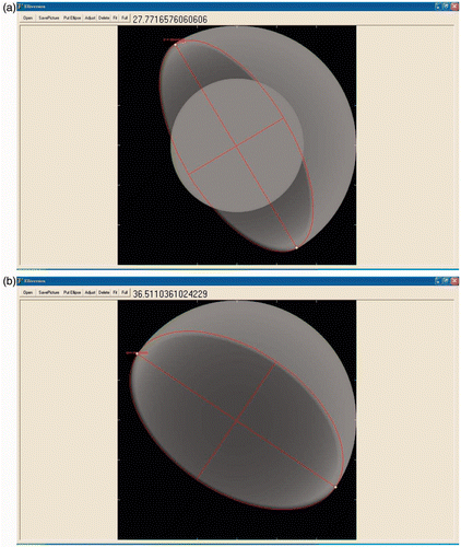
To verify the software, we synthesized 96 radiographs with random inclination and 5° to 52° version using total hip replacement (THR) Simulator Citation[13]. THR Simulator is software for synthesizing radiographs of THR with varying parameters, such as acetabular radiographic version, inclination, acetabular size, acetabular thickness, and head size. The software can be downloaded freely Citation[13]. In this study, half of the synthesized radiographs included the femoral head and half were without it ().
Figure 1. Synthetic radiographs with (a) and without (b) the femoral head. Acetabular version was measured using Elliversion. The user can read the version from the upper label.
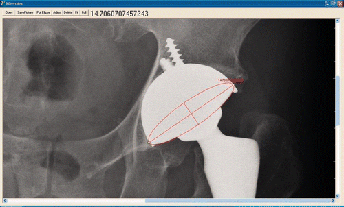
These synthetic radiographs and 28 real radiographs were each measured with both Elliversion and the trigonometric method. Measurements were performed twice by the same individual, with a one-week interval between measurements (). We then calculated the difference between the repeated measurements and the absolute error. If conditions were not well adjusted when taking the radiographs, we could only see the outer-lower part of the ellipse, with the inner-upper parts being invisible. This inability to see the entire ellipse may result in bigger differences between the measurements. We defined radiographs of this kind as poor-quality radiographs, and analyzed the data again after excluding the four poor-quality radiographs ().
Figure 2. Post-operative radiograph of a United right total hip arthroplasty. Acetabular version was measured using Elliversion.
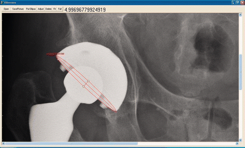
Figure 3. Post-operative radiograph of a United left total hip arthroplasty. Acetabular version was measured using Elliversion. In this case, half of the ellipse is not visible, and we could only assume the shape. This results in a bigger difference between the measurements. We defined radiographs of this kind as poor-quality radiographs.
Student's t-test was used for statistical analysis of the measuring error and inter-measurement difference.
Ethical approval
This study was approved by the Institutional Review Board of Tao-Yuan General Hospital (TYGH098019).
Results
presents the results of the precision study. For synthetic radiographs including the femoral head, the absolute error for the ellipse method ranged from 0° to 0.98°, with a mean ± SD of 0.28 ± 0.21°; for the trigonometric method, the range was 0° to 2.06°, and the mean was 0.73 ± 0.45°. There was a significant difference between the ellipse method and the trigonometric method in this group (p < 0.01). For synthetic radiographs without the femoral head, the absolute error of the ellipse method ranged from 0.01° to 1.24°, with a mean of 0.32 ± 0.21°; for the trigonometric method, the range was 0° to 1.02°, and the mean was 0.29 ± 0.19°. There was no significant difference between the ellipse method and the trigonometric method in this group (p = 0.19).
Table I. Absolute error of measurements for synthetic radiographs using the ellipse and trigonometric methods.
presents the results of the repeated measurement study. For synthetic radiographs including the femoral head, the absolute difference for the ellipse method between the two measurements ranged from 0.03° to 1.44°, with a mean of 0.33 ± 0.31°. For the trigonometric method, the absolute difference between the two measurements ranged from 0.06° to 2.19°, with a mean of 0.56 ± 0.43°. The absolute difference was significantly different between the two measurements (p = 0.001). For synthetic radiographs without the femoral head, the absolute difference for the ellipse method between the two measurements ranged from 0° to 0.88°, with a mean of 0.24 ± 0.19°. For the trigonometric method, the absolute difference between the two measurements ranged from 0.01° to 1.41°, and the mean was 0.26 ± 0.28°. There was no significant difference between the two measurements (p = 0.17). For real radiographs, the absolute difference between the two measurements using the ellipse method ranged from 0.01° to 1.89°, with a mean of 0.44 ± 0.43°. For the trigonometric method, the absolute difference between the two measurements ranged from 0.01° to 2.11°, and the mean was 0.58 ± 0.55°. There was no significant difference between the two measuring methods (p = 0.12). However, if we excluded the four plain radiographs of insufficient quality, there was a significant difference between the two measuring methods (p = 0.04).
Table II. Absolute difference for the repeated measurements.
Our results showed that the ellipse method had better precision than the trigonometric method for synthetic radiographs including the femoral head (p < 0.01). In synthetic radiographs without the femoral head, the accuracy of both methods showed no statistical difference (p = 0.19). The ellipse method also showed less inter-measurement difference on good-quality radiographs.
Discussion
Computer aided surgery has become increasingly prevalent in recent years. However, we believe that computer aided post-operative evaluation is as important as intra-operative introduction of artificial intelligence, if not more so, in improving clinical outcomes and long-term patient satisfaction.
The results of this study confirmed our hypothesis that the ellipse method offered better precision compared to the trigonometric method. In radiographs including the femoral head, the femoral head obscures the edge, and the ellipse method performed better in such cases. In radiographs without the femoral head, however, version could be measured directly using the trigonometric method, so the ellipse method offered no advantage.
For plain radiographs, there was no statistically significant difference between the two measuring methods, although there was a tendency for our ellipse method to have better precision. However, if we excluded the four plain radiographs with poor quality (), we could measure more accurately using our method (p = 0.04).
Some orthopaedic surgeons may question the value of precisely measuring the acetabular version. As mentioned earlier, if we have a precise method for measuring version, we may have a smaller least significant change for Liaw's version. We can detect tiny cup movements more easily, making earlier detection of cup loosening possible.
For precise measurement of a prosthesis, radiostereometric analysis (RSA) is a good option Citation[14], being able to achieve a precision of 30 µm. However, RSA requires the attachment of special tantalum markers to the prosthesis during the manufacturing process, and to the patient during the operation, which is impractical for everyday clinical use. RSA also requires special software which is expensive and not available in every country.
For digital radiograph systems, the vendors always offer orthopaedic suit software as an option for customers. This orthopaedic suit software usually provides the ellipse method for measuring acetabular version, as in the OrthoView™ software (OrthoView, Southampton, UK), and the ellipse method is thus readily accessible in most countries.
Conclusions
We developed our own ellipse method for measuring acetabular version. We have proven it to be more precise than the well-accepted trigonometric method for use with synthetic radiographs and good-quality plain radiographs. The method can be further used in measurements which require precision, such as determination of Liaw's version.
Declaration of interest
The authors thank United Orthopedic Corporation, Hsinchu, Taiwan, for providing us with postoperative radiographs of a U2 total hip arthroplasty. This study was supported by a grant from the National Science Council of Taiwan (NSC98-2314-B-087-001-MY2).
References
- Liaw CK, Hou SM, Yang RS, Wu TY, Fuh CS. A new tool for measuring cup orientation in total hip arthroplasties from plain radiographs. Clin Orthop Relat Res 2006; 18: 134–139
- Ackland MK, Bourne WB, Uhthoff HK. Anteversion of the acetabular cup. Measurement of angle after total hip replacement. J Bone Joint Surg Br 1986; 68(5–6)409–413
- Lewinnek GE, Lewis JL, Tarr R, Compere CL, Zimmerman JR. Dislocations after total hip-replacement arthroplasties. J Bone Joint Surg Am 1978; 60(2)217–220
- Pradhan R. Planar anteversion of the acetabular cup as determined from plain anteroposterior radiographs. J Bone Joint Surg Br 1999; 81(3)431–435
- Liaw CK, Yang RS, Hou SM, Wu TY, Fuh CS. Measurement of the acetabular cup anteversion on simulated radiographs. J Arthroplasty 2009; 24(3)468–474
- Fabeck L, Farrokh D, Tolley M, Descamps PY, Gebhart M, Delince P. A method to measure acetabular cup anteversion after total hip replacement. Acta Orthop Belg 1999; 65(4)485–491
- Widmer KH. A simplified method to determine acetabular cup anteversion from plain radiographs. J Arthroplasty 2004; 19(3)387–390
- Wan Z, Malik A, Jaramaz B, Chao L, Dorr LD. Imaging and navigation measurement of acetabular component position in THA. Clin Orthop Relat Res 2009; 467(1)32–42
- Liaw CK, Wu TY, Yang RS, Hsu YN, Wu TJ, Hou SM. Direct measurement of acetabular radiographic version using an ordinary goniometer: A precision study. Comput Aided Surg 2011; 16(4)196–201
- Olivecrona H, Weidenhielm L, Olivecrona L, Beckman MO, Stark A, Noz ME, Maguire GQ, Jr, Zeleznik MP, Svensson L, Jonson T. A new CT method for measuring cup orientation after total hip arthroplasty: A study of 10 patients. Acta Orthop Scand 2004; 75(3)252–260
- Liaw CK, Yang RS, Hou SM, Wu TY, Fuh CS. A simple mathematical standardized measurement of acetabulum anteversion after total hip arthroplasty. Computational and Mathematical Methods in Medicine 2008; 9(2)105–119
- McLaren RH. Prosthetic hip angulation. Radiology 1973; 107(3)705–706
- Wu TY, Yang RS, Fuh CS, Hou SM, Liaw CK. THR Simulator – the software for generating radiographs of THR prosthesis. BMC Musculoskelet Disord 2009; 10: 8
- Bragdon CR, Greene ME, Freiberg AA, Harris WH, Malchau H. Radiostereometric analysis comparison of wear of highly cross-linked polyethylene against 36- vs 28-mm femoral heads. J Arthroplasty 2007; 22(6 Suppl 2)125–129
Notice of Correction
The version of this article published online ahead of print on 25 Mar 2013 contained an error on page 1. The first author, Chen-Kun Liaw, should have been affiliatied with affiliations 1, 2 and 3 and the third affiliation should have read “Ming Chuan University, Tapei City, Taiwan”. The errors have been corrected for this version.