Abstract
Introduction: The aim of this study was to evaluate the association between frequency of the posterior cruciate ligament (PCL) mechanoreceptors and age in men.
Methods: Nineteen normal right knees harvested from human male cadavers were evaluated. Age ranged from 17 to 64 years with a mean of 35 years old. PCL was separated for sampling in femoral and tibial portions. Topographic distribution and frequency within the ligament texture were determined employing the Pro-Image digital analysis system. Mechanoreceptors were counted and classified according to the criteria proposed by Freeman & Wyke.
Results: A total of 1820 mechanoreceptors were found, Type II being the most frequent one. Analysis of the femoral portion of the ligament showed an equivalent predominance of Types II and IV mechanoreceptors. Tibial portion had a predominance of Type II mechanoreceptors, followed by Type IV. At this portion, receptors Types I and III were less commonly identified.
Conclusion: In the tibial portion of the PCL, there is predominance of Type II mechanoreceptors followed by Types IV, I and III mechanoreceptors, respectively. No relationship was found between the total number of mechanoreceptors and age in the femoral and tibial portions of the PCL.
Introduction
There are numerous studies linking the proprioceptive role of the posterior cruciate ligament (PCL) as a determinant for the use of knee prosthesis, and there are others that show comparable results using prosthesis models [Citation1–4]. Balancing and determining its proper tension becomes critical to good joint kinematics improvement [Citation5]. A previous study reported that patients undergoing total knee arthroplasty (TKA) with preservation of the PCL have greater range of motion, higher quadriceps strength, increasing stability and reducing stress at the interface bone-cement-prosthesis and inferring decrease in the wear of polyethylene [Citation1,Citation6]. Moreover, Stern & Insall [Citation4] reported 90.8% of non-surgical revision after 21 years undergoing TKA sacrificing PCL.
Based on this controversy, Del Valle et al. [Citation7] histologically analyzed the PCL in arthritic knees compared to normal knees in order to study the role of innervation, specifically mechanoreceptors and if these corpuscles are involved in neural degenerative process of osteoarthrosis [Citation7].
The mechanoreceptors are specialized nerve structures that transmit mechanical deformation through electrical signals to trigger the potential of a nerve, informing the positioning and motion. It acts as a trigger or alert, sending impulses to the central nervous system that triggers reflex mechanisms, which act as protection for the joint, preventing the inadequate movements and sensitizing the spatial orientation of the individual and activating the dynamic stabilizers muscle [Citation8–16].
Our group develops a line of research that investigates the joint proprioception through mechanoreceptors. Oliveira et al. [Citation17,Citation18] studied the nerve endings in the intervertebral discs of the human lumbar spine and Ejnisman et al. [Citation19,Citation20] evaluated the mechanoreceptors present in the inferior glenohumeral ligament. The development of this research motivated the study of mechanoreceptors in the PCL of normal human cadaveric knees using histological technique immunohistochemistry.
In this study, we aimed to study the mechanoreceptors of the PCL in normal human male cadaveric knees and analyze the distribution of different types according to age.
Methods
Material
We dissected 19 knees, the right side of fresh human cadavers from the Death Verification Service of our Institution. All procedures were approved by the Ethics Committee of the Universidade Federal de São Paulo (Protocol number 161/01).
It was used as an exclusion criterion knee with apparent external injury, as well as those that presented macroscopic changes of joint degeneration, using only the right side of male cadaver knees. The bodies were identified by age and color.
Inclusion criteria included human male cadavers with no history of orthopedic disorders that impairs the knee biomechanics.
Dissection technique
Through a median incision above the knee about 20 cm, addressing skin subcutaneous tissue performed the opening of the extensor apparatus in the medial and lateral dislocation of the patella with wide exposure of the femoral–tibial joint. We performed dissection and removal of both menisci and the identification of the anterior and PCLs. After removal of the PCL, we cleaned the adjacent synovial tissue and identified the femoral origin with a point of suture with Mononylon 5.0. The ligaments were placed in a container with a solution of 10% formalin and sent to the laboratory, where in a period of 48 h it was divided into PCL femoral and ligament cross-posterior tibial portion (). These two fragments were embedded in paraffin block properly identified and cataloged in a book of research in the Department of Pathology of our Institution.
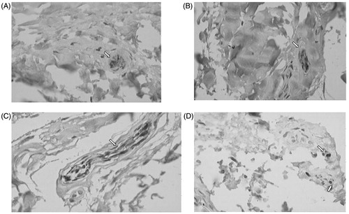
Figure 1. Type I mechanoreceptor (A), Type II mechanoreceptor (B), Type III mechanoreceptor (C) and Type IV mechanoreceptor (D). S100 protein (400×).
Methods immunohistochemistry
After fixation, the pieces were dehydrated in increasing concentrations of alcohol cleared in xylene and embedded in paraffin. Histological sections were made from paraffin block, with a maximum thickness of 4 μm in slides previously salinized (3-inopropylthriethoxsilane Sigma Chemical CO., Carpinteria, CA, code A3648). The slides, left in an oven at 60 °C for 24 h to increase adhesion of the sections were stained by immunohistochemistry of streptavidin-biotin-peroxidase for the polyclonal anti-S-100 protein (Dako, Carpinteria, CA, code Z0311, lot 026 1/5.000 dilution) [Citation19,Citation20].
We proceeded to count the nerve endings throughout the length of the PCL fragments, stained by immunohistochemistry, interactively with 400× magnification, with the aid of digital system Image-Pro image analysis, which consists of a video camera model TK-1180V, which transmits the captured image of the Olympus microscope (Model BX40 with plan-achromatic objective) to a Pentium MMX 233 MHz computer with 63 megabytes of RAM, equipped with a digitizer board and software Pro-image, working in a Windows environment (). The slides were evaluated in order to quantify the presence of mechanoreceptors found in portions of the PCL tibial and femoral and classify them according to the classification of Freeman & Wyke [Citation15].
Statistical analysis
The qualitative and quantitative variables were represented by absolute (n) and relative (%) numbers and continuous quantitative variables were presented as mean, standard deviation (SD), median, minimum and maximum values. The comparison between the four types of mechanoreceptors was made by the Friedman test for related samples. When the test result was statistically significant, the differences were identified by multiple comparison test of Newman–Keuls. The Wilcoxon test was applied for comparison between non-parametric paired samples. The presence of correlation between age and numbers of mechanoreceptors found was evaluated by the non-parametric Spearman correlation test. We adopted a significance level for p < 0.05 (α < 5%). We used the Software GraphPad StatMate version 2.00 (Windows, GraphPad Software, San Diego, CA, USA).
Results
The average age of the cadavers was 35 years old (SD = 16), ranging from 17 to 64 years old.
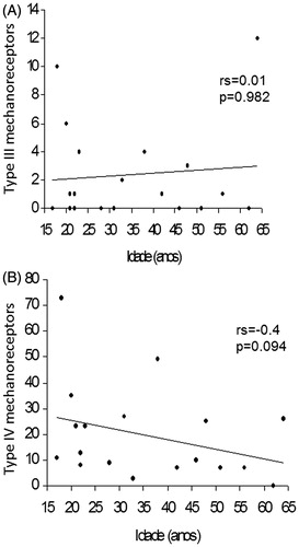
There was no statistically significant correlation between age and the number of Type I and Type II mechanoreceptors in the femoral portion of the PCL (). We also observed no statistically significant correlation between age and the number of Type III and Type IV mechanoreceptors in the femoral portion of the PCL ().
Figure 2. Correlation between age and the number of Type I (A) and Type II (B) mechanoreceptors in the femoral portion of the PCL.
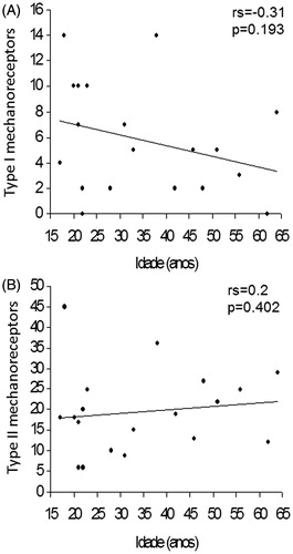
Figure 3. Correlation between age and the number of Type III (A) and Type IV (B) mechanoreceptors in the femoral portion of the PCL.
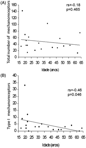
According to we observed no statistically significant correlation between age and the total number of mechanoreceptors in the femoral portion of the PCL. On the other hand, we found a statistically significant inverse correlation between age and the number of Type I mechanoreceptors in the tibial portion of the PCL (), indicating that the higher the age, the lower the number of Type I mechanoreceptors.
Figure 4. Correlation between age and the total number of mechanoreceptors in the femoral portion of the PCL (A) and correlation between age and the number of Type I mechanoreceptors (B).
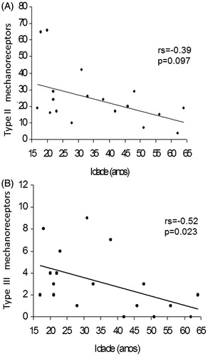
There was no statistically significant correlation between age and the number of Type II mechanoreceptors in the tibial portion of the PCL. Nevertheless, we found a statistically significant correlation between age and the number of Type III mechanoreceptors in the tibial portion of the PCL (), indicating that the higher the age, the lower the number of Type III mechanoreceptors.
Figure 5. Correlation between age and the number of Type II (A) and Type III (B) mechanoreceptors in the tibial portion of the PCL.
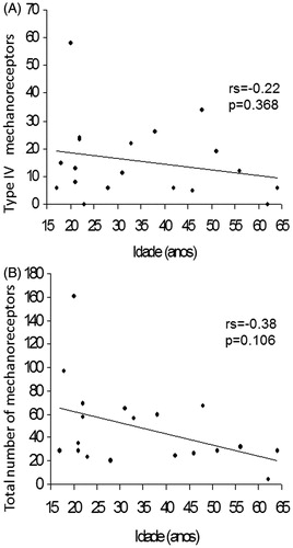
There was no statistically significant correlation between age and the number of Type IV mechanoreceptors and the total number of mechanoreceptors in the tibial portion of the PCL ().
Figure 6. Correlation between age and the number of Type IV mechanoreceptors (A) and correlation between age and the total number of mechanoreceptors (B) in the tibial portion of the PCL (B).
We found statistically significant differences between the types of mechanoreceptors (p < 0.001). The multiple comparison test showed that the Type III mechanoreceptors proved to be the least frequent and different from the others. Type I was more common than Type III and less frequent than the others while the Types II and IV did not differ ().
Table 1. Descriptive measures the number of Types I, II, III and IV mechanoreceptors of the femoral portion of the PCL.
We found statistically significant differences between the types of mechanoreceptors (p < 0.001). The multiple comparison test showed that Types I and III proved to be the least common, similar and different from the others. Type IV was more frequent than Types I and III and less frequent than Type II, Type II proved to be the most frequent ().
Table 2. Descriptive measures regarding Types I, II, III and IV mechanoreceptors of the tibial portion of the PCL.
We found no difference between the femoral and tibial portions regarding the total number of mechanoreceptors (p = 0.546) ().
Table 3. Descriptive measures the total number of mechanoreceptors of the femoral and tibial portions of the PCL of 19 knees.
Discussion
The main results of our study are the predominance of Types II and IV mechanoreceptors while Types I and III mechanoreceptors are less frequent in the femoral portion of the PCL, while in the tibial portion of the PCL there is an increased number of Type II mechanoreceptors followed by Types IV, I and III mechanoreceptors, respectively. The correlation between the presence of proprioceptive mechanoreceptors in the joint tissues (spine, shoulder, knee, ankle) has been studied extensively in animals and in humans [Citation1,Citation7,Citation9–11,Citation13–23]. We decided to focus attention on the relationship due to the PCL, always controversial regarding its replacement or not when choosing the type of total knee prosthesis to be implanted. Pagnano et al. [Citation5] published a review article on the role of the PCL in TKA relating it to various items, such as kinematics, wear, range of motion, stability and gait analysis. Current studies do not present necessary support to state that there is benefit from the model that preserves the PCL. Becker et al. [Citation24] conducted a comparative study in patients who underwent TKA in both knees with different models over the PCL and found no statistically significant difference in relation to functional outcome, given that 50% of patients could not know what was the best side and the other remaining equally divided on the preference.
The choice of S-100 protein specific dye as the neural structures was based on the studies published by Del Valle et al. [Citation7], Oliveira et al. [Citation17,Citation18] and Ejnisman et al. [Citation19,Citation20]. The tissue was performed with the aim of rescuing the image of all mechanoreceptors found, to measure its length and width and sort them according to size and morphology, according to the classification of Freeman & Wyke [Citation15]. This process was facilitated by the system of digital image analysis used. Most authors who have studied the mechanoreceptors hydrochloride used gold as a histological stain [Citation13–16,Citation25,Citation26]. Del Valle et al. [Citation15] demonstrated that immunohistochemical techniques are more sensitive and specific to the mechanoreceptors.
The results are not comparable to literature works because none of them made the count and differentiation of types of mechanoreceptors with age, this technique uses immunohistochemistry. In this context, Katonis et al. [Citation16] found mechanoreceptors of Types I, II and IV in the PCL of normal cadavers without quantifying or relating to age. The authors did not clarify the reason for the absence of Type III mechanoreceptors, perhaps not justified by histological technique as specific as the protein S-100. Del Valle et al. [Citation7] studied this technique with immunohistochemical analysis, the PCL knees of normal and osteoarthritis patients around 60 years old were described regarding the types of mechanoreceptors in a different way by proposing its own classification.
The predominance of Type II mechanoreceptor in our series reported in both the femoral and tibial portion was also observed in the anterior cruciate ligament, studied by Schutte [Citation25]. This type, also known as Paccini corpuscles, acts as a dynamic mechanoreceptor, being inactive in the joint property and assets in motion acceleration and deceleration. It presents the characteristic of quick adaptation and a low profile of response to mechanical stresses [Citation10,Citation11,Citation15,Citation27,Citation28]. Type IV mechanoreceptor, the second group in frequency in our study, is the articular nociceptive system. It is inactive during normal circumstances and becomes active when the joint is subjected to mechanical deformation under the effect of abnormal or inflammatory mediators, such as histamine, bradykinin and prostaglandins [Citation15,Citation29].
We observed a frequency close to blood vessels inferring a vasomotor function [Citation9,Citation25,Citation28]. The mechanoreceptors of Types I and III (also known as Ruffini and Golgi corpuscles, respectively), responsible for slow adaptation and low mechanical profile, were found in smaller quantities in human PCL. Previous studies [Citation7,Citation16,Citation30] found no Type III corpuscles, probably by using the first technique that is a less specific histological and immunohistochemical analysis compared with our study where the objective was to study the entire ligament.
Our results showed a statistical tendency of a decrease in the human PCL mechanoreceptors with increasing age in normal knees. However, we encourage future studies with a higher population to confirm this fact raised in our study. To infer that the PCL in elderly patients maintain its proprioceptive role a larger sample is needed. Moreover, continuing in this line of research it would be interesting to conduct a comparative study of mechanoreceptors in the PCL checking for deterioration or not the amount and distribution of types of mechanoreceptors, altering or interfering with the mechanism of the knee proprioception.
We observed negative results regarding the relationship between the mechanoreceptors with age in male. This is possible due to the low media found between the studied samples. We encourage further studies to better investigate this result.
Our study is the first to characterize the association between mechanoreceptors and age in men. No work was found in the literature data demonstrating a comparative study regarding sex or affected side. Regarding the material, we decided to use only human male fresh cadaveric knees (<24 h post-mortem), in order to decrease the bias of the survey (want to reduce bias in selection of cases).
Conclusion
There was no statistically significant difference between the femoral and tibial portions of the PCL regarding the total number of mechanoreceptors.
In the femoral portion of the PCL there is predominance of Types II and IV mechanoreceptors while Types I and III mechanoreceptors are less frequent.
In the tibial portion of the PCL there is predominance of Type II mechanoreceptors followed by Types IV, I and III mechanoreceptors, respectively.
No relationship was found between the total number of mechanoreceptors and age in the femoral and tibial portions of the PCL.
Declaration of interest
The authors report no conflicts of interest. The authors alone are responsible for the content and writing of this article.
References
- Dorr LD, Scott RD, Ranawat CS. Controversies of total knee arthroplasty. Part I. Importance of posterior cruciate ligament. In: Ranawat CS, ed. Total condylar knee arthroplasty. New York (NY): Springer-Verlag; 1985:197--201
- Warren PJ, Olanlokun TK, Cobb AG, Bentley G. Proprioception after knee arthroplasty – The influence of prosthetic design. Clin Orthop 1993;297:182–7
- Scott RD, Thornhill TS. Posterior cruciate supplementing total knee replacement using conforming inserts and cruciate recession: effect on range of motion and radiolucent lines. Clin Orthop 1994;309:146–9
- Stern SH, Insall JN. Total knee arthroplasty with posterior cruciate ligament substitution designs. Surgery of the knee. 2nd ed. New York (NY): Churchill Livingstone; 1994:829–67
- Pagnano MW, Cushner FD, Scott WN. Role of the posterior cruciate ligament in total knee arthroplasty. J Am Acad Orthop Surg 1998;6:176–87
- Andriacchi TP, Galante JO, Fermier RF. The influence of total knee-replacement design on walking and stair climbing. J Bone Joint Surg 1982;64-A:1328--35
- Del Valle ME, Harwin SF, Maestro A. Immunohistochemical analysis of mechanoreceptors in the human posterior cruciate ligament. J Arthroplasty 1998;13:916–22
- Polacek P. Receptors of the joints. Their structure, variability and classification [TESE]. Brno: Leharska Fakulta University; 1966
- Schultz RA, Miller DC, Kerr CS, Micheli L. Mechanoreceptors in human cruciate ligaments. J Bone Joint Surg 1984;66:1072–6
- Zimny ML, Schutte M, Dabezies E. Mechanoreceptors in the human anterior cruciate ligament. Anat Rec 1986;214:204–9
- Zimny ML. Mechanoreceptors in articular tissues. Am J Anat 1988;182:204--9
- Alexiades M, Scuderi G, Vigorito V, Scott WN. A histological study of the posterior cruciate ligament in the arthritic knee. Am J Knee Surg 1989;2:916--22
- McLain RF. Mechanoreceptor in human cervical facets joints. Spine 1994;19:495–501
- Michelson JD, Huntchins C. Mechanoreceptor in human ankle ligaments. J Bone Joint Surg 1995;77-B:219–24
- Freeman MAR, Wyke B. The innervation of the knee joint. An anatomical and histological study in cat. J Anat 1967;101:505–32
- Katonis P, Assimakopoulos A, Agapitos M, Exarchou E. Mechanoreceptors in the posterior cruciate ligament: a histological study on the cadaver knee. Acta Orthop Scand 1991;62:276–8
- Oliveira VM. Estudo das terminações nervosas dos discos intervertebrais da coluna lombar em humanos [Thesis]. São Paulo: Universidade Federal de São Paulo – Escola Paulista de Medicina; 2001
- Oliveira VM, Puertas EB, Alves MTS, et al. Study of nerve endings in the intervertebral discs of the lumbar spine in humans. Rev Bras Ortop 2002;37:195–200
- Ejnisman B. Immunohistochemical study of the inferior glenohumeral ligament mechanoreceptors in human cadavers [Thesis]. São Paulo: Universidade Federal de São Paulo – Escola Paulista de Medicina; 2001
- Ejnisman B, Faloppa F, Carrera EF, et al. Immunohistochemical study of the inferior glenohumeral ligament mechanoreceptors in human cadavers. Rev Bras Ortop 2002;37:289–98
- Kennedy JC, Alexander IJ, Hayes KC. Nerve supply of the human knee and its functional importance. Am J Sports Med 1982;10:329–35
- Hogervorst T, Brand RA. Current concepts review. Mechanoreceptors in joint function. J Bone Joint Surg 1998;80-A:1365–78
- Granata Jr GSM. Study of the innervation of the human meniscus [Thesis]. São Paulo: Universidade Federal de São Paulo – Escola Paulista de Medicina; 2003
- Becker MW, Insall JN, Faris PM. Bilateral total knee arthroplasty: one cruciate retaining and one cruciate substituting. Clin Orthop 1991;271:122–4
- Schutte MJ, Dabezies EJ, Zimny ML, Happel LT. Neural anatomy of the human anterior cruciate ligament. J Bone Joint Surg 1987;69-A:243–7
- Kleinbart FA, Bryk E, Evangelista J, et al. Histologic comparison of posterior cruciate ligaments from arthritic and age-matched knee specimens. J Arthroplasty 1996;11:726–31
- Skoglund, S. Anatomical and physiological studies of knee joint innervation in the cat. Acta Physiol Scand 1956;36:1--101
- Johansson H, Sjolander P, Sojka P. A sensory role for the cruciate ligaments. Clin Orthop 1991;268:161--78
- Grigg P, Schaible HG, Schmidt RF. Mechanical sensitivity of group II and IV afferents from posterior articular nerve in normal and inflamed cat knee. J Neurophysiol 1986;55:635--43
- Raunest J, Sager M, Burgener E. Proprioception of the cruciate ligaments: receptor mapping in an animal model. Arch Orthop Trauma Surg 1998;118:159–63