Abstract
Objectives: To evaluate the current practice in the approach to the painful hip in children. To determine the superiority of ultrasonographic techniques over plain X-ray and to provide guidance to primary care. Methods: We carried out a prospective cohort study of 55 patients with a painful hip. The management leading up to admission and diagnostic procedures were examined. We carried out ultrasound of all affected hips and repeated the scan every three days. Effusion on ultrasound was noted and was correlated to the symptoms. Results: In total 29/55 patients had plain X-rays taken before or at the initial presentation. No abnormalities were found. Ultrasound showed effusion in 48/55 children. The reduction of the effusion correlated with the improvement in symptoms. An effusion persisting longer than 26 days correlated well to those patients who went on to be diagnosed with a chronic condition (3/4 patients).
Conclusions: At present many practitioners use plain X-ray in the initial diagnosis. Ultrasound should be the initial diagnostic tool of choice. Repeat ultrasound at one month may be of use to determine if the child is likely to develop a chronic condition.
Keywords::
Introduction
The incidence of a limp in children has been reported as 1.8 in 1000 children (Citation1) and is a common symptom seen in the primary care setting. It is more common in males and has a mean presentation age of four to five years. Transient synovitis of the hip is the most common cause of hip pain in the child, with an incidence of 1.1 cases per 1000 children (Citation1–3). It is thought the real numbers may be higher as many cases of limping children are not given a final diagnosis (Citation4,Citation5).
The differential diagnosis includes septic arthritis, Perthes' disease, slipped upper femoral epiphysis (SUFE), juvenile rheumatoid arthritis (JRA), fracture and malignancy. The early diagnosis of the painful hip is often difficult but essential (Citation6,Citation7). A careful history and examination should be performed. There are various clinical pointers which should be used, for example, a SUFE tends to present in the overweight child over 10 years of age, sepsis or tumour may present with pyrexia and general malaise and juvenile rheumatoid arthritis may present with multiple joints involved.
When sepsis is present inflammatory markers are usually raised and the patient appears unwell, however in most other cases of a painful hip and even in some septic cases, laboratory findings can be non specific. Plain X-ray usually appears normal and in young children the characteristics of pain can be unclear. At present there are still different approaches to the early diagnosis (Citation9).
It is imperative to confirm or rule out sepsis in the early stages as this poses an immediate risk to the patient's life, and delays to joint washout can cause irreparable damage to articular cartilage leading to considerable morbidity.
Septic arthritis is relatively common in infancy and childhood, to the extent that all primary care physicians caring for children can expect to encounter this disease at some point; however, the incidence has fallen due to H. influenza vaccination (Citation1,Citation3,Citation8). Some MRI studies have shown that septic arthritis is more often associated with an underlying osteomyelitis.
We assessed the techniques employed in our local area in the initial assessment of the painful hip in children carried out in a primary care and primary care paediatric practices. In Poland the primary care system is formed by a two tier system where an initial general practitioner will refer first to a specialist musculoskeletal primary care practitioner. They will then refer the patient to hospital if needed.
Our objective was to describe the value of the use ultrasound in the painful hip, to show its superiority over X-ray and to provide clinical care plans which can be used both in primary care and hospital settings.
Materials and methods
We carried out a prospective descriptive cohort study with a five year follow-up. Local ethics approval was obtained.
Patients
We prospectively collected data from 55 children aged 2 to 12 (mean 4.5) who were referred to our hospital unit between November 2000 and May 2001 (Orthopaedic and Paediatric Rheumatology departments, Medical Academy Bydgoszcz). Patient selection took no account of demographics (age, weight, height, gender, etc).
Only patients with no clear history of injury were included in this study. Any patients with previous hip problems including Perthes' disease, JRA and sepsis were also excluded. All patients complained of hip pain, knee pain and/or limping. These symptoms presented between three and eight days prior to hospital admission.
Measurements
Clinical examination and routine blood tests with inflammatory markers (white cell count, C-reactive protein) were performed. Bed rest and skin traction were applied for pain relief and appropriate analgesics were given.
On the first day of admission, ultrasound scans were performed on both hip joints in all 55 patients. Ultrasound examination was repeated every three days. The examination was performed by one of the authors (SJ) as described by Terjesen et al. (Citation10). We used 5MHz Aloka or Siemens. The femoral head and neck and the joint capsule were visualised. The distance of the capsule to the femoral neck was measured. A difference of two or more millimetres between the painful and the asymptomatic joint was diagnosed as effusion.
In 7 children with suspicion of septic arthritis, (swinging pyrexia, high pain reaction on movement and joint effusion seen on ultrasound) diagnostic joint aspiration was performed and intravenous broad spectrum antibiotics were given.
A diagnosis of transient synovitis was given based on clinical findings, normal laboratory findings and ultrasound signs of joint effusion, after excluding septic arthritis.
Results
X-rays before or at admission
22 children had plain X-ray films taken prior to admission in the GP paediatric practice. Of these, 18 were AP hip projections only, while in 4 other cases an additional axial hip view was taken. In addition, 2 patients had knee X-rays taken. Finally, 5 children had their first X-ray taken at admission. This shows that in total 29 of 55 patients had plain X-rays during the initial presentation. None of the X-ray films taken at the initial admission revealed any abnormality.
Repeated ultrasound
In 48 of 55 patients the initial ultrasound examination revealed a capsular distension as sign of effusion in the symptomatic hip. The mean difference in capsule-neck distance was 5.5 mm (range: 4.0–10.0 mm). As the clinical picture improved, ultrasound evidence of capsular distension subsided on the repeat scans performed at three day intervals.
At seven days the distension had disappeared in six patients. Distension was visible between 8 and 26 days in 42 patients. In 4 patients, ultrasound signs of capsular distension persisted over 26 days. Among these patients, two cases of Perthes' disease and one juvenile rheumatoid arthritis were diagnosed.
In seven patients we found no ultrasound evidence of effusion. In the results of the ultrasound examinations are summarized.
Table I. Results of ultrasound study.
Aspiration
A diagnostic aspiration of the hip joint was performed in seven children with suspicion of septic arthritis, including joint effusion seen on ultrasound. In all patients there were no microbiological signs of inflammation and the culture was negative. No child required formal washout of the hip joint.
In one patient of this subgroup the clinical symptoms subsided and ultrasound imaging was normal at three-month follow-up. In other patients with positive effusion, five-year follow-up showed no complications.
Final diagnoses
In 7 children without ultrasound evidence of effusion, other causes of hip pain and limping were excluded. In these patients symptoms resolved in seven days and there were no complications in the five-year follow-up. These patients were given a diagnosis of non specific hip pain. Perthes’ disease was diagnosed using plain X-ray (done four to seven weeks after initial presentation) and JRA diagnosed clinically and with the presence of increased rheumatoid markers. Overall, transient synovitis was the most common established diagnosis. summarizes the final diagnoses of our study.
Table II. Final diagnosis.
Discussion
Key findings
More than 50% of the children in our study had plain X-rays taken as part of the initial workup. None revealed any abnormality. Repeated ultrasound proved to be a more useful tool in the initial diagnosis of children with hip pain. Transient synovitis was the most common cause of hip pain in children. In 7 children sepsis could be ruled out by joint aspiration. In 3/4 patients with persisting effusion after one month, a chronic cause was present, as was demonstrated by appropriate blood tests and imaging.
Strengths and limitations
This prospective study explored a patient population that would be relevant to most GP, paediatric and orthopaedic practices. As we examined a group of patients admitted with the problem, the population may be slightly different to that presenting initially to their GP. Many experienced GPs may feel confident enough not to refer patients without fever and only mild hip complaints. We expect those patients who are not referred will be relatively well children with short-lived symptoms (which may indeed increase the overall prevalence of transient synovitis and other non specific problems). However, the message of the study remains the same.
The use of plain X-rays
Pain is thought to be most commonly caused by capsular distension due to intravascular synovial effusion. Plain X-ray cannot visualize the soft tissue structures which are most commonly affected and the value of using X-ray for initial imaging is very limited (Citation13). We argue that X-ray imaging should not be the initial investigation of choice.
40% of the children in our study had a plain X-ray prior to admission without any clear reason. All these examinations showed no abnormality. This high rate of unnecessary investigation may be explained twofold.
First, general and paediatric practitioners in our area may be unaware of the superiority of other diagnostic techniques. Second, in very young children it may be difficult to rule out a history of trauma completely (where an X-ray is indicated).
We observed that two patients had an X-ray of the knee. This reminds us again that in children, hip problems may present with knee pain, and a thorough physical examination is required before any investigations.
Ultrasound
Both MRI and ultrasound will nicely visualise the soft tissue structures of the hip and any effusion (Citation10,Citation14,Citation15). Ultrasound is generally felt to be superior even though MRI is better at differentiating the underlying causes of the effusion. This is due to the fact that ultrasound is easy and quick to perform; and that it can be performed without any anaesthesia (which is commonly required for children in the MRI scanner) (Citation16). Ultrasound can also be easily used to monitor the effusion and its subsidence (Citation10).
We followed patients up for five years as part of the study, and while this length of follow up is not necessarily required in a primary care setting, certainly repeat ultrasound at perhaps one month or later might be useful to assess for persistent capsular distension, which we have shown may indicate a chronic problem.
Prognosis of transient synovitis
Our study confirmed transient synovitis as the commonest cause of hip pain in children (5). Transient synovitis is a self limiting disorder with no long term sequelae.
Briggs et al. traced 286 patients with a diagnosis of transient synovitis and in only one patient did the symptoms persist with an eventual diagnosis of Perthes' disease (Citation17). We agree with Mumme that only a very small number of patients with this diagnosis will eventually be re-diagnosed with a more chronic condition (Citation18).
However, Mallet et al. report almost a 33% rate of minor clinical and X-ray Perthes' like changes with longer follow up. Our patient population was comparable and did not show this (Citation19). Some reports suggest that diagnosis of transient synovitis in older children should be followed up with MRI to exclude Perthes' as pure transient synovitis is less common in this age group (Citation18). However, our five years of follow-up only revealed only 3 cases of chronic conditions, all of which were seen to have persistence effusion of the capsule. Monitoring was performed using ultrasound only and no cases were missed.
Sepsis
Sepsis is crucial to rule out but much less common then transient synovitis. Due to the number of children in this study, is not unrealistic we did not pick up any cases. Given the natural history of sepsis, we do not agree with the watchful waiting approach without diagnostic imaging (Citation3). Septic arthritis is suspected in the presence of Kocher's criteria (i.e.: non weight bearing on affected side, ESR > 40 mm/h, pyrexia > 38°C, and WBC > 12,000; the more criteria present, the greater the suspicion of sepsis) and an effusion picked up on ultrasound (as without effusion osteomyelitis might be more likely). From here, surgeons will swiftly proceed to aspiration of the joint and if a positive culture is found, joint arthrotomy and washout is required. It should be noted that septic arthritis in children over one year is uncommon (Citation20), and in our study this is shown by the fact that all aspirations carried out were negative for culture.
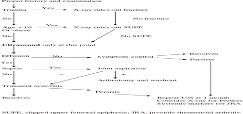
An algorithm for daily practice
Hip pain in the child is a common complaint. There are only a few studies showing the relevance of this topic to primary care (Citation2,Citation3,Citation6). The differential diagnosis of a painful hip is often difficult in the early stages due to non specific symptoms and investigations (Citation11,Citation12). Algorithms based on clinical and laboratory findings in children with a painful hip have been proposed (Citation11).
Still there is some confusion among practitioners and a need for clear guidelines. (Citation9) We have developed a graphic care plan which can be followed by practitioners for the initial diagnosis and follow up of these children (). This care plan is designed to state clearly the few indications for X-ray, thus decreasing the number of unnecessary exposures, and also to reinforce the superiority of ultrasound as the primary diagnostic tool.
Figure 1. Careplan for the painful hip in children
In conclusion, ultrasound is a more useful diagnostic tool in the initial assessment of hip pain in children than X-ray, despite current practice. X-ray may be indicated in specific presentations or the subsequent follow up, but not routinely.
Declaration of interest: The authors report no conflicts of interest. The authors alone are responsible for the content and writing of the paper.
References
- Fischer SU, Beattie TF. The limping child: epidemiology, assessment and outcome. J Bone Joint Surg [Br]. 1999; 81–B:1029–34.
- De Inocencio J. Epidemiology of musculoskeletal pain in primary care. Arch Dis Child 2004;89:431–4.
- Vijlbrief AS, Bruijnzeels MA, van der Wouden JC, van Suijlekom-Smit LW. Incidence and management of transient synovitis of the hip: a study in Dutch general practice. Br J Gen Pract. 1992;42:426–8.
- Do TT. Transient synovitis as a cause of painful limps in children. Curr Opin Pediatr 2000;12:48–51.
- Bosch R, Niedermeier C, Heimkes B. Value of ultrasound in differential diagnosis of paediatric hip joint effusion (Perthes disease, C. fugax, epiphysiolysis capitis femoris). Z Orthop Ihre Grenzgeb. 1998;136:412–9.
- Wittke R. Hip pain-a problem of differential diagnosis in the primary care practice. MMW Fortschr Med. 2005; 147:33–6.
- Konermann W, de Pellegrin M. The differential diagnosis of juvenile hip pain in the ultrasonographic picture. Transient coxitis. Legg-Calvé-Perthes disease, epiphysiolysis of the femur head. Orthopade 1993;22:280–7.
- De Inocencio J. Musculoskeletal pain in primary pediatric care: Analysis of 1000 consecutive general pediatric clinic visits. Pediatrics. 1998;102:E63.
- Zamzam MM. The role of ultrasound in differentiating septic arthritis from transient synovitis of the hip in children. J Pediatr Orthop B. 2006;15:418–22.
- Terjesen T, Osthus P. Ultrasound in the diagnosis and follow-up of transient synovitis of the hip. J Pediatr Orthop 1991;11:608–13
- Caird MS, Flynn JM, Leung YL, Millman JE, D'Italia JG, Dormans JP. Factors distinguishing septic arthritis from transient synovitis of the hip in children. A prospective study. J Bone Joint Surg Am. 2006;88:1251–7.
- Jung ST, Rowe SM, Moon ES, Song EK, Yoon TR, Seo HY. Significance of laboratory and radiological findings for differentiating between septic arthritis and transient synovitis of the hip. J Pediatr Orthop. 2003;23:368–72.
- Karmazyn B, Loder RT, Kleiman MB, Buckwalter KA, Siddiqui A, Ying J, . The role of pelvic magnetic resonance in evaluating non hip sources of infection in children with acute non traumatic hip pain. J Pediatr Orthop. 2007;27:158–64.
- Flynn JM, Widmann RF. The limping child: evaluation and diagnosis. J Am Acad Orthop Surg, 2001;9:89–98.
- Yang WJ, Im SA, Lim GY, Chun HJ, Jung NY, Sung MS, . MR imaging of transient synovitis: differentiation from septic arthritis. Pediatr Radiol. 2006;36:1154–8.
- Serafini G, Zadra N. Anaesthesia for MRI in the paediatric patient. Curr Opin Anaesthesiol. 2008;21:499–503.
- Briggs RD, Baird KS, Gibson PH. Transient synovitis of the hip joint. J R Coll Surg Edinb. 1990;35(1):48–50.
- Mumme T, Berkemeier E, Maus U, Bauer A, Wirtz DC. Coxitis fugax-the beginning of Perthes' disease? Z Orthop Ihre Grenzgeb. 2005;143:529–33.
- Mallet JF, Rigault P, Padovani JP, Touzet P. Transient synovitis 1 of the hip in childhood. “Observation hip”. Rev Chir Orthop Reparatrice Appar Mot. 1981;67:791–803.
- Rutz E, Brunner R. Septic arthritis of the hip—current concepts. Hip Int. 2009;19:9–12.