Myopia affects around 25% of people living in West Europe.Citation1 Although multiple twin studies and family studies confirm that genetics plays an important role in the formation and progression of myopia, the mechanism of this ocular disease is still a mystery despite some progress made through several genome-wide association studies (GWAS).Citation2–5 Unfortunately the single nucleotide polymorphisms (SNPs) or the genes suggested in these GWAS only have minor or moderate contributions to myopia. To have a more homogeneous phenotype of myopic case group might help to find more significant genes. Axial myopia is mainly the consequence of increased axial length while non-axial myopia is mainly due to malfunction of other ocular components such as the lens or cornea.Citation6 Thus the mechanisms of axial myopia and non-axial myopia are less likely to be the same. If mixed up, these two types of myopia, although it cannot be regarded as a phenotype error since most studies only concern the trait of myopia not axial myopia, the efficiency of these studies will be affected, notably by increasing the false positive rate. So far, there is no clear method to differentiate axial myopia from non-axial myopia.
The study recruited 491 French high myopic participants (see ) between 2007 and 2010 in Purpan Hospital, Toulouse, France. Only patients with a refractive error lower than −5D in both eyes were included. The biometric information of the right eye such as refractive error and axial length was selected for the linear regression analysis. SPSS15.0 was used in all the statistical procedures in this study:
TABLE 1 Ocular biometric information of the 491participants.
| (1) | Variables: the refractive error (RE) was set as the dependent value, and the axial length (AL), age, and sex as the independent values; then the multiple linear regression analysis was carried out (option stepwise). However, age and sex did not make sufficient contributions to the RE (p > 0.05). Thus these two variables were removed from the model. | ||||
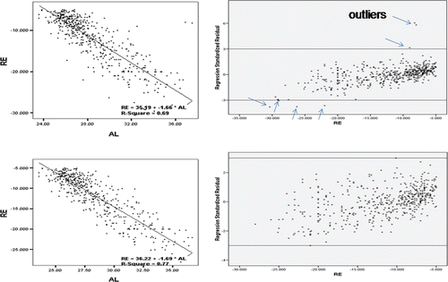
| (2) | Model generated: the refractive error (RE) was set as the dependent value and axial length (AL) as the independent value. A linear regression model was then generated. RE = 35.19 − 1.66 × AL, R2 = 0.69, R = 0.83 (see , top left). | ||||
| (3) | Outlier detection: in a linear regression model, residuals outside the region (−3<y<3, y = standardized residual) were considered to be outliers. After calculating residuals and plotting standardized residuals with RE, seven samples were found to be outside that region (see , top right). | ||||
| (4) | These seven outliers were removed and steps 3 and 4 were repeated until no more outliers were detected. | ||||
| (5) | Finally, only 478 individuals survived as axial myopic subjects. The linear regression model was RE = 36.22 − 1.69 × AL, R2 = 0.77, R = 0.88 (see , bottom left). All the residuals were within the region of −3<y<3 (see , bottom right). | ||||
| (6) | Reappraisal of the clinical records of the 13 outliers was undertaken to validate the method. New ocular examinations were applied to five of them. A careful subjective refraction detected the mistakes that refractive errors were overrated in two cases. In one case, B-sonography revealed that the axial length measurement was biased by the presence of a marked posterior staphyloma. In another two cases, forme frustre keratoconus was detected by elevation topography. Among the remaining eight patients, four patients had had cataract surgery in a different center to our center before being recruited into the cohort and the preoperative values of the refractive status were collected elsewhere. In a further four cases no clear explanation could be derived from the clinical history and no novel examination was currently conducted. | ||||
FIGURE 1 Left: Linear regression model before and after removing outliers. Right: Plotting standardized residuals with RE before and after removing outliers.
By definition, axial length plays a major role in the formation of axial myopia. Non-axial myopic patients are usually individuals with a long axial length but not very low refractive error or severe refractive error with short axial length. When searching myopia genes in case-control studies, since most randomly collected cases are axial myopes, the myopia genes, if suggested by results, are likely to be genes of axial myopia. Thus it may be an advantage to remove non-axial myopic subjects to have a more homogeneous phenotype in genetic studies of this disorder. Fewer cases are needed to achieve the same result and a lower false positive rate can be expected. Generally speaking, a 1mm elongation of AL is equivalent to a myopia shift of RE from −2D to −2.5D.Citation7 If greater than that number, then other factors contribute to the refractive error.
It is clear that AL and RE are strongly correlated in our high myopic group (R = 0.88). It is also obvious that after filtering non-axial myopia, the value of R2 is greater, which means more RE can be explained by the variance of AL. Recent data also suggested axial length and height might share common genes.Citation8 Altogether 13 samples were removed from our study, corresponding to 2.5% of the whole high myopic group. After rechecking the raw data of these 13 patients, an ophthalmological cause was found to explain the discrepancy between refractive error and axial length detected in nine patients. Thus this clearly suggests the pertinence of the method. However there is no explanation for the remaining four outliers. One reason could be a change of the dioptric power of the lens. In the near future, novel imaging methods such as OCT or ray-tracing will detect the lens curvature or refractive index modifications. Further work would be required to recheck those outlier patients. However, if the reason for being an outlier is mainly due to a posterior staphyloma, the patient can be added back into the axial myopic group with caution.
In conclusion, this study provides information on separating axial myopia from non-axial myopia. A more homogeneous phenotype would contribute to the identification of myopia genes.
ACKNOWLEDGMENTS
Declaration of interest: The authors report no conflicts of interest. The authors alone are responsible for the content and writing of the paper.
REFERENCES
- Kempen JH, Mitchell P, Lee KE, Tielsch JM, Broman AT, Taylor HR, Ikram MK, Congdon NG, O’Colmain BJ, Eye Diseases Prevalence Research Group. The prevalence of refractive errors among adults in the United States, Western Europe, and Australia. Arch Ophthalmol 2004;122:495–505.
- Nakanishi H, Yamada R, Gotoh N, et al. A genome-wide association analysis identified a novel susceptible locus for pathological myopia at 11q24.1. PLoS Genet 2009;5:e1000660.
- Solouki AM, Verhoeven VJ, van Duijn CM, et al. A genome-wide association study identifies a susceptibility locus for refractive errors and myopia at 15q14. Nat Genet 2010;42:897–901.
- Hysi PG, Young TL, Mackey DA, et al. A genome-wide association study for myopia and refractive error identifies a susceptibility locus at 15q25. Nat Genet 2010;42:902–905.
- Li Z, Qu J, Xu X, et al. A genome-wide association study reveals association between common variants in an intergenic region of 4q25 and high-grade myopia in the Chinese Han population. Hum Mol Genet 2011;20:2861–2868.
- Young TL, Metlapally R, Shay AE. Complex trait genetics of refractive error. Arch Ophthalmol 2007;125:38–48.
- Meng W, Butterworth J, Malecaze F, Calvas P. Axial length of myopia: a review of current research. Ophthalmologica 2010;225:127–134.
- Zhang J, Hur YM, Huang W, Ding X, Feng K, He M. Shared genetic determinants of axial length and height in children: the Guangzhou twin eye study. Arch Ophthalmol 2011;129:63–68.