Abstract
The ethanol leaf extract (ELE) of Cassia fistula Linn. (Caesalpinaceae) was evaluated for antiulcer activity against pylorus ligation-induced gastric ulcer. Ranitidine (30 mg/kg b.w.) and ELE at doses of 250, 500, and 750 mg/kg b.w. were administered orally in different groups of rats (n = 6), 1 h prior to pyloric ligation. Four hours after pyloric ligation, the gastric juice was collected for evaluation of various parameters. The antiulcer activity of ELE was evidenced by the significant attenuation of gastric volume, pH, free acidity, and total acidity in the gastric juice of pyloric-ligated rats in a dose-dependent manner, and this protective effect could be due to strengthening of the mucosal defense mechanism. ELE pre-treatment significantly attenuated the fall in status of sialic acid and fucose accompanied by an increase in hexose, hexosamine, total non-amino polysaccharide, total carbohydrate, and C:P ratio in the gastric juice of pylorus-ligated rats, and this effect could be due to protection of the mucosal barrier system. ELE pre-treatment significantly prevented the increase in LPO and SOD accompanied by a fall in CAT, in the gastric juice of pyloric-ligated rats. This protective ability of ELE against pylorus ligation-induced gastric ulcer could be attributed to its free radical scavenging and antioxidant properties. Higher doses of ELE (750 mg/kg b.w.) produced maximum antiulcer activity comparable to ranitidine treatment. In essence, the antiulcer activity of ELE could be attributed to (i) a decrease in gastric acid secretion, (ii) protection of the mucosal barrier and restoration of mucosal secretions, (iii) inhibition of free radical generation or prevention of lipid peroxidation, and (iv) free radical scavenging or antioxidant properties.
Introduction
Gastric ulcer is said to occur due to an imbalance between luminal acid synthesis and mucosal defense. Acid and pepsin components constitute the aggressive factors, and the mucous layer of mucin–bicarbonate secretion, prostaglandins, and other healing factors constitutes the defensive factors (CitationSanyal et al., 1983). Ulcer therapy is now mainly focused on limiting the deleterious effects of offensive acid secretion in the stomach (CitationSairam et al., 2003). Conventional treatment of ulcer comprises regular food and adequate rest, use of antacids, and avoidance of ulcerogenic foods (CitationAnoop & Jegadeesan, 2003). In addition, H2 receptor blockers and proton-pump inhibitors are advocated to reduce gastric acid secretion. The use of antiulcer agents, however, has been shown to induce a wide array of deleterious and adverse effects, leading to their withdrawal or cessation in clinical practice (CitationHoogerwerf & Pasricha, 2001). Hence, efforts are continuously being made to derive active principles from natural sources to suggest an alternative remedy for the treatment of gastric ulcer.
Cassia fistula Linn. (Caesalpinaceae) is a medium sized tree, widely cultivated throughout India as an ornamental and deciduous plant (CitationChatterjee & Pakrashi, 1992). In the Ayurvedic system of medicine, this plant is used for the treatment of hematemesis, pruritus, leucoderma, and diabetes (CitationAlam et al., 1990). Ethanol extracts of the pods and stem bark of Cassia fistula exhibit hypoglycemic, antiviral, and anticancer properties, and this plant is also used in the treatment of epilepsy, convulsions, delirium fibris, pimples, burns, syphilis, and dysuria. The leaves are said to be useful in ringworm infections and the flowers are reported to be effective in fungal infection (CitationChopra et al., 1992). This plant has a high therapeutic value, and it exerts antipyretic and analgesic effects (CitationPatel et al., 1965). In Sri Lanka, the plant is said to be useful in the treatment of skeletal fractures (CitationEkanayake, 1980). The hexane, chloroform, ethyl acetate, methanol, and water extracts of flowers of this plant are reported to exhibit antibacterial activity against Gram-positive organisms (CitationDuraipandiyan & Ignacimuthu, 2007). The hepatoprotective property of the hexane leaf extract of this plant against carbon tetrachloride- and paracetamol-induced hepatotoxicity was demonstrated in rats (CitationBhakta et al., 1999, Citation2001). Studies conducted in this laboratory have shown the protective properties of the ethanol leaf extract of Cassia fistula in post-treatment of carbon tetrachloride-induced liver damage in rats (CitationPradeep et al., 2005).
Cassia fistula plant parts are known to be an important source of secondary metabolites, notably phenolic compounds. Fistucacidin, an optically inactive leucoanthocyanidin (3,4,7,8,4′-pentahydroxyflavan), was first extracted from the heartwood (CitationPadmanabha Rao & Venkateswarlu, 1965). Kaempferol and a proanthocyanidin have been isolated from the acetone extract of the flower (CitationNarayanan & Seshadri, 1972). Besides phenolics and their derivatives, a certain amount of alkaloids have also been reported in the flowers (CitationAsseleih et al., 1990). An investigation that characterized the total phenolic, proanthocyanidin, and flavonoid contents in vegetative and reproductive organs of Cassia fistula found in Mauritius and harvested at different stages showed that among the vegetative organs, the young and old leaves exhibited the highest total phenolic, flavonoid, and proanthocyanidin contents (CitationLuximon-Ramma et al., 2002). The leaves of Cassia fistula were also shown to contain secondary metabolites, namely (–)-epiafzelechin, (–)-epiafzelechin-3-O-glucoside, (–)-epicatechin, procyanidin B2, biflavonoids, triflavonoids, rhein, rhein glucoside, sennoside-A, sennoside-B, chrysophanol, physcion, and rhamnetin-3-O-gentiobioside (CitationBahorun et al., 2005).
The present study was conducted to evaluate the antiulcer activity of the ethanol leaf extract of Cassia fistula against pylorus ligation-induced ulcer in rats, due to a paucity of data along these lines.
Materials and methods
Animals
Wistar albino male rats (180 ± 20 g), procured from the institutional animal house facility, were randomly divided into five groups with six animals in each. They were housed in polypropylene cages over husk bedding and provided with standard pellet feed and water ad libitum, unless otherwise indicated. The animals were maintained at 25 ± 2°C with a 12 h light and dark cycle. Animal experiments were performed after obtaining Institutional Animal Ethics Committee (IAEC) approval and in strict adherence to its guidelines.
Chemicals
Ranitidine was obtained as a gift from Madras Pharmaceuticals, Chennai. Malondialdehyde (MDA) was purchased from Sigma-Aldrich Chemicals, USA. All the other chemicals used in this study were analytical grade and were purchased locally.
Plant material
Fresh leaves of Cassia fistula were collected from Tamil Nadu Medicinal Plant and Herbal Corporation Limited (TAMPCOL), Chennai, during the months of August to November, 2007. The plant and its leaves were authenticated by Dr. S. Narayanappa, the Chief Botanist, TAMPCOL. Voucher specimens of the leaves were deposited in the herbarium of the Botanical Survey of India, Comibatore, Tamil Nadu, India (Herbarium No. BSI/C.F./001/2002).
Preparation of ethanol leaf extract
Immediately after collection, the leaves were washed in tap water twice and once in distilled water to remove the external dirt and unwanted materials. The leaves were shade dried for 72 h. Small bits of plant material, the petioles, midribs, and twigs, were removed after shade drying. The dried leaves were then crushed by hand into coarse powder. This powdered leaf material (100 g) was subjected to Soxhlet extraction using 95% ethanol. The Soxhlet extract was evaporated to dryness at 60°C over a water bath and the yield of this ethanol leaf extract (ELE) was 15–17%. ELE was sparingly soluble in water and, hence, it was suspended in distilled water for its administration to rats.
Evaluation of antiulcer activity of ELE
The antiulcer activity of ELE was evaluated in pylorus-ligated rats. Pylorus ligation was done as described by CitationOliveira et al. (2004). All the rats used in this study were placed over wire-mesh flooring in the polypropylene cages, to avoid coprophagy, and fasted for 48 h, allowing free access to water. Group I animals received distilled water (0.5 mL/kg b.w.; orally) and served as control. Group II rats received ranitidine (30 mg/kg b.w.; orally), and they served as the reference drug group for comparison. Groups III, IV, and V received ELE orally at 250, 500, and 750 mg/kg b.w., respectively. All the above treatments were made 1 h prior to pylorus ligation. Pylorus ligation was performed in all the above groups of rats under mild ether anesthesia. The animals were provided with water, but food was deprived during the postoperative period. Four hours after ligation, the animals were sacrificed by cervical decapitation and the stomach was removed quickly.
Collection of gastric juice
The stomach was cut along the greater curvature and the gastric juice was collected in clean centrifuge tubes. The juice was centrifuged and the volume of supernatant was recorded. The gastric juice pH was measured quickly using a pH meter, and the juice was then subjected to biochemical analysis.
Determination of free and total acidity
The free and total acidity of the gastric juice was determined by volumetric analysis as detailed by CitationHawk (1947). Briefly, 0.5 mL of gastric juice was pipetted into a 100 mL conical flask, 2–3 drops of Topfer’s reagent was added, and the mixture was titrated with 0.01 N NaOH (previously standardized with 0.01 N oxalic acid) until all traces of red color disappeared and it turned a yellowish orange. The volume of titrated alkali was noted, and this corresponded to the free acidity. Then, 2–3 drops of phenolphthalene solution was added and titration was continued until a definite red tinge reappeared. Again, the total volume of alkali added was noted. This volume corresponded to the total acidity. The free and total acidity of the gastric juice was derived by calculation.
Estimation of glycoproteins
The glycoproteins present in the gastric juice were extracted by the modified method of CitationAminoff (1961) as described by CitationNiebes (1972). Briefly, 0.1 mL of the gastric juice was mixed with 5 mL of 95% ethanol and centrifuged for 30 min at 3000 rpm. The supernatant was discarded to obtain a precipitate of glycoproteins, which was used for the estimation of mucopolysaccharides, i.e. sialic acid, hexose, fucose, and hexosamine.
Assay of sialic acid
The glycoprotein precipitate of the gastric juice was hydrolyzed using 0.1 N H2SO4. The hydrolysate was subsequently reduced using periodic acid and treated with thiobarbituric acid to liberate a color complex, which was extracted with butanol and measured at 550 nm (CitationNiebes, 1972).
Assay of total hexose
The glycoprotein precipitate of the gastric juice was treated with orcinol–H2SO4 reagent and heated at 80°C for 15 min to yield a color, which was measured at 540 nm (CitationNiebes, 1972).
Fucose
The glycoprotein precipitate of the gastric juice was initially treated with H2SO4 and subsequently with cystein hydrochloride. The difference in absorbance read at 393 nm and 440 nm was taken as an index of the measurement of fucose content of the sample (CitationNiebes, 1972).
Assay of hexosamine
The glycoprotein precipitate was digested with HCl. This hydrolysate was treated with acetylacetone and subsequently colored with Erlich reagent and ethanol, which was measured at 530 nm (CitationNiebes, 1972).
Assay of total non-amino polysaccharide
Initially, 0.1 mL of the gastric juice was precipitated with ethanol. The precipitate was then treated with bromosulfuric acid reagent and tryptophan and kept in a water bath for development of color, which was measured at 520 nm (CitationNiebes, 1972).
Estimation of lipid peroxidation
Lipid peroxidation (LPO) in the gastric juice was determined by the method of CitationOhkawa et al. (1979). Briefly, 100 µL of gastric juice and standard tubes containing MDA taken at a concentration range of 3–12 nM were mixed with 0.2 mL of 8.1% sodium dodecyl sulfate, 1.5 mL acetic acid, and 1.5 mL of thiobarbituric acid. This mixture was made up to 4 mL with distilled water and kept in a boiling water bath at 90°C for 1 h. After cooling to room temperature, 1 mL of distilled water was added and the pink color formed was measured at 532 nm against a reagent blank.
Estimation of superoxide dismutase activity
The activity of superoxide dismutase (SOD) (EC 1.15.1.1) in gastric juice was estimated as detailed by CitationMarklund and Marklund (1974). A mixture containing 2.5 mL of Tris-HCl buffer, 0.1 mL of ethylenediamine tetraacetic acid (EDTA), and 0.5 mL of diethylenetriamine pentaacetic acid (DTPA) was prepared. To this mixture, 0.5 mL of pyrogallol was added and the increase in absorbance was read at 420 nm for 3 min, to determine the rate of autooxidation of pyrogallol. To 0.1 mL of the gastric juice taken in a separate tube, 2.5 mL of Tris-HCl buffer, 0.1 mL of EDTA, and 0.5 mL of DTPA was added. To this mixture, 0.5 mL of pyrogallol was added and the increase in absorbance was read at 420 nm for 3 min. This measurement constituted the rate of inhibition of autooxidation of pyrogallol brought about by the gastric juice.
Estimation of catalase activity
The activity of catalase (CAT) (EC 1.11.1.6) in gastric juice was estimated by the method of CitationSinha (1972). To 0.1 mL of gastric juice, 1 mL of phosphate buffer was added. Then 0.5 mL of H2O2 was added to initiate the reaction. The reaction was arrested at 0, 30, and 60 s intervals by the addition of 2 mL of dichromate–acetic acid reagent. The reagent blank was prepared by the addition of 1.6 mL of buffer and 2 mL of dichromate–acetic acid reagent, taken in separate tubes. The blank and the test tubes were heated in a boiling water bath for 10 min to develop color. The tubes were cooled to room temperature and their color intensity was measured at 570 nm against the blank.
Estimation of protein
The protein content in the gastric juice of pylorus-ligated rats was measured as described by CitationLowry et al. (1951).
Statistical analysis
The data were subjected to one-way analysis of variance (ANOVA). Tukey’s multiple comparison test was done to evaluate the significance of differences of means between various treatment groups, using an SPSS statistical package (Version 7.5). The values are presented as mean ± SEM and a p value <0.05 was considered significant.
Results
Effect of ELE on gastric volume, pH, and free and total acidity
The gastric volume, pH, and free and total acidity levels in the gastric juice of pylorus-ligated rats were significantly decreased by the standard drug, i.e. ranitidine (group II), as compared to the saline-treated control (group I). Pre-treatment with ELE prior to pylorus ligation (groups III, IV, and V) caused a fall in the levels of all the above parameters in a dose-dependent manner () in the gastric juice. The administration of 750 mg/kg of ELE (group V) produced a fall in all the above parameters comparable to ranitidine.
Table 1. Effect of ethanol leaf extract (ELE) of Cassia fistula on acid secretory parameters in gastric juice of pylorus-ligated rats.
Effect of ELE on glycoproteins in the gastric juice
The effect of ELE and ranitidine pre-treatments on the status of glycoproteins in the gastric juice of pylorus-ligated rats is presented in . The data show a fall in the status of sialic acid and fucose in ranitidine-treated rats (group II) as compared to the positive control (group I). In contrast, a highly significant increase in the status of total hexose, hexosamine, total non-amino polysaccharide, and total carbohydrate was observed in group II rats when compared to group I. Pre-treatment with ELE at 250, 500, and 750 mg/kg (groups III, IV, and V, respectively) also caused a highly significant decrease in sialic acid and fucose accompanied by an increase in all the other glycoproteins in a dose-dependent manner, when these groups were compared to control. Treatment of rats at a higher dose of ELE (group V) produced an effect comparable to that in ranitidine-treated rats in the status of all the glycoproteins investigated in the gastric juice of pylorus-ligated rats.
Table 2. Levels of glycoproteins in gastric juice of pylorus-ligated rats treated with ELE of Cassia fistula.
Effect of ELE treatment on C:P ratio
While the total protein was decreased highly significantly by about 40%, the carbohydrate:protein (C:P) ratio was elevated by about two-fold in the gastric juice of ranitidine pre-treated rats (group II) when they were compared to control (group I). Although pre-treatment with ELE at 250 and 500 mg/kg (groups III and IV) also produced an effect similar to that in rantidine-treated rats, their efficacy was not comparable to this group. On the other hand, pre-treatment with ELE at 750 mg/kg (group V) produced an effect comparable to that in ranitidine pre-treated rats ().
Table 3. Status of total carbohydrate (C), total protein (P), and C:P ratio in gastric juice of pylorus-ligated rats treated with ELE of Cassia fistula.
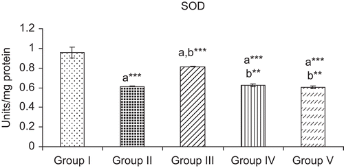
Effect of ELE on LPO and SOD
Ranitidine pre-treatment (group II) significantly decreased the levels of both LPO and SOD in the gastric juice of pylorus-ligated rats when this group was compared to positive control (group I). Pre-treatment with ELE produced a dose-dependent decrease in both LPO and SOD in the pylorus-ligated rats (groups III, IV, and V), and at the highest dose (group V) the decrease in the above parameters was comparable to that in ranitidine-treated animals ( and ).
Figure 1. Effect of ethanol leaf extract (ELE) of Cassia fistula on lipid peroxidation (LPO) in the gastric juice of pylorus-ligated rats. Group I was treated with saline. Group II was pre-treated with ranitidine (30 mg/kg). Groups III, IV, and V were pre-treated with ELE at 250, 500, and 750 mg/kg, respectively. Results are given as mean ± SEM of six numbers of animals in each group. a, group I compared with groups II–V; b, group II compared with groups III–V. *p < 0.05; **p< 0.01; ***p < 0.001.
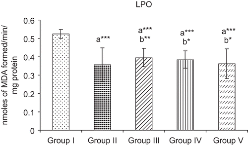
Figure 2. Effect of ELE of Cassia fistula on superoxide dismutase (SOD) activity in the gastric juice of pylorus-ligated rats. Group I was treated with saline. Group II was pre-treated with ranitidine (30 mg/kg). Groups III, IV, and V were pre-treated with ELE at 250, 500, and 750 mg/kg, respectively. Results are given as mean ± SEM of six numbers of animals in each group. a, group I compared with groups II–V; b, group II compared with groups III–V. **p< 0.01; ***p < 0.001.
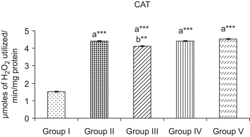
Effect of ELE on CAT
Ranitidine pre-treatment (group II) caused a more than two-fold increase in the activity of CAT in the gastric juice of pylorus-ligated rats when they were compared to the control (group I). A similar increase in the activity of CAT was also observed in rats pre-treated with ELE in a dose-dependent manner (groups III, IV, and V), and at the highest dose (group V) this increase was comparable to that in ranitidine-treated rats ().
Figure 3. Effect of ELE of Cassia fistula on catalase (CAT) activity in the gastric juice of pylorus-ligated rats. Group I was treated with saline. Group II was pre-treated with ranitidine (30 mg/kg). Groups III, IV, and V were pre-treated with ELE at 250, 500, and 750 mg/kg, respectively. Results are given as mean ± SEM of six numbers of animals in each group. a, group I compared with groups II–V; b, group II compared with groups III–V. **p< 0.01; ***p < 0.001.
Discussion
Ulcer is associated with an imbalance between protective and aggressive factors, and inflammation is the leading cause of this imbalance (CitationYuan et al., 2006). Several mechanisms such as an increase in acid secretion, pepsin activity, reduction in mucus and bicarbonate secretion, and reduction in gastric mucosal blood flow are said to be the fundamental cause of gastric ulceration (CitationGalunska et al., 2002). Pylorus ligation-induced ulcer is said to be due to autodigestion of gastric mucosa and breakdown of the gastric mucosal barrier (CitationSairam et al., 2001).
In the present study, pre-treatment of pylorus-ligated rats with ranitidine (group II) and 750 mg/kg of ELE (group V) produced a comparable antiulcer activity, as evidenced by a reduction in the increase in gastric volume, pH, and free and total acidity (). The methanol bark extract of Mimusops elengi L. (Sapotaceae) (CitationShah et al., 2003) and flower extract of Hemidesmus indicus R. Br. (Asclepiadaceae) (CitationAnoop & Jegadeesan, 2003) were reported to reduce total acidity and volume of gastric acid secretion in gastric ulcer-induced rats, and this antiulcer activity was attributed to strengthening of the mucosal defense mechanism by these plant extracts. It is said that an increase in bicarbonate ion concentration plays an important role in protecting the gastric and duodenal mucosa against hydrochloric acid (CitationSuzuki & Ishii, 1996), and the mucosal defense mechanism may be due to the epithelial cells of gastric mucosa, which are impermeable to hydrogen ions, thereby forming a physical barrier (CitationDavenport et al., 1964). Additionally, protection against experimental ulcers may be due to the effect of prostaglandins in the parietal cells (CitationSumangala et al., 1998), as they enhance the mucosal resistance, perhaps by increasing the secretion of mucus and bicarbonates and strengthening the mucosal barrier (CitationSzabo et al., 1981). In the light of these reports, it is suggested that the antiulcer activity of ELE observed in the present study could be due to strengthening of the mucosal defense mechanism.
One of the essential criteria to determine the status of the mucosal resistance/barrier is the state of mucus secretion in the stomach. Higher molecular weight glycoproteins or mucins are mainly responsible for the gel-forming characteristics of mucus. Increased mucus secretion by gastric mucosa can inhibit gastric ulceration by preventing back-diffusion of H+ ions and by buffering of the acid gastric juice (CitationVenables, 1986). The C:P ratio is a direct index of the dissolved muco-substances in the gastric juice. An increase in C:P ratio is a reliable index of an effective mucosal barrier (CitationSanyal et al., 1983), and this ratio is a reflection of mucin activity (CitationJain et al., 1994). The importance of mucus secretion as a response to gastric mucosal trauma has long been recognized (CitationEzer, 1988). The greater is the production of mucus, the lower is the degree of ulceration. Mucus also protects the mucosa and submucosa from inflammatory reaction. The higher is the mucin content, the lower is the free acidity. In the case of duodenal ulcers, CitationBardhan (1989) suggested mucosal defense agents to be a new dimension in the treatment of gastroduodenal diseases. In addition, measurement of hexosamine concentration is said to be an index of mucin content in the gastric juice (CitationPillai & Santakumari, 1984). In the present study, the restoration of hexosamine and the C:P ratio in pylorus-ligated ELE pre-treated rats ( and ) is a clear demonstration of the production of mucin activity and mucosal resistance by ELE.
Several plant extracts have been shown to increase hexosamine and hexose accompanied by a reduction in the concentration of sialic acid and fucose followed by an increase in the C:P ratio in pylorus-ligated or gastric ulcer-induced rats (CitationPillai & Santakumari, 1984; CitationAnoop & Jegadeesan, 2003; CitationShah et al., 2003; CitationRao et al., 2004), and it is reported that these extracts exhibit antiulcer activity by protection of the mucosal barrier system leading to more production of mucus, resulting in less degree of ulceration. It is postulated that the higher is the mucus production, the higher will be the mucin content and the lower will be the free acidity (CitationAnoop & Jegadeesan, 2003). In this study, ELE pre-treatment caused a dose-dependent reversal toward a fall in status of sialic acid and fucose accompanied by an elevation in the status of hexose, hexosamine, total non-amino polysaccharide, and total carbohydrates (). ELE treatment also caused a fall in protein and increase in the C:P ratio in the gastric juice of pylorus-ligated rats (). At a higher dose level (750 mg/kg), ELE produced a protective effect comparable to that of the standard drug ranitidine. In view of these observations, it may be suggested that the antiulcer activity of ELE observed in this study could be due to the protection of the mucosal barrier system of gastric mucosa in pylorus-ligated rats.
The role of free radicals in gastric ulceration is well documented (CitationCochran et al., 1983; CitationRichards & Sharma, 1991), and O2•–, H2O2−, and OH• radicals are important reactive oxygen species (ROS), which have been reported to cause peroxidation of lipids. The increased LPO in the gastric juice of pylorus-ligated rats could be attributed to the generation of the above reactive oxygen species, and the increased SOD activity observed in pylorus-ligated rats could be due to oversynthesis of this enzyme in decreasing the increase in generation of free radicals. It is a known fact that SOD scavenges superoxide radicals, which are responsible for lipid peroxidation (CitationFridovich, 1986). This reaction leads to increased generation of H2O2−, which is also capable of producing more oxidative damage. Apart from acid- and pepsin-related factors, ulceration-induced oxidative stress is due to the involvement of ROS (CitationMiller, 1987). During stress, LPO and SOD are significantly increased and CAT levels are decreased. The increase in SOD is due to increased ROS generation during mucosal damage. This leads to increased generation of H2O2 and its accumulation due to the decrease in CAT level (CitationSairam et al., 2003). Inactivation of gastric peroximes during stress may also aggravate mucosal damage due to lipid peroxidation (CitationBoyd et al., 1981). The ROS scavenging activity of SOD is effective only when it is followed by the action of CAT, because the dismutase activity of SOD generates H2O2, which needs to be scavenged by CAT for its conversion to O2 and H2O (CitationDas et al., 1997). In the present study, the decrease in activity of CAT in pylorus-ligated rats could be due to the overutilization of this enzyme toward scavenging of free radicals.
In this investigation, pre-treatment with ELE in pylorus-ligated rats prevented the increase in LPO and SOD accompanied by a decrease in CAT in the gastric juice in a dose-dependent manner (–). It is likely that ELE prevents gastric ulcer by markedly decreasing LPO and subsequent oxidative damage. Further, ELE might also restore the balance between free radicals and their scavenging enzymes, i.e. SOD and CAT, in the gastric mucosa effectively by counteracting free radicals generated by a cascade of reactions initiated by lipid peroxidation. In brief, ELE might decrease lipid peroxidation by quenching free radicals in the gastric mucosa of pylorus-ligated rats and thus exhibit antiulcer activity. Several plant extracts have been shown to decrease the increase in LPO and SOD accompanied by an increase in CAT in various models of ulcer induced in rats, and their antiulcer activities were attributed to free radical scavenging and antioxidant properties (CitationSairam et al., 2003; CitationRao et al., 2004). Our present results are in agreement with these reports.
Ranitidine pre-treatment caused effective protection in the status of all the acid secretory parameters, glycoproteins, LPO, SOD, and CAT in the gastric juice of pylorus-ligated rats (–, –). It is reported that ranitidine is an H2 receptor blocker, and this effect could be the cause for the restoration of gastric mucosa and subsequent inhibition of gastric acid secretion and maintenance of mucus secretion (CitationHoogerwerf & Pasricha, 2001) in pylorus-ligated rats.
In conclusion, ELE exhibited antiulcer activity comparable to the standard drug, i.e. ranitidine, at its highest dosage level (750 mg/kg) of administration. The antiulcer activity of ELE could be attributed to (i) a decrease in gastric acid secretion, (ii) protection of the mucosal barrier and restoration of mucosal secretions, (iii) inhibition of free radical generation or prevention of lipid peroxidation, and (iv) free radical scavenging or antioxidant properties.
Declaration of interest
The authors thank the UGC-UPE research project (Project No.HS-43) for providing financial assistance to do this study.
References
- Alam MM, Siddiqui MB, Hussian W (1990): Treatment of diabetes through herbal drugs in rural India. Fitoterapia 61: 240–242.
- Aminoff D (1961): Methods for the quantitative estimation of N-acetylneuraminic acid and their application to hydroxylates of sialomucoids. Biochem J 81: 384–392.
- Anoop A, Jegadeesan M (2003): Biochemical studies on the anti-ulcerogenic potential of Hemidesmus indicus R. Br. Var. J Ethnopharmacol 84: 149–156.
- Asseleih LMC, Hernandez OH, Sanchez JR (1990): Seasonal variation in the content of sennosides in leaves and pods of two Cassia fistula populations. Phytochemistry 29: 3095–3099.
- Bahorun T, Neergheen VS, Aruoma OI (2005): Phytochemical constituents of Cassia fistula. Afr J Biotechnol 4: 1530–1540.
- Bardhan KD (1989): Omeprazole in the management of refractory duodenal ulcer. Scand J Gastroenterol 24: 63–73.
- Bhakta T, Banerjee S, Mandal SC, Maity TK, Saha BP, Pal M (2001): Hepatoprotective activity of Cassia fistula leaf extract. Phytomedicine 8: 220–224.
- Bhakta T, Mukherjee PK, Mukherjee K, Banerjee S, Mandel SC, Maity TK, Pal M, Saha BP (1999): Evaluation of hepatoprotective activity of Cassia fistula leaf extract. J Ethnopharmacol 66: 277–282.
- Boyd SC, Sasame HA, Boyd MR (1981): Gastric glutathione depletion and acute ulcerogenesis by diethylmalate given subcutaneously to rats. Life Sci 28: 2987–2992.
- Chatterjee A, Pakrashi SC (1992): The Treatise on Indian Medicinal Plants, Vol. 2. New Delhi, CSIR, pp. 41–42.
- Chopra RN, Nayar SL, Chopra IC (1992): Glossary of Indian Medicinal Plants. New Delhi, Publication and Information Directorate, CSIR, pp. 1201–1202.
- Cochran T, Stefanko J, Moore C, Saik R (1983): Dimethylsulphoxide production against gastric stress ulceration. Curr Surg 40: 435–437.
- Das D, Bandyopadhyay D, Bhattacharya M, Banarjee RK (1997): Hydroxyl radical is the major causative factor in stress-induced gastric ulceration. Free Radic Biol Med 23: 8–18.
- Davenport HW, Warner HA, Code CF (1964): Functional significance of gastric mucosal barrier to sodium. J Gastroenterol 47: 142–152.
- Duraipandiyan V, Ignacimuthu S (2007): Antibacterial and antifungal activity of Cassia fistula L: an ethanomedicinal plant. J Ethnopharmacol 112: 590–594.
- Ekanayake DT (1980): Plants used in the treatment of skeletal fractures in the indigenous system of medicine in Sri Lanka. Sri Lankan Forester 14: 145–152.
- Ezer E (1988). Novel method for producing standard subacute gastric ulcer in rats and for the quantitative evaluation of the healing process: effect of several drugs on healing. J Pharmacol Methods 20: 279–291.
- Fridovich I (1986): Biological effects of superoxide radical. Arch Biochem Biophys 247: 1–11.
- Galunska B, Marazova K, Yankova T (2002): Effects of paracetamol and propacetamol on gastric mucosal damage and gastric lipid peroxidation caused by acetyl salicylic acid (ASA) in rats. Pharamcol Res 46: 141–148.
- Hawk S (1947). Hawk’s Physiological Chemistry. New York, McGraw-Hill Book Company, pp. 147–148.
- Hoogerwerf WA, Pasricha PJ (2001): Agents used for control of gastric acidity and treatment of peptic ulcers and gastro esophageal reflux disease. In:Hardman JG, Limbird LE, Gilman AG, eds., Goodman and Gilman’s the Pharmacological Basis of Therapeutics. New York, McGraw-Hill, pp. 1005–1020.
- Jain SM, Parmar NS, Santani DD (1994): Gastric anti-ulcer activity of calcium channel blockers in rats. Indian J Pharmacol 26: 29–34.
- Lowry OH, Rosebrough NJ, Farr AL, Randall RJ (1951): Protein measurement with the Folin phenol reagent. J Biol Chem 193: 265–275.
- Luximon-Ramma A, Bahorun T, Soobrattee MA, Aruoma OI (2002): Antioxidant activities of phenolic, proanthocyanidins and flavonoid components in extracts of Cassia fistula. J Agric Food Chem 50: 5042–5047.
- Marklund S, Marklund G (1974): Involvement of superoxide anion radical in the autooxidation of pyrogallol and a convenient assay for superoxide dismutase. Eur J Biochem 47: 469–474.
- Miller TA (1987): Mechanisms of stress-related mucosal damage. Am J Med 83: 8–14.
- Narayanan V, Seshadri TR (1972): Proanthocyanidins of Cassia fistula. Indian J Chem 10: 379–381.
- Niebes P (1972): Determination of enzymes and degradation products of glycosaminoglycan metabolism in the serum of healthy and varicose subjects. Clin Chim Acta 42: 399–408.
- Ohkawa H, Ohishi N, Yagi K (1979): Assay for lipid peroxides in animal tissues by thiobarbituric acid reaction. Anal Biochem 95: 351–358.
- Oliveira FA, Vieira GM Jr, Chaves MH, Almeida FRC, Florencio MG, Lima RCP Jr, Silva RM, Santos FA, Rao VSN (2004): Gastroprotective and anti-inflammatory effects of resin from Protium heptaphyllum in mice and rats. Pharmacol Res 49: 105–111.
- Padmanabha Rao TV, Venkateswarlu V (1965): Fistucacidin from the bark and heartwood of Cassia fistula Linn. Bull Nat Inst Sci India 31: 28–33.
- Patel D, Karbhari D, Gulati D, Gokhale D (1965): Antipyretic and analgesic activities of Aconatum spicatum and Cassia fistula. Pharm Biol 157: 22–27.
- Pillai NR, Santakumari G (1984): Effects of nimbidin on acute and chronic gastro-duodenal ulcer models in experimental animals. Planta Med 2: 143–146.
- Pradeep K, Victor Raj Mohan C, Gobianand K, Karthikeyan S (2005): Effect of pretreatment of Cassia fistula Linn. leaf extract against subacute CCl4 induced hepatotoxicity in rats. Indian J Exp Biol 43: 526–530.
- Rao CV, Ojha SK, Radhakrishnan K, Govindarajan R, Rastogi S, Mehrotra S, Pushpangadan P (2004): Anti-ulcer activity of Utleria salicifolia rhizome extract. J Ethnopharmacol 91: 243–249.
- Richards RT, Sharma HM (1991): Free radicals in health and disease. Indian J Clin Pract 2: 15–23.
- Sairam K, Priyambada S, Aryya NC, Goel RK (2003): Gastroduodenal ulcer protective activity of Asparagus racemosus: An experimental, biochemical and histological study. J Ethnopharmacol 86: 1–10.
- Sairam K, Rao CV, Dora Babu M, Goel RK (2001): Prophylactic and curative effects of Bocapa monnierian gastric models. Phytomedicine 8: 423–430.
- Sanyal AK, Mitra PK, Goel RK (1983): A modified method to estimate dissolved mucosubstances in gastric juice. Indian J Exp Biol 21: 78–80.
- Shah PJ, Gandhi MS, Shah MB, Goswami SS, Santani D (2003): Study of Mimusops elengi bark in experimental gastric ulcer. J Ethnopharmacol 89: 305–311.
- Sinha AK (1972): Colorimetric assay of catalase. Anal Biochem 47: 389–394.
- Sumangala PR, Dilip MK, Shivanand NB, Sathiamoorthy SS (1998): Prostaglandin mediated acid secretion inhibitory effect as a possible mechanism for the antiulcer effect of angiotension converting enzyme inhibitor (Captopril) in pylorus ligated rats. Indian J Pharmacol 30: 385–389.
- Suzuki M, Ishii H (1996): Pathophysiology of Helicobacter pylori induced gastric mucosal injury. Asian Med J 39: 186–191.
- Szabo S, Trier JS, Frankel PW (1981): Sulfhydryl compounds may mediate gastric cytoprotection. Science 214: 200–202.
- Venables CW (1986): Mucus pepsin and peptic ulcer. Gut 27: 233–238.
- Yuan Y, Padol IT, Hunt RH (2006): Peptic ulcer disease today. Gastroenterol Hepatol 3: 80–89.