Abstract
Objectives. The association between aortic atherosclerosis and neurological damage during cardiac surgery is well recognized. The purpose was here to analyze the size distribution of particles produced at cross-clamp manipulation of the ascending aorta. Design. A human cadaveric aortic perfusion model of retrograde design was applied (n=27). With this model, washout samples were collected from the pressurized ascending aorta during cross clamp manipulation. Before the experiment, the aorta was flushed to remove debris and with a baseline sample collected. The cross-clamp was opened to collect ten repeated aliquots with dislodged particles. Collected washout samples were evaluated by digital image analysis and microscopy. Results. Cross-clamping produced a significant output of particles, which was seen for size intervals of 1 mm and smaller (p=0.002 to p=0.022). In all size intervals the particle output correlated with the degree of overall aortic calcification (p=0.002 to p=0.025). The model generated substantially more small-size particles than large debris (p<0.010). Conclusions. Aortic clamping was here verified to dislodge aortic debris which correlated with the degree of observed calcification. Macroscopic particles were few. In contrast, cross-clamping produced substantial numbers of small-size particles. These findings emphasize microembolic risks associated with cross-clamping of atherosclerotic vessels.
Embolization caused by aortic manipulation is a matter of concern both in cardiac surgery and vascular surgery. Cerebral embolization may have devastating effects in terms of stroke (Citation1). The relationship between aortic atherosclerosis and stroke is well described (Citation1,Citation2). An additional issue within cardiac surgery is the frequent occurrence of neurocognitive decline in the postoperative period (Citation3), a phenomenon that in part may reflect microembolization. It has been demonstrated that all patients undergoing cardiac surgery has detected microemboli when measured by transcranial Doppler (Citation4,Citation5). In general, Doppler measurement reports the number of recorded signals whereas the size and nature of these particles are less well described. In the present study, the size distribution of particles produced at aortic cross-clamp manipulation was analyzed. Our hypothesis suggests that aortic manipulation is not limited to macroembolization but also microemolization, and further, that embolization is proportional to the degree of aortic calcification. Emboli material was reviewed from a cadaveric perfusion model, as previously described (Citation6,Citation7).
Material and methods
Subjects
This study analyzed the material from two previous studies (Citation6,Citation7), including 27 consecutive subjects undergoing autopsy. These two studies (n=10 and n=17 subjects) analyzed particle embolization at aortic cross clamping and shared the same cadaveric perfusion model. Collected particle deposits were here merged and analyzed in a new fashion. The study had approval from the Umeå University review board (Dnr 01-142).
Aortic Perfusion Model
The model here referred used cadaveric aorta with retrograde perfusion. The model is previously described and to which reference is made (Citation6,Citation7). In brief; the aortic specimen (ascending aorta, arched, and part of the descending aorta) was prepared, branches and small vessels sealed, and a perfusion cannula was connected to the descending part. Perfusion medium consisted of saline (9 g/L NaCl). The aorta was flushed to remove debris. The procedure followed a standardized protocol, identical between the two referred studies. Prior to manipulation a baseline sample was collected in a 50 ml test tube from the ascending aorta. The ascending aorta was then clamped according to routine surgical principles (standard 70-mm aortic cross-clamp, Pilling Co, Fort Washington, PA, USA). The clamp jaws were sutured to the aortic adventitia to allow repeated clamping at the same location. The cross-clamp was then opened and closed under pressure to collect a new sample of the perfusate. This procedure was repeated ten cycles with additional samples collected. Any dislodged particles followed the washout, which was here analyzed.
Washout samples were centrifuged, the supernatant aspirated and the particle deposits were resuspended in de-ionized and filtered water to lyze remaining erythrocytes. After a repeated centrifugation the deposits were fixed for ten minutes in 4% formaldehyde in phosphate buffer. Fixation was ceased by another set of centrifugation and water resuspension, followed by staining and two additional washing/centrifugation cycles. The deposits were spread on a microscope slide, left to dry at room temperature, and stored until further analysis. If heavily condensed, the sample was separated onto additional slides to enable reliable image analysis.
After the perfusion experiment the aorta was cut open. The magnitude of overall calcifications was graded using a semi quantitative scale (0=normal to 4=porcelain aorta). The wall thickness was measured and presence of ulcerative plaques was recorded.
Image analysis
Slides with deposits were here reevaluated according to a new protocol and with the aim to subdivide particles by size. Particles were analyzed both macroscopically (Sony DXC-101P, Tokyo, Japan) and microscopically (Olympus CK40-F200, Olympus Optical Company Ltd, Tokyo, Japan). The principle is described in . The analysis was similar to that previously described (Citation7). However, in the present study threshold settings were tuned to avoid cellular debris to instead focus on solid particles with an embolic potential only. Moreover, results from the two scanning methods (e.g., macroscopic and microscopic) were here merged to form a continuous diameter span from microscopic to macroscopically identified particles.
Figure 1. Schematic illustration of the used evaluation methods; macroscopic (left panel) and microscopic (right panel), respectively. With the macroscopic approach, the entire deposit was scanned in one view for measurement of particle count and size. With the microscopic approach, three randomly selected views were scanned and measured. The total particle output was extrapolated from the known area of the microscopic view in relation to the entire area of the deposit.

In brief, at macroscopic evaluation the entire deposit was scanned in one view. When washout samples were deposited onto more than one slide, each slide was scanned and the particle results were merged. The microscopy analysis did not cover the entire deposit. Three views were randomly selected by arbitrarily changing the coordinates of the microscope stage. This routine reduced the influence from unevenly distributed particles within the deposit. From these three measurements the overall particle output was extrapolated. This procedure was made possible by measuring the area of the particle deposit on each slide, in relation to the area of the three recorded microscopic views. The type of sample was unknown to the observer during analysis to avoid bias. Microscope and image-analyzer settings were adjusted to avoid duplicate measurements between the two approaches.
The image analyzer (Zeiss KS 300, version 3.0, Carl Zeiss Vision GmbH, Hallbergmoss, Germany) recorded particles and their maximum diameter. A computer was programmed to categorize recorded particles into diameter intervals. This protocol had a geometric interface between the macroscopic and microscopic approaches. Macroscopically observed particles less than 0.5 mm were merged with microscopically observed particles above 100 μm. With this method there is a small risk that particles were duplicated between the two methods. However, this phenomenon was less likely to have occurred because macroscopically observed particles would not have fit inside the microscopic view.
Statistical analysis
Because of a skewed distribution of particle counts, both within and between subjects, data were logarithmically transformed (Log10). Differences were analyzed by Wilcoxon matched pairs test. Spearman's test was used for correlation analyses. To analyze the effect of repeated clamping, linear regressions were applied to calculate particle slope values against repetitive maneuvers. Differences from horizontal slope (0 values) were analyzed by Wilcoxon matched pairs test. A few samples at macroscopic analysis contained no visible particles. At logarithmic transformation these observations were replaced by the nearest zero value of 0.1 that logarithmically equals −1. This routine generated a close to normal distribution after logarithmic transformation and the results are therefore presented as mean values ± standard error (sem), unless otherwise stated. Demographic data showed a reasonable normal distribution. SPSS version 16.0 was used for statistical calculations. A p-value above 0.05 was considered non-significant.
Results
Demographic details
The demographic profile of the merged subject cohorts included 74% men, a mean age of 71.6±2.4 years, a history of previous stroke in 30%, diabetes mellitus in 19%, and hypertension in 22%, respectively. The subjects had many characteristics in common with routine cardiac surgery patients. At autopsy, coronary disease was diagnosed among 74% of the subjects and 52% had valvular disease. In 35% signs of a previous myocardial infarction were found. The diameter of the ascending aorta at the clamp site was 32.1±0.6 mm with a wall thickness of 1.7±0.1 mm. The overall magnitude of aortic calcification was 2.0±0.2 according to a semi quantitative scale. One subject was without overt signs of aortic calcification, and none had porcelain characteristics. Ulcerative calcifications were identified in 59% of the cases.
Particle analysis
Particles generated at aortic cross-clamping varied between subjects. Without respect to size intervals the median number of dislodged particles (n × 1000) was 81.7 with a quartile range from 7.2 to 302.8 particles. These values denote the specific effect of clamping with the baseline value subtracted. The corresponding figures for debris larger than 0.5 mm showed a median of three particles with a quartile range of 0 to 12. Because of this variability between size groups and between subjects, particle counts were logarithmically transformed.
In further analysis, particles were separated into diameter intervals. Particles induced by cross-clamping varied in counts between size groups. shows the particle output during repeated manipulations. Referring to the specific clamp effect (first cross-clamp versus baseline) significant outputs of particles were observed in the smaller diameter intervals, starting with 0.5–1 mm (). There was a significant correlation, in all diameter intervals, between the overall magnitude of aortic calcifications and the number of particles. In contrast, no such correlations were observed for baseline particle recordings ().
Figure 2. Logarithmically transformed particle output produced at aortic cross-clamping with subdivision into diameter intervals. Mean of Log10 values with standard errors indicated (n=27).
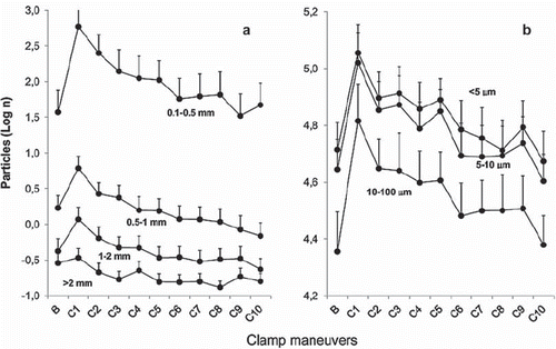
Table I. Statistical output.
Repeated clamping resulted in a sustained particle effect (). The significance against baseline level remained during the second and third clamp maneuver for small-size particles (<0.5 mm). This was exemplified for the 10–100 μm interval (p=0.043, p=0.030 referring the second and third maneuver, respectively). During further clamping particle counts approach that for baseline. This was evaluated by linear regression, with a negative slope that differed from horizontal level, which yielded significance for intervals ≤2 mm (p=0.003 – p<0.001).
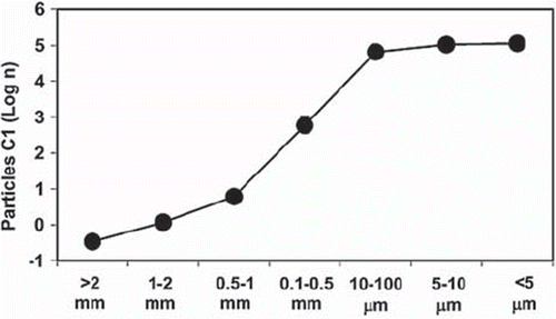
Large particles were few in comparison with the counts in the small-size groups (). This was statistically confirmed by a significant correlation between particle numbers and size (p<0.010). This phenomenon was also true for baseline counts of particles. The latter issue illustrates the background noise seen with this model.
Figure 3. Logarithmically transformed particle numbers as a function of particle size. Mean of Log10 values are presented (n=27). Standard error bars are indicated but escape detection due to small spread. Spearman's rank correlation test was applied (p<0.010).
Discussion
Neurological complications in association with cardiac surgery reflect a complex and multifactorial problem. This problem covers a wide spectrum from stroke to diffuse brain damage. According to the American Heart Association and American Collage of Cardiology, adverse cerebral outcome after cardiac surgery may be subdivided in two categories. Type I deficit includes major focal neurological deficits, stupor and coma, among which stroke is the typical example. Type II deficit is an injury without a detectable focal lesion, instead being associated with memory deficits, deterioration in intellectual function, confusion, agitation, and/or seizures (Citation8). The reported incidence of type-I deficit ranges from about 1.5% to 5% (Citation9–12). Type-II injury is generally considered to be substantially more frequent. Newman (Citation3) reported a 53% occurrence of neurocognitive decline after on-pump cardiac surgery, although many questions remain to be answered in this context (Citation13).
Embolization is an important mechanism behind neurological damage in both cardiac and carotid surgery. It is intuitively understood that a large-size particle dislodged from the atherosclerotic aorta or carotid arteries may cause stroke. To what extent microscopic particles from the manipulated vessel contribute to neurological deficits is less clarified. In all patients undergoing cardiac surgery, microemboli are detected by transcranial Doppler (Citation14,Citation15). Microembolization has been observed at both aortic cannulation and during aortic cross-clamping (Citation15). Doppler-detected microemboli are also described during cardiac catheterizations (Citation16), percutaneous coronary interventions (Citation17), and pulmonary-vein ablation (Citation18). However, Doppler ultrasound is associated with technical limitations, both in terms of distinguishing between gaseous versus solid particles but also to particle-size characteristics. In general, ultrasound is more sensitive to gaseous emboli compared with solid material (Citation19). The ultrasound methodology is complex and undergoes continuous technical refinements (Citation20).
In a study by Bergman et al. (Citation21) particle embolization was explored by means of intra-aortic filters used during coronary artery bypass grafting. They reported an average of 10.5 captured particles. Dislodged material was found in all filters. These filters had a pore size of 120 μm. In our experimental study, we categorized particles into different size intervals. With a 0.5 mm cut-off we recorded somewhat fewer particles than described above. However, our particle count referred to clamp-specific effects only which must be considered. In comparison, the microscopic debris was substantial in our analysis, a finding not possible to measure in a clinical setting.
The present study focused on experimental aortic cross clamping. A cadaveric perfusion model designed at our institution (Citation6,Citation7) was here reevaluated to explore particle-size characteristics of dislodged material. It was obvious, and as previously identified, that aortic cross-clamping generated a substantial output of particles. In this study the size distribution of particles was explored. The effect from cross clamping was identified for all particle size intervals of less than 1 mm diameter. The particle numbers varied markedly between size groups. Of particular interest, in all size intervals the number of dislodged particles correlated with the overall magnitude of aortic calcifications. There was an inverse logarithmic relationship between particle number and size. Our findings highlight the microembolic potential of cross clamping and possible type-II brain damage. A correlation is known to occur between cognitive decline and particle numbers identified by transcranial Doppler (Citation22). This correlation is in consonance with our observations, with the assumption that the Doppler technique is sensitive to particles of microembolic type.
An intriguing question addresses to what extent multiple small-size emboli may contribute to identifiable ischemic lesions. Rapp et al. described this question in different animal models by injecting atherosclerotic debris into the carotid artery (Citation23). They concluded that both particle size and numbers are of relevance for ischemic lesions (Citation23,Citation24). Brain infarction, identified by magnetic resonance and histology, was seen with injected particles as small as 60 to 100 μm (Citation24).
Another finding in our study was that particle output decreased with repeated clamping. However, several clamp maneuvers were required until the clamp site was free from particles and the output reached baseline levels. This underscores the importance for the surgeon to avoid repetitive clamp maneuvers of the aorta and supports benefits with single-clamp techniques.
The described perfusion model has obvious limitations, as previously addressed (Citation6,Citation7). In this analysis we were unable to identify a significant occurrence of large-sized particles with obvious stroke potential. Our study does not talk against this mechanism. This shortcoming must be seen in perspectives of the low stroke rate among cardiac surgery patients versus the limited number of subjects in our study. A background particle noise was evident in the model. However, this noise did not correlate with the recorded degree of aortic calcification, a correlation seen for the clamp-specific findings. The aortic calcification referred to the overall specimen and not the clamp site per se, which addresses a possible limitation. The model used cadaveric aorta which may have contributed to the noise problem. Another limitation with the cadaveric model is to what extent the reported results express post mortem changes rather than clamp effects. It is important to emphasize that the influence from noise or cadaveric effects was avoided by pair-wise comparisons between clamping and baseline samples. Moreover, it is known that the histology of blood vessels is less affected by degenerative changes in the early post mortem period than other tissues (Citation25). Our study analyzes a clinical problem with an experimental approach. Generalization of our findings to the complex surgical situation must be considered with care.
In conclusion, aortic cross-clamping was found associated with particle embolization which correlated to the degree of calcification. In our study, macroscopic particles were few. Instead, the present findings emphasize the importance of numerous small-size particles and microembolization. Aortic cross-clamping should be included among mechanisms behind diffuse forms of brain dysfunction after cardiac surgery.
Acknowledgements
The authors would like to acknowledge Mrs Anne-Marie Österdahl, Mr Dan Nylund, Dr Eva Lundin and Dr Karin Sixtensdotter Graffmo at the Department of Clinical Pathology, Umeå University Hospital. This work was supported by Swedish Society for Medical Research, funds of the Medical Faculty, Umeå University Hospital, the Heart Foundation of Northern Sweden, and the Swedish Stroke Foundation.
Declaration of interest: The authors report no conflicts of interest. The authors alone are responsible for the content and writing of the paper.
References
- Filsoufi F, Rahmanian PB, Castillo JG, Bronster D, Adams DH. Incidence, topography, predictors and long-term survival after stroke in patients undergoing coronary artery bypass grafting. Ann Thorac Surg. 2008;85:862–70.
- van der Linden J, Hadjinikolaou L, Bergman P, Lindblom D. Postoperative stroke in cardiac surgery is related to the location and extent of atherosclerotic disease in the ascending aorta. J Am Coll Cardiol. 2001;38:131–5.
- Newman MF, Kirchner JL, Phillips-Bute B, Gaver V, Grocott H, Jones RH, : Neurological Outcome Research Group and the Cardiothoracic Anesthesiology Research Endeavors Investigators. Longitudinal assessment of neurocognitive function after coronary-artery bypass surgery. N Engl J Med. 2001;344:395–402. Erratum in: N Engl J Med 2001; 344: 1876.
- Braekken SK, Russel D, Brucher R, Abdelnoor M, Svennevig JL. Cerebral microembolic signals during cardiopulmonary bypass surgery. Frequency, time of occurrence, and association with patient and surgical characteristics. Stroke. 1997; 28:1988–92.
- Abu-Omar Y, Balacumaraswami L, Pigott DW, Matthews PM, Taggart DP. Solid and gaseous cerebral microembolization during off-pump, on-pump, and open cardiac surgery procedures. J Thorac Cardiovasc Surg. 2004;127:1759–65.
- Boivie P, Hansson M, Engstrom KG. Embolic material generated by multiple aortic crossclamping: A perfusion model with human cadaveric aorta. J Thorac Cardiovasc Surg. 2003;125:1451–60.
- Boivie P, Hansson M, Engstrom KG. Intraluminal aortic manipulation by means of intra-aortic filter, cannulation, and external clamp maneuvers evaluated versus dislodged embolic material. J Thorac Cardiovasc Surg. 2006;131:283–9.
- Eagle KA, Guyton RA, Davidoff R, Edwards FH, Ewy GA, Gardner TJ, . American College of Cardiology; American Heart Association. ACC/AHA 2004 guideline update for coronary artery bypass graft surgery: a report of the American College of Cardiology/American Heart Association Task Force on Practice Guidelines (Committee to Update the 1999 Guidelines for Coronary Artery Bypass Graft Surgery). Circulation. 2004;110:340–437.
- Roach GW, Kanchuger M, Mangano CM, Newman M, Nussmeier N, Wolman R, . Adverse cerebral outcomes after coronary bypass surgery. Multicenter Study of Perioperative Ischemia Research Group and the Ischemia Research and Education Foundation Investigators. N Engl J Med. 1996;335:1857–63.
- Bucerius J, Gummert JF, Borger MA, Walther T, Doll N, Onnasch JF, . Stroke after cardiac surgery: A risk factor analysis of 16,184 consecutive adult patients. Ann Thorac Surg. 2003;75:472–8.
- Charlesworth DC, Likosky DS, Marrin CA, Maloney CT, Quinton HB, Morton JR, . Northern New England Cardiovascular Disease Study Group. Development and validation of a prediction model for strokes after coronary artery bypass grafting. Ann Thorac Surg. 2003;76:436–43.
- Boivie P, Edstrom C, Engstrom KG; Side differences in cerebrovascular accidents after cardiac surgery: A statistical analysis of neurologic symptoms and possible implications for anatomic mechanisms of aortic particle embolization. J Thorac Cardiovasc Surg. 2005;129:591–8.
- Rosengart TK, Sweet J, Finnin EB, Wolfe P, Cashy J, Hahn E, . Neurocognitive functioning in patients undergoing coronary artery bypass graft surgery or percutaneous coronary intervention: Evidence of impairment before intervention compared with normal controls. Ann Thorac Surg. 2005;80:1327–34.
- Braekken SK, Russel D, Brucher R, Abdelnoor M, Svennevig JL. Cerebral microembolic signals during cardiopulmonary bypass surgery. Frequency, time of occurrence, and association with patient and surgical characteristics. Stroke. 1997; 28:1988–92.
- Abu-Omar Y, Balacumaraswami L, Pigott DW, Matthews PM, Taggart DP. Solid and gaseous cerebral microembolization during off-pump, on-pump, and open cardiac surgery procedures. J Thorac Cardiovasc Surg. 2004;127:1759–65.
- Lund C, Nes RB, Ugelstad TP, Due-Tønnessen P, Andersen R, Hol PK, . Cerebral emboli during left heart catheterization may cause acute brain injury. Eur Heart J. 2005;26: 1269–75.
- Bahrmann P, Werner GS, Heusch G, Ferrari M, Poerner TC, Voss A, Figulla HR. Detection of coronary microembolization by Doppler ultrasound in patients with stable angina pectoris undergoing elective percutaneous coronary interventions. Circulation. 2007;115:600–8.
- Kilicaslan F, Verma A, Saad E, Rossillo A, Davis DA, Prasad SK, . Transcranial Doppler detection of microembolic signals during pulmonary vein antrum isolation: Implications for titration of radiofrequency energy. J Cardiovasc Electrophysiol. 2006;17:495–501.
- Wikstrand V, Linder N, Engström KG. Evaluation of the Doppler technique for fat emboli detection in an experimental flow model. J Extra Corpor Technol. 2008;40:175–83.
- Russel D, Brucher R. Online automatic discrimination between solid and gaseous cerebral microemboli with the first multifrequency transcranial Doppler. Stroke. 2002;33: 1975–80.
- Bergman P, Hadjinikolaou L, van der Linden J. Aortic atheroma is related to number of particulates captured by intra-aortic filtration in CABG. Eur J Cardiothorac Surg. 2002;22:539–44.
- Clark RE, Brillman J, Davis DA, Lovell MR, Price TR, Magovern GJ. Microemboli during coronary artery bypass grafting. Genesis and effect on outcome. J Thorac Cardiovasc Surg. 1995;109:249–57.
- Rapp JH, Pan XM, Sharp FR, Shah DM, Wille GA, Velez PM, . Atheroemboli to the brain: Size threshold for causing acute neuronal cell death. J Vasc Surg. 2000;32:68–76.
- Rapp JH, Pan XM, Yu B, Swanson RA, Higashida RT, Simpson P, . Cerebral ischemia and infarction from atheroemboli <100 microm in Size. Stroke. 2003;34:1976–80.
- Yla-Herttula S, Nikkari T. Effects of post-mortem time on the biochemical composition of coronary arteries. Atherosclerosis. 1985;56:1–10.