Abstract
Objective. Pectoral muscle flaps (PMF) are effective in terminating protracted sternal wound infections (SWI) but long-term outcome remains uncertain. Therefore, the aim of this study was to evaluate long-term outcome in patients treated with PMF. Design. Thirty-four of 263 patients revised because of deep SWI from 1991–2005 were treated with PMF. Of the 21 patients alive, 11 had left-sided, two right-sided and eight bilateral procedures. Sternal debridement without closure of the sternum was done in 17 patients. Nineteen of 21 patients responded to a questionnaire. Results. At follow-up on average 5.9 years (range 1.9–14.8 years) after surgery 63% (12/19) experienced unstable chest. Two thirds (12/18) reported problems carrying a grocery bag and 37% (7/19) had problems putting on a coat. Reduction of power and mobility was more common in the right arm and shoulder even in patients with left-sided PMF. Thirty-two percent (6/19) would have preferred alternative treatment if possible to avoid sternal instability even if healing had been substantially delayed. Conclusions. Surgery with PMF and sternal debridement was associated with long-term disability, which appeared to be significant in one third of the patients. The function of the right arm and shoulder was affected more often despite the majority of procedures being left-sided suggesting that loss of skeletal continuity of the chest wall is more disabling than loss of pectoral muscle function.
Deep sternal wound infections remain a major cause of morbidity and mortality after cardiac surgery and are costly for the health care system (Citation1–6). Sternal wound infections also imply a considerable psychological burden for the patient (Citation7). The incidence of sternal infection after sternotomy has been reported to vary between 0.25–9% (Citation3,Citation6,Citation8–13). Post-discharge surveillance up to three months appears essential for reliable assessment of the true incidence of sternal wound infections (Citation9,Citation14).
Until the 1980s the standard treatment of sternal wound infections was debridement and open granulation with secondary closure or closed catheter irrigation, but failure of treatment was common with mortality rates as high as 37.5% (Citation15). The use of pectoral muscle flaps to treat severe and life-threatening sternal wound infections was introduced by Jurkiewicz et al. in 1980 (Citation16). Delaying closure of the wound until systemic signs of infection have subsided and the wound has healthy granulation tissue led to a significant decrease in complications related to muscle flap closure (Citation17). Over the last years vacuum-assisted closure (VAC) has been used to accelerate this process with successful results facilitating both primary closure and closure with muscle flaps (Citation18,Citation19).
Many reports show that vigorous sternal debridement and the use of muscle flaps are effective in terminating infections (Citation20–22). Short-term studies assessing functional implications suggest potential long-term consequences for the patients (Citation23,Citation24). However, available reports on the long-term consequences for patients treated with pectoral muscle flaps are few and conflicting (Citation25–27). As it was our impression that the use of pectoral muscle flaps had increased at our institution we wanted to assess the use of pectoral muscle flaps and to evaluate the long-term outcome in these patients.
Material and methods
Patients
The University Hospital in Linköping is the only referral center in the southeast region of Sweden, serving a population of approximately one million. By examining our operation register we found a total of 263 patients who underwent surgical revision due to deep sternal wound infection between January 1991 and September 2005.
Of the 263 patient, 34 patients were treated with unilateral or bilateral pectoral muscle advancement flaps. Twenty-one of these patients were alive in September 2006. Eleven of the patients had a left-sided procedure, two a right-sided procedure and eight had bilateral procedures. Nineteen of 21 patients responded to a questionnaire. The questionnaires were followed up by telephone contact when complementary information was needed. In spite of this, minor variations in the response rate to different questions remain explaining variations in the numbers and percentages presented. Seventeen patients responded to a complementary questionnaire assessing whether the patients were right- or left-handed. The average follow-up time was 5.6 ± 3.3 (range 1.9–14.8) years after the original surgery.
Demographic and clinical data relevant to wound healing were retrieved from a database and from the patients’ medical records. The study was performed according to the Helsinki Declaration of Human Rights and ethical approval waived by the Regional Ethical Review Board in Linköping according to the update of the Ethical Review Act (2003:460).
Questionnaire
None of the standardized “Quality of Life forms” was found particularly useful in order to address the specific problems of pectoral muscle flap surgery. Hence, the query was based on consultation with our physiotherapists and the limited data on short-term outcome available in the literature (Citation24). The patients were asked to answer a questionnaire with 34 questions assessing sternal stability, cosmetic satisfaction, pain and ability to open a door, carry a grocery bag and to put on a coat. Patients were requested to grade disability into four grades: No disability, mild disability, significantly disabled but can manage by myself, severely disabled and in need of assistance. The questionnaire also assessed the role of postoperative chest problems in relation to other limiting comorbidities, patients overall satisfaction with the surgery and postoperative care, whether the postoperative complications made them regret the surgery and whether they would have preferred an alternative treatment even if it would have delayed healing by up to three months.
Operative technique
The pectoral muscle flaps were with few exceptions dissected out by plastic surgeons according to a modified standard technique. The modification allowed harvest of enough muscular surface to cover the sternal defect by the left pectoral muscle in most cases. The left pectoral was elevated first and planned to be pedicled on the thoracoacromial vessels as the left side is unsafe to use on parasternal perforators if the left internal thoracic artery has been harvested.
Dissection was first conducted on the superficial side of the muscle, separating skin and subcutaneous tissue from the muscle. The endpoint of this dissection was the diagonal lateral border of the muscle, which was dissected caudally until the abdominal rectus was reached. A few centimetres of the rectus above the costal margin was included in the flap, which after the caudal transection was dissected free along the sternal border. The muscle was “rolled” cranially and the thoracoacromial vessels identified and followed on the deep side. The muscular origin on the clavicle was transected taking care that only the muscle origin on the anterior surface of the clavicle was severed, leaving the posteriorly located fibrofatty tissues. Transection was carried all the way laterally. Thereafter, the thick muscular convergence towards humerus was transacted allowing the flap to be pulled forward. By using a slightly rotating pull, the flap was able to reach the entire vertical length of the sternal cavity where it was sutured into place with resorbable sutures. If the left pectoral muscle did not suffice, the right flap was elevated, either antegrade or retrograde. The undermined skin flaps on both sides were closed in the midline.
Statistical analysis
SPSS 14.0 (SPSS Inc, Chicago, Illinois, USA) was used for descriptive statistical analyses. Cumulative long-term survival was assessed with Kaplan-Meier analysis. The results are presented as numbers, percentages or mean ± standard deviation unless otherwise stated.
Results
Twelve thousand one hundred and twenty-five surgical operations with cardiopulmonary bypass were performed at our institution over the time period studied, 263 (2.2%) patients developed a deep sternal wound infection that was treated with surgical revision.
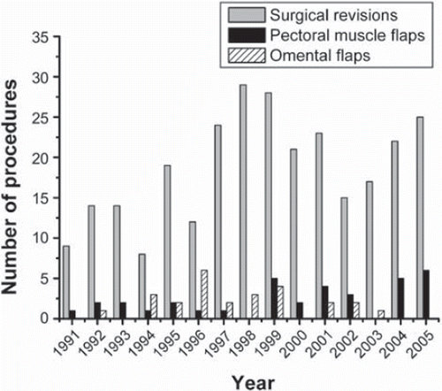
Over the time period 1991–2000, 9.1% of the patients with a deep sternal wound infection that required surgical revision ended up with pectoral muscle flaps at our institution. Between 2001 and 2005 the corresponding figure was 17.6%. The use of omental flaps decreased over the same period of time ().
Overall 34 (12.9%) of these patient ended up with pectoral muscle flaps. None of the patients died within 30 days from the primary procedure and Kaplan-Meier cumulative 5-year survival was 75.0%.
Twenty-one of the patients were alive at follow-up on average 5.6 ± 3.4 years after the pectoral muscle flap procedure. In one patient a stable sternum was covered by a pectoral muscle flap because of recurrent osteitis and in another patient partial stability of sternum was found. Furthermore, attempts to stabilize the sternum with PDS-suture (size 1 and 0) during surgery were made on two patients of whom one stated that the chest was stable in the questionnaire.
The demographic characteristics of the 21 patients included in this study are presented in and . All but four patients had undergone coronary artery bypass grafting with a concomitant valve procedure in two patients.
Table I. Demographic characteristics in association with the original surgical procedure of the 21 patients with pectoral muscle flaps who were alive at follow-up.
Table II. Presentation of individual patients and main outcome with regard to original procedure, duration to and type of pectoral muscle flap procedure, attempt to stabilize sternum or pectoral muscle flap procedure performed in a patient with partially stable sternum (PS), duration of follow-up and limitations in relation to comorbidity. M, male; F, female; (NR), did not respond to questionaire. *Indicates patient who would have preferred alternative treatment if possible even if healing of the wound infection had been delayed up to three months. PS**, sternum partially stable. Arthralgia refers to pain in joints not specified by the patients; LLCP***, lower limb circulatory problem not specified by the patient; COPD, chronic obstructive pulmonary disease.
The most common pathogenic agents found among these 21 patients were Coagulase-negative staphylococci (13/21) and Staphylococcus aureus (4/21). Escherichia coli (1/21), Proteus mirabilis (1/21), Enterococcus faecalis (1/21) and Klebsiella pneumonia (1/21) were found in the remaining cases.
Sternal stability and functional result
Sixty-eight percent (13/19) reported sensation of a clicking sternum and 63% (12/19) of the patients perceived that their chest was unstable. Seventy-five percent (9/12) of the patients that reported unstable chest described this disability as mild and 25% (3/12) reported it to be of a moderate degree. Although no one reported it to be of a severe degree 37% (7/19) reported that they felt limited in their daily life to some extent because of unstable chest.
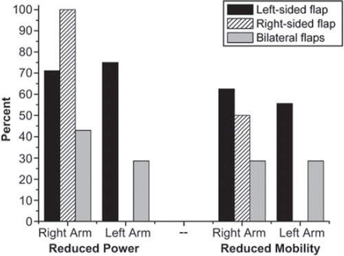
All patients who responded regarding the dominant hand were right-handed (17/17). The prevalence of self assessed functional disability in the upper extremities related to left-sided, right-sided and bilateral flaps are shown in .
Figure 2. Self assessed functional disability in the upper extremities with regard to reduction in power and active mobility of the shoulder.

Problems from the right side were more common although 90% (19/21) of the pectoral muscle flaps involved either left side or bilateral flaps whereas right sided or bilateral flaps were used in 48% (9/19) of the cases. Sixty-three percent (10/16) of the patients experienced a reduction of power of the right arm and 47% (8/17) a reduction of power of the left arm ().
Forty-seven percent (8/17) of the patients stated that their mobility of the right shoulder was reduced and 39% (7/18) that the mobility of left shoulder was reduced.
Regarding questions on the impact of daily life 67% (12/18) of the patients reported that they had problems carrying a grocery bag. Twenty-eight percent (5/18) required assistance and another 11% (2/18) reported significant difficulty although they managed without assistance.
Thirty-seven percent (7/19) experienced problems putting on a coat and 16% (3/19) reported significant difficulties. No one required assistance.
Twenty-two percent (4/18) had problems opening a door and 11% (2/18) reported significant difficulties. No one required assistance.
Dyspnea
Eighty-nine percent (16/18) of the patients were bothered by dyspnea prior to the original cardiac surgery, 75% (12/16) stated that they were less bothered at follow-up whereas 18% (3/17) expressed more problem with dyspnea at follow-up.
Pain
Overall 83% (15/18) of the patients stated that they experienced chest pain and 37% (7/19) reported that they were limited in their daily life because of this. Chest pain was initiated by movements of the chest in 40% (6/15) and by physical effort in 60% (9/15).
Of the patients that reported pain 78% (12/15) had it only infrequently and 60% (9/15) never used analgetic drugs. Twenty percent (3/15) of the patients used analgetic drugs more than once weekly. Of the patients that reported pain two of 15 regarded it to be angina pectoris and one of them reported complete pain relief by sublingual nitroglycerine.
Cosmetic result
Seventy-two percent (13/18) of the patients reported that they were satisfied with the cosmetic results after the pectoral muscle flap procedure, 17% (3/18) were not completely satisfied and 11% (2/18) were dissatisfied.
Overall satisfaction and the role of comorbidities
At follow-up the mean age was 68 ± 12 years. Apart from the limitations reported above the patients also expressed limitations due to other causes presented in . However, one-third (5/15) of the patients were mainly limited by postoperative chest problems and 44% (7/16) reported that they had ceased with recreational activities because of these problems. Although, 68% (13/19) expressed overall satisfaction with the original surgical procedure and postoperative care, 32% (6/19) would have preferred alternative treatment if possible even if healing of the wound infection had been delayed up to three months (). None of the patients regretted undergoing cardiac surgery, despite their chest problems.
Discussion
Overall problems with unstable chest, reduced mobility and power of the shoulder, and pain were common at long-term follow-up in our study. Although most patients reported their discomfort as mild it is noteworthy that the problems affected activities of daily life such as carrying a grocery bag in two thirds or putting on a coat in approximately one third of the patients. This also corresponded to overall satisfaction with the original procedure with the majority being satisfied but almost one third that would have preferred an alternative treatment even it would have prolonged the healing of the wound up to three months.
Our impression that the use of pectoral muscle flaps had increased was confirmed and they were used in 18% during the period 2001–2005. However, this appeared to be explained by a reduced use of omental flaps and thus not by a true increase in the use of flaps in general (). There is little data in the literature regarding the need for flap procedures in association with sternal wound infections. Klesius et al. (Citation23) reported a 19% incidence in patients with deep sternal wound infections, which is on level with our results despite markedly different bacteriological findings. The dominating pathogens in our study were Coagulase-negative staphylococci, which is in accordance with other studies (Citation6,Citation9,Citation11).
In contrast to previous studies, the majority of the patients in our study had problems with unstable chest and a clicking sternum. Ringelman et al. (Citation27) and Yuen et al. (Citation26) reported that 42.5% and 45% respectively perceived an unstable chest compared to 63.2% in our study. Most of our patients also reported a reduction of power and mobility of the arm and shoulder at either one or both sides (). Interestingly, the patients with left-sided flaps, either unilateral or bilateral, reported reduction of power and mobility more frequently on the right side. This might partly be explained by the fact that all patients that responded regarding favored hand were right-handed. Patients with bilateral flaps reported less problems on both sides than the patients with left-sided flaps. Only two patients had unilateral right-sided flaps and both of them reported reduced power of the right arm and one of them decreased mobility of the right shoulder. None of them had problems from the left side. By and large these findings suggest that loss of sternal stability and skeletal continuity of the chest wall is more disabling than loss of pectoral muscle function. Efforts to achieve a stable chest should thus have priority in the treatment of sternal infections.
A high success rate has been reported using VAC treatment as a single-line therapy followed by rewiring without the use of soft tissue flaps (Citation4). Encouraging short-term results have also been reported combining bilateral pectoral muscle flaps with early surgical debridement and sternal closure employing rigid fixation principles (Citation21). Sternal debridement and use of pectoral muscle flaps without sternal fixation should be considered a bail out procedure for life-threatening infections or protracted infections that cannot be terminated by a more conservative approach.
Ringelman et al. (Citation27), Yuen et al. (Citation26), and Francel et al. (Citation25) found a substantially lower prevalence of arm or shoulder weakness (41%, 25% and 20%, respectively). Some of the discrepancy might be explained by the way questions were posed. In our study the patients were asked about reduced power and it is conceivable that a lesser proportion would have reported weakness than reduced power, as the majority of patients described their symptoms as mild.
The proportion of patients that reported pain was larger in our study (83%) compared to that found by Yuen et al. (Citation26) and Ringelman et al. (Citation27) (43% and 51%, respectively). However, the discomfort caused by pain in our patients appeared mild as 78% of the patients with pain only had it infrequently and 60% of them never used analgetic drugs.
The impact of pectoral muscle flaps on pulmonary function was not objectively assessed in our study but has been investigated previously. Cohen et al. (Citation28) compared the pulmonary function prior to and after reconstruction with pectoralis major and rectus abdominis flaps and found that the objective pulmonary function was significantly improved after reconstruction with muscle flaps, preferably pectoral muscle flaps.
Cosmetic problems appeared to be of lesser magnitude for the patients in our study with only 27.8% expressing dissatisfaction with the cosmetic result. In contrast, cosmetic problems appeared to overshadow problems from the shoulder and arm in the study by Yuen et al. (Citation26) who reported that 56% of the patients complained about noticeable and bothersome chest wall contour irregularity. The cosmetic result may vary depending on how it is assessed and by the surgical technique employed. Our patients were not examined by us and it is appreciated that individual perception, particularly of elderly patients, and findings on examination may differ. Ringelman et al. (Citation27) found abnormal contour on examination in 85% of the patients.
The study included all patients undergoing pectoral muscle flap procedures within an area of one million inhabitants and, hence, no referral selection bias should be present. In the absence of preoperative data we believe that functional testing would have added little and furthermore the patients’ perception of the situation is what matters for the patient.
Our conclusion is that although sternal debridement combined with pectoral muscle advancement flap can be life-saving or terminate protracted sternal wound infections, this method should be used on strict indications given that one third of the patients have significant long-term disability. The finding that function of the right arm and shoulder was affected more often despite the majority of procedures being left-sided suggests that loss of skeletal continuity of the chest wall may be more disabling than loss of pectoral muscle function.
Acknowledgements
The authors are indebted to Anette Brostedt, physiotherapist at the Department of Cardiothoracic Surgery, Linköping University Hospital for professional advice when compiling the questionnaire. The authors report no conflicts of interest. The authors alone are responsible for the content and writing of the paper.
Declaration of interest: The authors report no conflicts of interest. The authors alone are responsible for the content and writing of the paper.
References
- El Oakley RM, Wright JE. Postoperative mediastinitis: Classification and management. Ann Thorac Surg. 1996; 61:1030–6.
- Friberg O, Dahlin LG, Levin LA, Magnusson A, Granfeldt H, Kallman J, . Cost effectiveness of local collagen-gentamicin as prophylaxis for sternal wound infections in different risk groups. Scand Cardiovasc J. 2006;40:117–25.
- Toumpoulis IK, Anagnostopoulos CE, Derose JJ, Jr., Swistel DG. The impact of deep sternal wound infection on long-term survival after coronary artery bypass grafting. Chest. 2005;127:464–71.
- Mokhtari A, Sjogren J, Nilsson J, Gustafsson R, Malmsjo M, Ingemansson R. The cost of vacuum-assisted closure therapy in treatment of deep sternal wound infection. Scand Cardiovasc J. 2008;42:85–9.
- Risnes I, Abdelnoor M, Almdahl SM, Svennevig JL. Mediastinitis after coronary artery bypass grafting risk factors and long-term survival. Ann Thorac Surg. 1502;89:1502–9.
- Steingrimsson S, Gottfredsson M, Kristinsson KG, Gudbjartsson T. Deep sternal wound infections following open heart surgery in Iceland: A population-based study. Scand Cardiovasc J. 2008;42:208–13.
- Swenne CL, Skytt B, Lindholm C, Carlsson M. Patients’ experiences of mediastinitis after coronary artery bypass graft procedure. Scand Cardiovasc J. 2007;41:255–64.
- Group TPMS. Risk factors for deep sternal wound infection after sternotomy: A prospective, multicenter study. J Thorac Cardiovasc Surg. 1996;111:1200–7.
- Jonkers D, Elenbaas T, Terporten P, Nieman F, Stobberingh E. Prevalence of 90-days postoperative wound infections after cardiac surgery. Eur J Cardiothorac Surg. 2003;23:97–102.
- Sharma M, Berriel-Cass D, Baran J, Jr. Sternal surgical-site infection following coronary artery bypass graft: Prevalence, microbiology, and complications during a 42-month period. Infect Control Hosp Epidemiol. 2004;25:468–71.
- Wilson AP, Weavill C, Burridge J, Kelsey MC. The use of the wound scoring method ‘ASEPSIS’ in postoperative wound surveillance. J Hosp Infect. 1990;16:297–309.
- Stahle E, Tammelin A, Bergstrom R, Hambreus A, Nystrom SO, Hansson HE. Sternal wound complications – incidence, microbiology and risk factors. Eur J Cardiothorac Surg. 1997;11:1146–53.
- Gummert JF, Barten MJ, Hans C, Kluge M, Doll N, Walther T, . Mediastinitis and cardiac surgery – an updated risk factor analysis in 10,373 consecutive adult patients. Thorac Cardiovasc Surg. 2002;50:87–91.
- Friberg O, Svedjeholm R, Soderquist B, Granfeldt H, Vikerfors T, Kallman J. Local gentamicin reduces sternal wound infections after cardiac surgery: A randomized controlled trial. Ann Thorac Surg. 2005;79:153–61.
- Grossi EA, Culliford AT, Krieger KH, Kloth D, Press R, Baumann FG, . A survey of 77 major infectious complications of median sternotomy: A review of 7,949 consecutive operative procedures. Ann Thorac Surg. 1985;40:214–23.
- Jurkiewicz MJ, Bostwick J, 3rd, Hester TR, Bishop JB, Craver J. Infected median sternotomy wound. Successful treatment by muscle flaps. Ann Surg. 1980;191:738–44.
- Lindsey JT. A retrospective analysis of 48 infected sternal wound closures: Delayed closure decreases wound complications. Plast Reconstr Surg. 2002;109:1882–5.
- Sjogren J, Malmsjo M, Gustafsson R, Ingemansson R. Poststernotomy mediastinitis: A review of conventional surgical treatments, vacuum-assisted closure therapy and presentation of the Lund University Hospital mediastinitis algorithm. Eur J Cardiothorac Surg. 2006;30:898–905.
- Immer FF, Durrer M, Muhlemann KS, Erni D, Gahl B, Carrel TP. Deep sternal wound infection after cardiac surgery: Modality of treatment and outcome. Ann Thorac Surg. 2005;80:957–61.
- Pairolero PC, Arnold PG, Harris JB. Long-term results of pectoralis major muscle transposition for infected sternotomy wounds. Ann Surg. 1991;213:583–9.
- El Gamel A, Yonan NA, Hassan R, Jones MT, Campbell CS, Deiraniya AK, . Treatment of mediastinitis: Early modified Robicsek closure and pectoralis major advancement flaps. Ann Thorac Surg. 1998;65:41–6.
- Jones G, Jurkiewicz MJ, Bostwick J, Wood R, Bried JT, Culbertson J, . Management of the infected median sternotomy wound with muscle flaps. The Emory 20-year experience. Ann Surg. 1997;225:766–76.
- Klesius AA, Dzemali O, Simon A, Kleine P, Abdel-Rahman U, Herzog C, . Successful treatment of deep sternal infections following open heart surgery by bilateral pectoralis major flaps. Eur J Cardiothorac Surg. 2004; 25:218–23.
- Netscher DT, Eladoumikdachi F, McHugh PM, Thornby J, Soltero E. Sternal wound debridement and muscle flap reconstruction: Functional implications. Ann Plast Surg. 2003;51:115–22.
- Francel TJ, Kouchoukos NT. A rational approach to wound difficulties after sternotomy: Reconstruction and long-term results. Ann Thorac Surg. 2001;72:1419–29.
- Yuen JC, Zhou AT, Serafin D, Georgiade GS. Long-term sequelae following median sternotomy wound infection and flap reconstruction. Ann Plast Surg. 1995;35:585–9.
- Ringelman PR, Vander Kolk CA, Cameron D, Baumgartner WA, Manson PN. Long-term results of flap reconstruction in median sternotomy wound infections. Plast Reconstr Surg. 1994;93:1208–14.
- Cohen M, Yaniv Y, Weiss J, Greif J, Gur E, Wertheym E, . Median sternotomy wound complication: The effect of reconstruction on lung function. Ann Plast Surg. 1997; 39:36–43.
