Abstract
Objective. To investigate if earlier reported retrospectively derived criteria for predicting absence of infective endocarditis (IE) on transthoracic echocardiography could be prospectively confirmed or improved with transoesophageal echocardiography (TOE). Design. Prospective analysis of the relationship between predefined clinical IE features and findings on TOE in 708 IE suspected patients. Results. The previously reported criteria were rejected as 1/10 of our confirmed IE patients fulfilled criteria for predicting absence of IE. However, our study generated another model of low probability of IE: This disease was absent in 99.4% of patients with negative blood cultures and absence of vascular phenomena and predisposing cardiac conditions. Such patients accounted for 25% of our population of patients suspected of IE. Conclusions. The utility of earlier reported clinical criteria for predicting absence of IE proved insufficient. Instead the study generated new simpler criteria of low probability of IE. However, these included negative blood cultures, but echocardiography must not be postponed while awaiting the results of blood cultures. Therefore the proposed new criteria only apply to patients with documented negative blood cultures when the suspicion of IE arises, in our study only 10% of the population. Accordingly, the study documented the essential role of early echocardiography in suspected IE.
Trial registration: ClinicalTrials.gov identifier: NCT00524212.
Key words::
Infective endocarditis (IE) remains a diagnostic challenge and irreparable intracardiac injury and serious systemic complications may develop during diagnostic delays (Citation1–3).
Widespread consensus favour that clinical suspicion of IE promptly should lead to repeated blood cultures and echocardiography (Citation2,Citation4–6). However, the burden on the echocardiographic service is increasing and it has been recommended that the decision on ordering echocardiographic examination should take the pre-test probability of IE into account (Citation2,Citation7–10). To avoid overuse of echocardiography this was recently stressed by the European Association of Echocardiography (Citation4). Judicious use of echocardiography is repeatedly advised and it has been stated that patients with a very low probability of IE do not benefit from echocardiography (Citation11).
Nevertheless, a definition of low probability IE is missing, and few studies have attempted to identify clinical criteria predicting that echocardiography will be negative for IE (Citation7,Citation8). Yet, in a retrospective Australian study of 500 patients suspected of IE five clinical essential features of IE were identified (Citation12). These were: vasculitis/embolic phenomena, a recent history of intravenous drug abuse, presence of central venous access, prosthetic valve, and positive blood cultures. If these criteria all were absent in a given patient the likelihood of revealing findings of IE by means of transthoracic echocardiography (TTE) was nil (Citation12). In this prospective study we investigated if this model could be confirmed or improved with transoesophageal echocardiography (TOE).
Material and methods
Patients
Between March 2007 and May 2009, 852 consecutive adult patients were referred to our tertiary centre for combined clinical and echocardiographic evaluation of possible IE. The patients came from our own university hospital (situated at three different locations in the city) and from all other hospitals in the region. For IE our centre has a catchment area including approximately 1 500 000 citizens.
The study was approved by the institutional review board and all included patients gave informed consent to participate. Of the 852 patients 43 eligible patients were, however, not included because of febrile confusion or cerebral ischemia making these patients incapable of giving oral and written consent. Another 20 patients refused to participate and 30 patients were not included for various logistic reasons, e.g. foreigners with language barrier. In 51 patients, the data set was incomplete and these patients had to be excluded from the analysis. Thus, our study included the remaining 708 patients clinically suspected of IE. The study was reported to ClinicalTrials.gov (identifier: NCT00524212).
Echocardiography and diagnosis of infective endocarditis
According to the European guidelines TOE is not indicated in patients with a good-quality negative TTE and low clinical suspicion of IE (Citation2,Citation4). For logistic reasons, however, our centre performs TTE and immediate TOE in all patients suspected of IE because the majority of our patients comes from far and near hospitals. Earlier on, when we acted as recently suggested by the European Society of Cardiology and the European Association of Echocardiography (Citation2,Citation4), patients frequently recurred for TOE because the clinical suspicion of IE was revived or persisted in the referring department. Therefore, from 1 January 2007 we decided to perform TTE and immediate TOE to avoid unnecessary diagnostic doubt and delay and unnecessary interhospital transportation of weak patients. TTE is performed for overview, for the identification of major valvular lesions, and for quantification of volume overload, pressure gradients, pulmonary hypertension, valve areas and regurgitant orifice areas. TOE is used for identification or exclusion of specific IE-related findings (vegetations, abscesses, pseudoaneurysms and dehiscence of prosthetic valves). All examinations are performed or supervised by senior cardiologists who determine if the examination shows signs of IE or not. As it is recognised that TOE has a greater sensitivity than TTE for revealing findings of IE, we solely focused on the TOE findings in this study.
In our daily clinical practice the echocardiographic examiner also reports important clinical features such as disease duration, predisposing conditions, available laboratory findings, etc. However, the final diagnosis of IE is established or rejected by a multidisciplinary team including specialists from each of the departments of cardiology, infectious diseases, clinical microbiology, and heart surgery. In this study the diagnoses of IE were later validated according to the Duke criteria (Citation13,Citation14).
Statistics
Univariate analysis was carried out for the continuous and the dichotomous variables. Student's t-test was used for the normally distributed continuous variables and reported as mean ± SD. Chi-square (χ2) test was carried out for binary variables. Multivariate analysis was undertaken using logistic regression to determine independent predictors of IE findings on TOE and to identify independent predictors of a final diagnosis of IE eliciting regular treatment for IE. A p-value < 0.05 was considered significant. Data were analysed using STATA software package (STATA for Windows, version 10.0, Texas, USA).
Results
Findings of IE were made by means of TOE in 21% (n = 149) of the 708 patients (). The multidisciplinary team agreed that 145 patients had IE (). Of the 145 patients 76% fulfilled the Duke criteria of definite IE and 24% the criteria of possible IE (Citation13,Citation14).
Figure 1. Circles indicating patients with findings of infective endocarditis (IE) on transoesophageal echocardiography (TOE) and patients with a final diagnosis of IE made by our multidisciplinary team of specialists, respectively. In seven patients the TOE suspicion of IE could not be maintained and three patients were considered to have IE (on prosthetic valves) despite negative TOE.
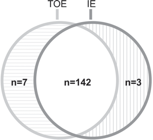
Table I. Clinical features of 708 patients suspected of infective endocarditis (IE) in relation to signs of IE on transoesophageal echocardiography (TOE).
Of the 149 patients with IE findings according to TOE, the native aortic valve was involved in 39 (26%), the mitral valve in 25 (17%), both valves in four (3%), and prosthetic valves in 39 (26%). Among the prosthetic valves, 56% were tissue valves and 44% mechanical prostheses. Right-heart IE was found by TOE in 42 (29% of the IE patients) and two thirds of these (n = 27) were related to pacemaker or ICD leads.
shows the distributions of patients with IE findings on TOE and of those with a final diagnosis of IE related to the number of the prospectively tested five clinical IE criteria, which in their joint absence were assumed to indicate absence of IE (Citation12). Forty percent (n = 280) of the IE suspected patients belonged to a group with none of the five tested clinical IE criteria. However, among these 280 patients 16 had findings of IE on TOE and 13 of these were given a diagnosis of IE by our multidisciplinary team. Thus, absence of all five tested clinical IE criteria had a predictive value of absence of IE of (267/280)*100% = 95%. However, 16 of our 149 patients with findings of IE on TOE (11%) and 13 of 145 with a final diagnosis of IE (9%) had none of the five tested IE criteria.
Table II. Distribution of the tested five clinical criteria of infective endocarditis (IE) (Citation12) related to findings of IE on transoesophageal echocardiography (TOE) and to the final diagnosis of IE made by a multidisciplinary team of specialists.
The clinical characteristics of our patients () also underwent multivariate analysis for identification of significant independent clinical markers of IE (). The two strongest independent markers of IE were: positive blood culture and vascular phenomena (embolism or vasculitis) followed by the four predisposing heart conditions listed in . Among our 708 patients, 175 (25%) did not exhibit any of our clinical IE markers listed in and 174 of these patients (99.4%) also had a TOE negative for IE as shown in . The remaining patient with none of the clinical IE markers of presented with intravenous drug abuse, continuous fever, and large tricuspid valve vegetations on TOE. The multidisciplinary team obviously established a diagnosis of IE in this patient ().
Table III. Independent predictors of positive findings of infective endocarditis (IE) on transoesophageal echocardiography in 708 patients suspected of IE.
Table IV. Distribution of the clinical markers of infective endocarditis (IE) listed in related to the occurrence of signs of IE on transoesophageal echocardiography (TOE) and related to the occurrence of final diagnoses of IE.
Discussion
In the original report on predicting absence of IE findings on TTE, no patient had such findings if all of five retrospectively identified clinical IE criteria were absent (Citation12). Our prospective study could not confirm this model. Although the negative predictive value of the model was high (95%) when applied to our population, 11% of our patients with IE findings on TOE and 9% of our patients with a final diagnosis of IE had none of the five risk factors (). Thus, if the original model had been applied to our patients, about 1/10 of our patients with IE would not have passed the threshold for receiving an echocardiogram. Omission of echocardiography in IE patients may indeed be deleterious. Not only is it important to diagnose the disease but just as decisive to identity life-threatening intracardiac destructions needing urgent heart surgery (Citation2,Citation15).
In the previous retrospective report (Citation12), TTE was used as reference while our patients had supplementary TOE which is recognised to improve IE diagnostics (Citation9,Citation16–19). TOE is considered superior to TTE in IE but TOE is neither 100% sensitive nor completely specific for IE (Citation4,Citation20). For the sake of comparison with the previous study on predicting absence of IE on TTE (Citation12) we found it relevant to relate the retrospectively derived five clinical IE criteria (Citation12) to TOE, our echocardiographic reference. However, as there is not necessarily identity between signs of IE on TOE and a final diagnosis of IE () it was also important to relate absence of the tested five clinical IE criteria to the final integrated diagnosis made by our multidisciplinary team. Among our 13 confirmed IE patients with none of the five tested clinical criteria of IE (), TOE findings categorised as major criteria according to the Duke classification were found in all 13 (Citation13,Citation14). Seven had native valve IE and six device related IE. All seven patients with native valve IE had large mobile vegetations. Five had IE confirmed at open-heart surgery. One patient refused any treatment because of advanced cancer and another patient with tricuspid valve IE achieved rapid remission on antibiotic treatment. The six device-related IE patients (three with ICD and three with DDD-pacemakers) all had continuous fever and large mobile lead and/or tricuspid valve vegetations on TOE. Lead extraction was performed in all six patients and three had superficial defects of the lead surface with segmental discoloration. The vegetations were not visible on the extracted leads but probably scraped off by the extraction catheter. All six patients were treated with antibiotics for some time before the lead extraction and cultures from the leads revealed bacterial growth in one patient only (enterococci).
The diverging results of our study and the original study (Citation12) on predicting absence of IE on echocardiography may partly be caused by differences between reference method (TTE versus TOE) and partly because of differing study populations. However, the proportion of patients with none of the tested five clinical criteria of IE was almost the same in the two studies (48% in the original study and 40% in our series) (Citation12). Yet, 20% of all our patients had a final diagnosis of IE compared to 9% in the original study (Citation12). This may suggest a higher referral threshold for the evaluation of possible IE in our centre. More selected patients in our series may also have contributed to the high proportion of 20% of IE patients in our study. However, only 3.5% of the patients were referred to our university clinic for further treatment of an already established definite diagnosis of IE.
Although our prospective study could not confirm the usefulness of the original clinical criteria for exclusion of IE we identified a new even simpler model. One fourth of our patients did not have any of the clinical IE markers: positive blood culture, vascular phenomena, or cardiac conditions predisposing to IE (). More than 99% of these patients had neither TOE findings of IE nor a confirmed diagnosis of IE (). However, one patient with none of our clinical IE markers of had typical echocardiographic findings of IE in terms of large tricuspid valve vegetations. This patient also had intravenous drug abuse and this combined with a temperature of 40°C would probably provoke an echocardiogram anytime despite absence of all our clinical risk features (Citation21).
Echocardiography is often requested in patients with fever despite a low clinical probability of IE although there is no general agreement on a definition of low probability IE (Citation7,Citation8,Citation11,Citation12). The new criteria derived from our analysis suggested that up to 25% of IE referrals to echocardiography may be patients with low probability IE ( and ). According to the results of our study it seemed possible to predict absence of IE by clinical judgement alone with a likelihood of 99.4% (95% confidence interval: 96.2–99.9%, 174/175, ). In view of echocardiographic priority assignment with regard to other heart diseases and cost-benefit it might seem relevant to consider simple clinical criteria before referral to echocardiography for possible IE (Citation22). However, echocardiography must not be postponed until the results of blood cultures are available (Citation4). The model is only applicable to patients in whom negative blood cultures are documented before a suspicion on IE is raised and the echocardiogram ordered. This is obviously a serious limitation of any model that includes negative blood cultures as a criterion of low probability of IE. It may take up to a week or more to exclude bacterial growth in a blood culture and many severe complications can take place during this period if echocardiography is postponed while awaiting the results of blood cultures. At the time of echocardiography, documented negative blood cultures existed in only 40% of the 25% of our patients who otherwise fulfilled the proposed new criteria for predicting absence of IE. Thus, at best our model could only save about 10% of echocardiograms in our IE suspected patients.
Limitations
Of the total study population of 852 patients clinically suspected of IE, 144 patients (17%) escaped analysis for various reasons. Patients who refused to participate in the study, patients excluded for logistic reasons, and patients with no complete data set hardly deviated from those included in the analysis. However, 43 patients could not be included because of cerebral incapacity. It would indeed be deleterious for our model of low probability of IE if some of these very ill patients actually suffered from IE but were denied echocardiography. Fortunately this was not the case as 40 of the 43 patients had at least one of the risk factors listed in . The remaining three patients had none of our clinical risk factors and none of them had a diagnosis of IE. Thus, if the 43 patients could have been included in the study this would not have impaired our proposed new clinical criteria of low probability of IE.
We could not confirm the usefulness of the previously reported criteria for omitting echocardiography in patients with suspected IE (Citation12). However, we cannot entirely rule out that the earlier reported criteria may be useful in a setting different from ours, e.g. in populations with more drug addicts and fewer device related IEs. After all, there is no decisive difference between the original criteria (Citation12) and our criteria of low probability of IE. The two models share important features such as negative blood culture, absence of embolism, and absence of valve prostheses.
It should also be emphasised that a less precisely defined patient selection already had taken place when the patients were referred to our department although we tried to describe this selection by classification of the main clinical referral reason in each individual patient (). Furthermore, we cannot entirely rule out that over- and underdiagnosis of IE took place in isolated cases despite meticulous discrimination between presence and absence of the disease based on integrated clinical and echocardiographic judgement of the expert panel of specialists. The diagnoses were also tested against the Duke criteria, and moreover no patient who initially was deemed negative of IE returned within the period of data collection or preparation of the manuscript with IE that had been overlooked in the first place.
Finally, it should be stressed that despite our study was prospective, the clinical criteria of low probability of IE were not predefined and should in principle be confirmed in prospective studies.
Conclusions
Our study showed that previously suggested clinical criteria proved insufficient for predicting absence of echocardiographic findings of IE. Using these criteria about 1/10 of our patients with confirmed IE would not have qualified for echocardiography.
Instead, our study generated a new model based on three simple clinical markers indicating low probability of IE if all three were absent in an individual patient. Thus, the likelihood of absence of IE was more than 99% in case of negative blood cultures, absence of vascular phenomena, and absence of recognised cardiac conditions predisposing to IE. In our study this included 25% of all patients. However, the model does not imply that echocardiography should be postponed until the results of blood cultures are available. It only applies to patients in whom negative blood cultures are documented when the suspicion on IE is raised and the echocardiogram is ordered. This is a serious limitation of our model of identification of low probability of IE. In our population only 40% of the 25% who otherwise fulfilled our criteria of low probability of IE had blood cultures that were known to be negative at the time of echocardiography. Thus, the model could only save about 10% of echocardiograms in our patients suspected of IE. Inasmuch as the most important clinical features of IE were included in our multivariate analysis it may seem questionable whether any superior clinical criteria of low probability of IE may appear. Accordingly, our study documented the irreplaceable role of early echocardiography in patients suspected of IE.
Acknowledgements
This work was supported by the Danish Heart Foundation, the Foundation of the Central Region of Denmark, and the foundations of King Christian den Tiende, Helga and Peter Korning, Aase and Ejnar Danielsen, Arvid Nilsson, Grosserer Valdemar Foersom, Carl and Ellen Hertz, the Institute of Clinical Medicine, Aarhus University, and the Research Council of Aarhus University Hospital, Skejby. The authors report no conflicts of interest. The authors alone are responsible for the content and writing of the paper.
Declaration of interest: The authors report no conflicts of interest. The authors alone are responsible for the content and writing of the paper.
References
- Habib G. Management of infective endocarditis. Heart. 2006;92:124–30.
- Habib G, Hoen B, Tornos P, Thuny F, Prendergast B, Vilacosta I, . Guidelines on the prevention, diagnosis, and treatment of infective endocarditis (new version 2009): The Task Force on the Prevention, Diagnosis, and Treatment of Infective Endocarditis of the European Society of Cardiology (ESC). Eur Heart J. 2009;30:2369–413.
- Knudsen JB, Fuursted K, Petersen E, Wierup P, Molgaard H, Poulsen SH, . Infective endocarditis: A continuous challenge. The recent experience of a European tertiary center. J Heart Valve Dis. 2009;18:386–94.
- Habib G, Badano L, Tribouilloy C, Vilacosta I, Zamorano JL, Galderisi M, . Recommendations for the practice of echocardiography in infective endocarditis. Eur J Echocardiogr. 2010;11:202–19.
- Westling K, Aufwerber E, Ekdahl C, Friman G, Gardlund B, Julander I, . Swedish guidelines for diagnosis and treatment of infective endocarditis. Scand J Infect Dis. 2007;39:929–46.
- Bonow RO, Carabello BA, Kanu C, de LA, Jr., Faxon DP, Freed MD, . ACC/AHA 2006 guidelines for the management of patients with valvular heart disease: A report of the American College of Cardiology/American Heart Association Task Force on Practice Guidelines (writing committee to revise the 1998 Guidelines for the Management of Patients With Valvular Heart Disease): Developed in collaboration with the Society of Cardiovascular Anesthesiologists: Endorsed by the Society for Cardiovascular Angiography and Interventions and the Society of Thoracic Surgeons. Circulation. 2006;114:e84–e231.
- Lindner JR, Case RA, Dent JM, Abbott RD, Scheld WM, Kaul S. Diagnostic value of echocardiography in suspected endocarditis. An evaluation based on the pretest probability of disease. Circulation. 1996;93:730–6.
- Kuruppu JC, Corretti M, Mackowiak P, Roghmann MC. Overuse of transthoracic echocardiography in the diagnosis of native valve endocarditis. Arch Intern Med. 2002;162:1715–20.
- Evangelista A, Gonzalez-Alujas MT. Echocardiography in infective endocarditis. Heart. 2004;90:614–7.
- Flachskampf FA, Daniel WG. Role of transoesophageal echocardiography in infective endocarditis. Heart. 2000;84:3–4.
- Robles P. Judicious use of transthoracic echocardiography in the diagnosis of infective endocarditis. Heart. 2003;89:1283–4.
- Greaves K, Mou D, Patel A, Celermajer DS. Clinical criteria and the appropriate use of transthoracic echocardiography for the exclusion of infective endocarditis. Heart. 2003;89: 273–5.
- Durack DT, Lukes AS, Bright DK, Alberts MJ, Bashore TM, Corey GR, . New criteria for diagnosis of infective endocarditis – utilization of specific echocardiographic findings. Am J Med. 1994;96:200–9.
- Li JS, Sexton DJ, Mick N, Nettles R, Fowler VG, Jr., Ryan T, . Proposed modifications to the Duke criteria for the diagnosis of infective endocarditis. Clin Infect Dis. 2000;30:633–8.
- Tingleff J, Egeblad H, Gotzsche CO, Baandrup U, Kristensen BO, Pilegaard H, . Perivalvular cavities in endocarditis: Abscesses versus pseudoaneurysms? A transesophageal Doppler echocardiographic study in 118 patients with endocarditis. Am Heart J. 1995;130:93–100.
- Kini V, Logani S, Ky B, Chirinos JA, Ferrari VA, John Sutton MG, . Transthoracic and transesophageal echocardiography for the indication of suspected infective endocarditis: Vegetations, blood cultures and imaging. J Am Soc Echocardiogr. 2010;23:396–402.
- Reynolds HR, Jagen MA, Tunick PA, Kronzon I. Sensitivity of transthoracic versus transesophageal echocardiography for the detection of native valve vegetations in the modern era. J Am Soc Echocardiogr. 2003;16:67–70.
- Chirillo F, Pedrocco A, De Leo A, Bruni A, Totis O, Meneghetti P, . Impact of harmonic imaging on transthoracic echocardiographic identification of infective endocarditis and its complications. Heart. 2005;91:329–33.
- Roe MT, Abramson MA, Li J, Heinle SK, Kisslo J, Corey GR, . Clinical information determines the impact of transesophageal echocardiography on the diagnosis of infective endocarditis by the duke criteria. Am Heart J. 2000;139: 945–51.
- Roberts WC, Buchbinder NA. Healed left-sided infective endocarditis: A clinicopathologic study of 59 patients. Am J Cardiol. 1977;40:876–88.
- Weisse AB, Heller DR, Schimenti RJ, Montgomery RL, Kapila R. The febrile parenteral drug user: A prospective study in 121 patients. Am J Med. 1993;94:274–80.
- Heidenreich PA, Masoudi FA, Maini B, Chou TM, Foster E, Schiller NB, . Echocardiography in patients with suspected endocarditis: A cost-effectiveness analysis. Am J Med. 1999;107:198–208.