Abstract
Objectives. To evaluate the efficacy and safety of rituximab in Japanese patients with systemic lupus erythematosus (SLE) and lupus nephritis (LN) who are refractory to conventional immunosuppressive therapy.
Methods. Eligible patients received rituximab at a dose of 1,000 mg at days 1, 15, 169, and 183, and were followed for 53 weeks after the first dose of rituximab. Overall disease activity was assessed monthly using a British Isles Lupus Assessment Group activity index. Patients with LN (Upr/Ucr ≥ 1.0 at study entry) were identified and their renal responses were evaluated according to the criteria proposed by the American College of Rheumatology (ACR) and the Lupus Nephritis Assessment with Rituximab (LUNAR) study.
Results. A total of 34 patients were enrolled and received at least one dose of rituximab. Decrease in disease activity was achieved in 16 (76.5%) out of 34 patients. In 17 patients with LN, response rates of 58.8% and 52.9% by ACR and LUNAR criteria, respectively, were seen. Successful steroid tapering was achieved in association with disease remission. Rituximab was well tolerated, and most adverse drug reactions were grade 1–2 in severity.
Conclusions. Rituximab is effective for treatment of Japanese patients with SLE and LN refractory to conventional therapy.
Introduction
Systemic lupus erythematosus (SLE) is a chronic autoimmune disease with variable manifestations. Lupus nephritis (LN) is one of the most serious complications of SLE and develops in 50–60% of SLE patients within the first 10 years of disease onset [Citation1]. LN is one of the main causes of morbidity and mortality in SLE patients and significantly reduces life expectancy to 88% at 10 years [Citation2]. Current treatments for SLE include immunosuppressive drugs such as corticosteroids and cytostatics; however, the disease may become refractory to conventional treatments over time. Long-term life expectancy and renal survival of SLE patients with LN have progressively increased with the introduction of newer treatments, earlier referral, and improved diagnostic criteria [Citation3].
A number of observational, open-label trials revealed that B-cell depletion therapy with rituximab, a chimeric anti-CD20 antibody, either as monotherapy or in combination with immunosuppressive medications, was safe and effective for the treatment of SLE and LN [Citation4–8]. In contrast, two large-scale, double-blind, placebo-controlled phase III clinical studies to examine the efficacy and safety of rituximab in patients with active SLE (Exploratory Phase II/III SLE Evaluation of Rituximab [EXPLORER]) [Citation9] or SLE with active proliferative LN (Lupus Nephritis Assessment with Rituximab [LUNAR]) [Citation10] did not demonstrate superiority of rituximab over placebo. However, a greater number of subjects in the rituximab arms of the EXPLORER and LUNAR studies achieved remission and showed serologic improvement in serum complement C3 and C4 levels, and reduced anti-double-stranded DNA (dsDNA) antibody levels than did those receiving placebo [Citation10,Citation11].
To date, rituximab has not been approved for the treatment of LN by the US Food and Drug Administration or the European Medicines Agency; nevertheless, the American College of Rheumatology (ACR) [Citation12] and European League Against Rheumatism and European Renal Association-European Dialysis and Transplant Association (EULAR/ERA-EDTA) guidelines [Citation13] recommend rituximab for the treatment of patients with LN who are refractory to conventional immunosuppressive therapy including cyclophosphamide (CYC) and mycophenolate mofetil (MMF).
To examine the safety and efficacy of rituximab in Japanese SLE patients, we have conducted a multicenter, open-label, phase II clinical trial of rituximab in patients with SLE refractory to conventional immunosuppressive therapy. To evaluate the applicability of the ACR and EULAR/ERA-EDTA guidelines to Japanese SLE patients with LN, a post-hoc analysis of the safety and efficacy of rituximab was done in a subset of LN patients from the study.
Materials and methods
Study design and patients
An open-label, multicenter, phase II study of rituximab in Japanese patients with refractory SLE was conducted between July 2007 and May 2010 in seven centers across Japan. The study was performed in accordance with the principles of the Declaration of Helsinki and Japanese GCP requirements for conducting clinical trials. The study was approved by the local ethics committees of all seven participating institutions and was registered in the University Hospital Medical Information Network database (UMIN000000763).
Patients underwent an initial screening visit and eligible patients were enrolled after giving informed consent. Inclusion criteria included age, 16–75 years; a history of meeting the 1997 ACR criteria for SLE [Citation14] with positive antinuclear antibodies (ANAs); and active disease in any organ system at screening despite at least 2 weeks of treatment with prednisolone > 0.75 mg/kg/day. Patients were excluded if they had a history of cancer or serious recurrent or chronic infection; uncontrolled medical disease; previous treatment with B-cell-targeted therapy; aspartate aminotransferase or alanine aminotransferase levels > 2.5-fold of the upper limit of normal (ULN); amylase or lipase level > 2-fold of the ULN; neutrophil counts < 1.0 × 10³ μL; positive results of hepatitis B or hepatitis C serology; hemoglobin concentration < 7 gm/dL (unless caused by hemolytic anemia due to SLE); platelet counts < 10,000/μL; and serum creatinine levels > 2.5 mg/dL.
Treatment
Patients continued to receive corticosteroid and any concomitant immunosuppressants at the same dose used before study entry. For those with high disease activity at screening, a dose increase in corticosteroid was allowed at the investigator's discretion, and should have started at least 7 days before initiation of rituximab treatment. Rituximab was administered at a dose of 1,000 mg given 2 weeks apart (days 1 and 15), which was repeated after 6 months from the first rituximab administration (days 169 and 183), similar to the EXPLORER and LUNAR studies (a total of four doses). To reduce or avoid infusion-related reactions associated with rituximab administration, acetaminophen (400 mg, po), chlorpheniramine maleate (2 mg, po), and methylprednisolone (100 mg, iv) were administered before each rituximab infusion. Once clinical improvement was observed, the dose of corticosteroid was tapered by 20% every 2–4 weeks and not allowed to re-increase once tapered.
Assessments and endpoints
Patients were followed for 53 weeks after the first rituximab administration on a monthly basis. Systemic disease activity was evaluated by the British Isles Lupus Assessment Group (BILAG) activity index [Citation15]. Periodic laboratory testing of the levels of circulating B cells; complements C3, C4, and CH50; and anti-dsDNA antibodies was also performed.
Overall disease activity was assessed in accordance with the approach reported by Dr. Isenberg of University College of London [Citation6]. Disease remission was defined as “a change from BILAG A or B score to a BILAG C or D score in every organ system.” Partial remission was defined as “a change from a BILAG A or B score to a C or D score in at least one organ system, but with presence of one BILAG A or B score in another organ system.” No improvement was defined as “a BILAG A or B score that remained unchanged at week 53.” For patients with involvement of only one organ, remission was a change from a BILAG A or B score to C or D score, and partial remission was a change from a BILAG A score to B score.
Renal responses were graded as complete renal response (CRR), partial renal response (PRR), or no response (NR) based on the criteria used in the LUNAR study [Citation10] and ACR guidelines [Citation16]. The overall renal response rate (ORR) was defined as “the sum of the CRR and PRR.” Based on the LUNAR study, CRR was defined as “normal serum creatinine levels” if abnormal at baseline, or serum creatinine levels ≤ 115% of baseline if normal at baseline; inactive urinary sediment (< 5 red blood cells (RBCs)/high-power field (hpf) and absence of RBC casts); and urinary protein to urinary creatinine (Upr/Ucr) ratio < 0.5. PRR was defined as “serum creatinine levels ≤ 115% of baseline; RBCs/hpf ≤ 50% above baseline with no RBC casts; and ≥ 50% decrease in Upr/Ucr ratio to < 1.0 (if baseline Upr/Ucr ratio ≤ 3.0) or to ≤ 3.0 (if baseline Upr/Ucr ratio > 3.0).” Based on the ACR guidelines, CRR was defined as “a ≥ 25% increase in estimated glomerular filtration rate (eGFR) if baseline values were abnormal; inactive urinary sediment (< 5 RBCs/hpf and absence of RBC casts); and at least a 50% decrease in Upr/Ucr ratio to 0.2.” PRR was defined as “stable eGFR (at least 75% of baseline value); inactive urinary sediment; and at least a 50% decrease in Upr/Ucr ratio to 0.2–2.0.” Patients were classified as NR if CRR or PRR criteria were not met. Patients who received any additional therapies for disease control, including dose increase of steroid and/or immunosuppressants, were also classified as NR.
An adverse event (AE) was defined as “any untoward medical occurrence (e.g., sign, symptom, disease, syndrome, concurrent illness, or clinically significant abnormal laboratory finding) that newly emerged or worsened during the study period relative to pretreatment baseline, regardless of the suspected cause.” AEs were graded according to the National Cancer Institute Common Terminology Criteria for Adverse Events (CTCAE) Version 3.0. Adverse drug reactions (ADRs) were defined as “any AEs for which an association with rituximab could not be completely ruled out.” Infusion reactions were defined as “any ADRs occurring during or within 24 h following the completion of rituximab infusion.” Serious ADRs were defined as “those that resulted in death, were life threatening, or required prolonged inpatient hospitalization.”
Statistical analyses
The differences in median values of BILAG activity score, SLE biomarkers (i.e., C3, C4, CH50, anti-DNA antibodies, urinalysis, and eGFR), and steroid doses at screening and at week 53 were examined by Wilcoxon's matched pairs signed-rank test to determine statistical significance. All statistical analyses were carried out using SAS software (Cary, NC, USA). Patients with Upr/Ucr ratio ≥ 1.0 at screening were included in the post-hoc analysis and percentages and 95% confidence intervals of subjects who met the response criteria were calculated. For those who dropped out of the study before week 53 for any reason, clinical data obtained at the last observation were used for analyses applying the last observation carried forward approach.
Results
Patient characteristics
A total of 34 patients were enrolled in this phase II study. Baseline demographic and clinical characteristics are summarized in . Of the 34 patients enrolled in the phase II study, 17 had renal involvement with Upr/Ucr ≥ 1.0 at screening and were included in the post-hoc analysis; of these, 10 had class III/IV LN based on the International Society of Nephrology or ISN/Renal Pathology Society or RPS classification [Citation17], one had class IIb LN, one had class VI LN, and five had never undergone a histopathological examination. Eight patients dropped out of the study prematurely before week 53: two patients for SLE flare, two were lost to follow-up, and one patient each for use of prohibited medication for treatment of concurrent illness other than SLE, withdrawal of informed consent, an AE not related to rituximab, and an ADR related to rituximab. Of the two patients who dropped out because of SLE flare, one was a 30-year-old female patient with highly active class IV LN who presented with massive proteinuria (Upr/Ucr = 10.0) and hematuria (> 50 RBCs/hpf) at study entry. She had LN for 7 months and was previously treated with steroid pulse, plasma exchange, and hemodialysis. High disease activity persisted even after the fourth dose of rituximab, and she was removed from the study to receive another intentional therapy. The other patient was a 22-year-old female patient with newly diagnosed class III LN. She presented at study entry with remarkably high levels of serum autoantibodies: ANA = 1,280 IU/mL, anti-dsDNA = 24 U/mL, anti-Sm = 500 U/mL, anti-RNP = 500 U/mL, and anti-SS-A/Ro = 18.7 U/mL. She was removed from the study after two doses of rituximab as per the investigator's judgment to receive plasma exchange.
Table 1. Baseline characteristics of the enrolled patients (n = 34).
Clinical efficacy
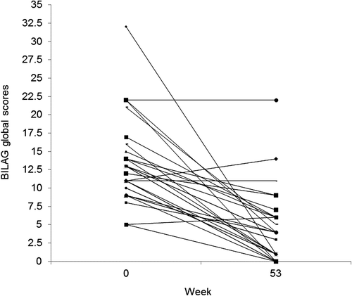
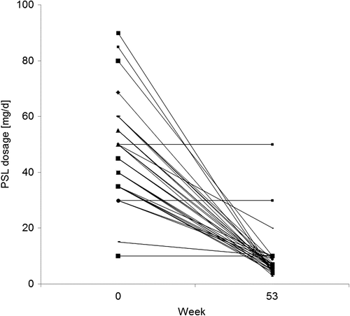
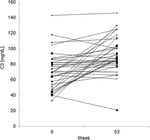
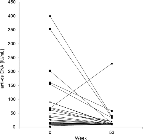
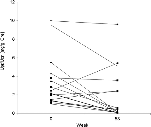
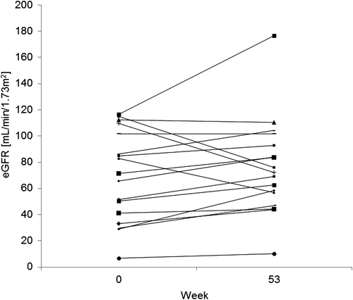
Peripheral B cells were depleted rapidly after the first course of rituximab treatment in all 34 patients (). Overall disease activity as measured by the BILAG index improved after rituximab treatment. A total of 26 of 34 patients (76.5%) responded to rituximab therapy at week 53; of these, 16 (47.1%) achieved remission and 10 (29.4%) achieved partial remission. BILAG global score in 34 patients decreased significantly from a median of 12.5 (interquartile range [IQR]: 10.0–14.0) at baseline to 3.5 (IQR: 1.0–6.0) at week 53 (P < 0.0001) (). A significant dose reduction in concomitant prednisolone was achieved, from 45.0 mg/day (IQR: 35.0–55.0) at baseline to 6.0 mg/day (IQR: 5.0–8.9) at week 53 (P < 0.0001) (). Serologic improvements were also observed, with a significant increase in C3 levels (69.0 mg/dL [IQR: 48.8–82.0] at baseline vs. 88.5 mg/dL [IQR: 81.5–103.8] at week 53; P < 0.0001; ); C4 (16.5 mg/dL [IQR: 8.0–322.0] at baseline vs. 22.0 mg/dL [IQR: 18.0–28.0] at week 53; P < 0.0001, data not shown); CH50 (31.2/mL [IQR 14.7–39.4] at baseline vs. 39.0/mL [IQR: 34.0–46.7] at week 53; P = 0.0027, data not shown); and anti-dsDNA antibody levels (20.5 IU/mL [IQR: 10.0–67.8] at baseline vs. 10.0 IU/mL [IQR: 10.0–12.8] at week 53; P < 0.0001; ). In 17 patients with renal involvement, the median value of Upr/Ucr decreased from 2.2 (IQR: 1.4–3.8) at baseline to 0.4 (IQR: 0.10–2.44) at week 53 (P = 0.0068; ). eGFR remained stable, with a median value of 71.3 mL/min/1.73 m2 (IQR: 41.2–101.5) at baseline versus 72.3 mL/min/1.73 m2 (IQR: 56.8–93.0) at week 53 (P = 0.1928; ). The renal response rates in accordance with LUNAR and ACR criteria for all 17 patients with LN and for the 10 patients with histologically confirmed class III/IV LN are presented in . Response rate was higher in the 10 patients with class III/IV LN than in all 17 LN patients. While the exact reason for this is not clear, patients with class III/IV LN had shorter disease duration (median: 16 months vs. 53 months), although the difference was not significant because of the small sample size. Only one patient had class VI LN, which is defined as advanced-stage LN with ≥ 90% of glomeruli globally sclerosed without residual activity. Patients with class VI LN are not expected to respond to drug therapies. Therefore, we speculate that the class III/IV LN population enrolled in the study could have had reversible lesions that contributed to their apparent response rate. No pre-study patient characteristics were found to be associated with response in this study, because of the small sample size.
Figure 1. B-cell response to rituximab. (a) CD19 + cells. (b) CD20 + cells. Patients received rituximab at a dose of 1,000 mg for a total of four doses at weeks 1, 3, 25, and 27.
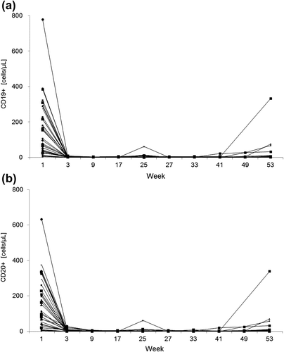
Table 2. Renal response to rituximab treatment.
Safety
A total of 154 ADRs with a suspected relationship to rituximab were observed. Most of the ADRs were mild to moderate with grade 1 or 2 severity, and only ten were grade 3 or 4 (). One patient who developed grade 3 cerebral infarction had presented at study entry with neuropsychiatric lupus with continuous disabling headache. This patient dropped out of the study as per the investigator's judgment. Grade cholecystitis, endometritis, and hypoferric anemia were observed in one patient who had presented at study entry with concurrent cholelithiasis, endometrial hyperplasia, and moderate hypoferric anemia. In both cases, the AEs were considered to most likely be associated with the underlying diseases or concomitant illnesses; however, the relationship to rituximab was not completely ruled out by the treating investigators. All of the ADRs were reversible and resolved by supportive treatments. There were 14 infusion-related ADRs, all of which were mild to moderate with grade 1 or 2 severity (). All of the symptoms cleared spontaneously and none required treatment discontinuation. None of the patients who participated in this study died.
Table 3. Grade 3/4 ADRs.
Table 4. Infusion reactions.
Discussion
The outcome of this Japanese study demonstrates that rituximab is effective for disease control and enables a reduction of corticosteroid dose in a cohort of Japanese SLE patients including a subset with LN who were refractory to conventional immunosuppressive therapy. Rituximab was also well tolerated, and all ADRs observed were previously known and mild to moderate in severity. The efficacy of rituximab in the current study is consistent with the findings from a retrospective analysis at University College London Hospital, in which 89% of patients with SLE (N = 50) achieved remission or partial remission with rituximab [Citation6]. A prospective registry study in France showed a 77% ORR with rituximab in 113 evaluable SLE patients [Citation18]. In another prospective registry of rituximab use in Europe, a 67% ORR was achieved in 164 patients with biopsy-proven LN [Citation19]. It is interesting that these registry studies were intended to evaluate the usefulness of rituximab off-label use in actual practice settings, and found favorable responses and acceptable tolerability for treatment of SLE and LN. A recent systematic analysis of 26 studies of rituximab in 300 patients with LN revealed effective remission, with a 74% ORR in patients not sufficiently controlled with standard treatment [Citation20]. Two other systematic reviews of off-label use of rituximab in SLE also suggested the usefulness of rituximab, not only for LN but also for other clinical manifestations of SLE [Citation21,Citation22]. Taken together, rituximab is worth considering as a therapeutic option for treatment of Japanese patients with SLE and LN refractory to conventional therapy. Further, we showed that the treatment guidelines from ACR and EULAR/ERA-EDTA are also applicable to Japanese patients.
Previous randomized, double-blind, placebo-controlled trials of rituximab in patients with moderate and severe extra-renal SLE (EXPLORER) [Citation9] or with class III or IV LN (LUNAR) [Citation10] failed to show statistically significant improvements with rituximab over placebo. These randomized controlled trials addressed the hypothesis that the addition of rituximab to the standard of care was superior to standard of care alone in controlling SLE activity. Several reasons have been considered to explain the failure of these trials; the aggressive background immunosuppressive therapy, including high-dose corticosteroids and full-dose MMF, may have masked any significant clinical benefit of rituximab [Citation23]. It is noteworthy that, despite the results of these studies, much attention is still paid to rituximab as an option for treatment of SLE [Citation24,Citation25].
In our study, significant dose reduction of concomitant steroid was achieved with rituximab treatment. It is interesting to note that this steroid-tapering effect of rituximab has been reported previously, not only in LN [Citation26,Citation27] but also in other diseases such as anti-neutrophil cytoplasmic antibody or ANCA-associated vasculitis [Citation28], pemphigus [Citation29], and steroid-dependent nephrotic syndrome [Citation30,Citation31]. Long-term steroid use is associated with clinically significant systematic AEs, and therefore steroid-avoiding treatment protocols consisting of rituximab and MMF have been studied in LN in an investigator-initiated clinical study (Rituxilup). Preliminary results seem promising [Citation32], and a prospective, international, multicenter, randomized controlled study is now underway [Citation33]. Another investigator-initiated, international, multicenter study named RING: Rituximab for Lupus Nephritis with Remission as a Goal, in which rituximab therapy is repeated every 6 months for a total of four courses, is also ongoing [Citation34]. Further studies are needed to clarify the optimal use of rituximab for the treatment of LN, and it is highly expected that the international cooperative studies of rituximab, which have been carefully designed based on the lessons learned from previous studies, may give us new insights into rituximab use for treatment of LN.
Acknowledgments
This study was sponsored by Zenyaku Kogyo Co., Ltd. We acknowledge the medical writing support of Dr. Stacey Tobin and Dr. J. Ludovic Croxford of Edanz Group Ltd.
Conflict of interest
K. Endo and N. Mashino are employees of Zenyaku Kogyo Co., Ltd. Y. Tanaka has received consulting fees, speaking fees, and/or honoraria from Abbvie, Daiichi-Sankyo, Chugai, Takeda, Mitsubishi-Tanabe, Bristol-Myers, Astellas, Eisai, Janssen, Pfizer, Asahi-kasei, Eli Lilly, GlaxoSmithKline, UCB, Teijin, MSD, and Santen, and has received research grants from Mitsubishi-Tanabe, Takeda, Chugai, Astellas, Eisai, Taisho-Toyama, Kyowa-Kirin, Abbvie, and Bristol-Myers. The other authors have no conflicts of interest to declare.
References
- Dooley MA, Aranow C, Ginzler EM. Review of ACR renal criteria in systemic lupus erythematosus. Lupus 2004; 13(11):857–60.
- Cervera R, Khamashta MA, Font J, Sebastiani GD, Gil A, Lavilla P, et al; European Working Party on Systemic Lupus Erythematosus. Morbidity and mortality in systemic lupus erythematosus during a 10-year period: a comparison of early and late manifestations in a cohort of 1,000 patients. Medicine (Baltimore). 2003;82(5):299–308.
- Moroni G, Quaglini S, Gallelli B, Banfi G, Messa P, Ponticelli C. Progressive improvement of patient and renal survival and reduction of morbidity over time in patients with lupus nephritis (LN) followed for 20 years. Lupus. 2013;22(8):810–8.
- Tanaka Y, Yamamoto K, Takeuchi T, Nishimoto N, Miyasaka N, Sumida T, et al. A multicenter phase I/II trial of rituximab for refractory systemic lupus erythematosus. Mod Rheumatol. 2007;17(3):191–7.
- Leandro MJ, Edwards JC, Cambridge G, Ehrenstein MR, Isenberg DA. An open study of B lymphocyte depletion in systemic lupus erythematosus. Arthritis Rheum. 2002;46:2673–7.
- Lu TY, Ng KP, Cambridge G, Leandro MJ, Edwards JC, Ehrenstein M, Isenberg DA. A retrospective seven-year analysis of the use of B cell depletion therapy in systemic lupus erythematosus at University College London Hospital: the first fifty patients. Arthritis Rheum. 2009;61(4):482–7.
- Jonsdottir T, Gunnarsson I, Mourao AF, Lu TY, van Vollenhoven RF, Isenberg D. Clinical improvements in proliferative vs membranous lupus nephritis following B-cell depletion: pooled data from two cohorts. Rheumatology (Oxford). 2010;49(8):1502–4.
- Melander C, Sallee M, Trolliet P, Candon S, Belenfant X, Daugas E, et al. Rituximab in severe lupus nephritis: early B-cell depletion affects long-erm renal outcome. Clin J Am Soc Nephrol. 2009;4(3):579–87.
- Merrill JT, Neuwelt CM, Wallace DJ, Shanahan JC, Latinis KM, Oates JC, et al. Efficacy and safety of rituximab in moderately-to-severely active systemic lupus erythematosus: the randomized, double-blind, phase II/III systemic lupus erythematosus evaluation of rituximab trial. Arthritis Rheum. 2010;62(1):222–33.
- Rovin BH, Furie R, Latinis K, Looney RJ, Fervenza FC, Sanchez-Guerrero J, et al. Efficacy and safety of rituximab in patients with active proliferative lupus nephritis: the lupus nephritis assessment with rituximab study. Arthritis Rheum. 2012;64(4):1215–26.
- Tew GW, Rabbee N, Wolslegel K, Hsieh H-J, Monroe JG, et al. Baseline autoantibody profiles predict normalization of complement and anti-dsDNA autoantibody levels following rituximab treatment in systemic lupus erythematosus. Lupus. 2010;19(2):146–57.
- Hahn BH, McMahon MA, Wilkinson A, Wallace WD, Daikh DI, Fitzgerald JD, et al. American College of Rheumatology guidelines for screening, treatment, and management of lupus nephritis. Arthritis Care Res. 2012;64(6):797–808.
- Bertsias GK, Tektonidou M, Amoura Z, Aringer M, Bajema I, Berden JHM, et al. Joint European League Against Rheumatism and European Renal Association-European Dialysis and Transplant Association (EULAR/ERA-EDTA) recommendations for the management of adult and paediatric lupus nephritis. Ann Rheum Dis. 2012;71(11):1771–82.
- Hochberg MC. Updating the American College of Rheumatology revised criteria for the classification of systemic lupus erythematosus. Arthritis Rheum 1997;40(9):1725.
- Hay EM, Bacon PA, Gordon C, Isenberg DA, Maddison P, Snaith ML, et al. The BILAG index: a reliable and valid instrument for measuring clinical disease activity in systemic lupus erythematosus. Q J Med. 1993;86(7):447–58.
- Renal Disease Subcommittee of the American College of Rheumatology Ad Hoc Committee on Systemic Lupus Erythematosus Response Criteria. The American College of Rheumatology response criteria for proliferative and membranous renal disease in systemic lupus erythematosus clinical trials. Arthritis Rheum. 2006;54(2): 421–32.
- Markowitz GS, D’Agati VD. The ISN/RPS 2003 classification of lupus nephritis: an assessment at 3 years. Kidney Int. 2007;71(6):491–5.
- Terrier B, Amoura Z, Ravaud P, Hachulla E, Jouenne R, Combe B, et al. Safety and efficacy of rituximab in systemic lupus erythematosus: results from 136 patients from the French AutoImmunity and Rituximab registry. Arthritis Rheum. 2010;62(8):2458–66.
- Díaz-Lagares C, Croca S, Sangle S, Vital EM, Catapano F, Martínez-Berriotxoa A, et al.; UK-BIOGEAS Registry. Efficacy of rituximab in 164 patients with biopsy-proven lupus nephritis: pooled data from European cohorts. Autoimmun Rev. 2012;11(5):357–64.
- Weidenbusch M, Rommele C, Schrottle A, Anders HJ. Beyond the LUNAR trial. Efficacy of rituximab in refractory lupus nephritis. Nephrol Dial Transplant. 2013;28(1):106–11.
- Murray E, Perry M. Off-label use of rituximab in systemic lupus erythematosus: a systematic review. Clin Rheumatol. 2010;29(7):707–16.
- Ramos-Casals M, Soto MJ, Cuadrado MJ, Khamashta MA. Rituximab in systemic lupus erythematosus: A systematic review of off-label use in 188 cases. Lupus. 2009;18(9):767–76.
- Ready V, Jayne D, Close D, Isenberg D. B-cell depletion in SLE: clinical and trial experience with rituximab and ocrelizumab and implications for study design. Arthritis Res Ther. 2013;15(Suppl 1):S2.
- Murphy G, Lisnevskaia L, Isenberg D. Systemic lupus erythematosus and other autoimmune rheumatic diseases: challenges to treatment. Lancet. 2013;382:809–18.
- Lisnevskaia L, Murphy G, Isenberg D. Systemic lupus erythematosus. Lancet. 2014;384:1878–88.
- Pepper R, Griffith M, Kirwan C, Levy J, Taube D, Pusey C, et al. Rituximab is an effective treatment for lupus nephritis and allows a reduction in maintenance steroids. Nephrol Dial Transplant. 2009;24(12):3717–23.
- Ezeonyeji A, Isenberg D. Early treatment with rituximab in newly diagnosed systemic lupus erythematosus patients: a steroid-sparing regimen. Rheumatology 2012;51(3):476–81.
- Stone J, Merkel P, Spiera R, Seo P, Langford C, Hoffman G, et al. Rituximab versus Cyclophosphamide for ANCA-associated vasculitis. N Eng J Med. 2010;363(3):221–32.
- Joly P, Mouquet H, Roujeau JC, D'lncan M, Gilbert D, Jacquot S, et al. A single cycle of rituximab for treatment of severe pemphigus. N Eng J Med. 2007;357(6):545–52.
- Iijima K, Sako M, Nozu K, Mori R, Tuchida N, Kamei, K, et al. Rituximab for childhood-onset, complicated, frequently relapsing nephrotic syndrome or steroid-dependent nephrotic syndrome: a multicentre, double-blind, randomized, placebo-controlled trial. Lancet 2014;384(9950):1273–81.
- Takei T, Itabashi M, Moriyama T, Kojima C, Shiohira S, Shimizu A, et al. Effect of single-dose rituximab on steroid-dependent minimal-change nephrotic syndrome in adults. Nephrol Dial Transplant. 2013;28(5):1225–32.
- Condon M, Ashby D, Pepper R, Cook H, Levy J, Griffith M, et al. Prospective observational single-centre cohort study to evaluate the effectiveness of treating lupus nephritis with rituximab and mycophenolate mofetil but no oral steroids. Ann Rheum Dis. 2013;72(8):1280–6.
- Imperial College London. Trial of Rituximab and Mycophenolate Mofetil Without Oral Steroid for Lupus Nephritis (RITUXILUP). NCT01773616. Available from: www.ClinicalTrials.gov.
- Houssiau FA, on behalf of European Working Party on Systemic Lupus Erythematosus Lupus Nephritis Trial Network. RING: rituximab for lupus nephritis with remission as a goal. NCT01673295. Available from: www.ClinicalTrials.gov.

