Abstract
Background: Accelerometry is increasingly used to assess physical activity (PA) in patients with chronic obstructive pulmonary disease (COPD). It is not known how the relationship of PA to clinical results depends on the position of the PA sensor. Methods: We assessed the effect of monitor position by measuring lower extremity (ankle), upper extremity (wrist) and total body movement (hip) in 52 patients with severe COPD (mean [± SD] age, 62 ± 10 years; FEV1, 38 ± 12% predicted) undergoing long-term oxygen therapy with and without walkers during a pulmonary rehabilitation (PR) program. Sensors were worn 8.5 ± 3.1 days and data was compared to the BODE score and the 6-minute walk distance (6MWD) assessed at the beginning and end of the PR. Results: Mean ankle PA was moderately related to the 6MWD, irrespective of patients being equipped with a walker or not (p < 0.05). Mean PA values were considerably lower in COPD patients with walker compared to patients without for all sensor positions. No significant association was observed between mean hip PA data and 6MWD; however, hip and ankle PA data were moderately related in walker-free and strongly related in walker patients (p < 0.01). In a multivariate regression model only ankle activity was significantly associated with the BODE score (p < 0.01). Conclusion: The sensor position had a significant impact on the association between PA recordings and the 6MWD in very severe COPD. In our setting, ankle measurement seemed to best reflect the clinical state of patients.
| Abbreviations | ||
| 6MWD | = | 6-minute walk distance |
| BMI | = | body mass index |
| CI | = | confidence interval |
| FEV1 | = | forced expiratory volume in 1s |
| IQR | = | interquartile range |
| LTOT | = | long-term oxygen therapy |
| MMRC | = | modified Medical Research Council dyspnea scale |
| PA | = | physical activity |
| PR | = | pulmonary rehabilitation |
| SD | = | standard deviation |
| VC | = | vital capacity |
| VMU | = | vector magnitude units. |
Introduction
Physical activity (PA) is an important measure in COPD, and a reduction in PA is currently hypothesized to be an early, though unspecific, symptom (Citation1). Reduced PA as assessed by subjective means is related to lung function decline in smokers (Citation2) and is linked with an increased risk of hospitalization and mortality in COPD (Citation3, 4). Accelerometers have the potential to improve the assessment of PA in COPD on the basis of objective measures (Citation5). By means of motion sensors and/or questionnaires it has been shown that the level of PA is progressively diminishing across different stages of the disease, reaching the lowest level in the most severely ill patients undergoing long-term oxygen therapy (LTOT) (Citation6–11, Citation14). However, the relationship between PA, COPD severity and clinical characteristics has not been satisfactorily evaluated, particularly with regard to methodological issues. For example, ambiguous results were reported for the relationship between objectively assessed PA and airway obstruction or the 6MWD across a number of studies (Citation8, Citation12, Citation13, Citation15–21). The activity monitors used and their placement varied between studies.
Using an uniaxial accelerometer at the ankle, Walker and colleagues found a weak correlation between mean activity and 6MWD (Citation21). Watz et al. reported moderate correlations between 6MWD and PA level as well as time in moderate activity using a biaxial sensor worn at the upper arm (Citation8). Belza, Steele and colleagues found strong correlations between 6MWD and daily activity measured by triaxial accelerometers worn at the hip (Citation16, Citation20). Likewise did Pitta et al. for walking and standing time (Citation6). It is not clear whether the inconsistency in findings regarding the relationship between PA and 6MWD is a function of study design, patient numbers or the device and position used to measure activity. The study goes on to attempt to answer this question.
Possibly the different results are partially due to the use of different sensors or differences in the investigated populations.
To which extent associations between clinical characteristics and PA are affected by a specific monitor placement has not been systematically assessed. Only few results are available regarding the comparison of sensor data acquired at different body locations (Citation21–23), although this seems to be a key question in order to compare recordings, especially in older patients who often suffer from disorders limiting their mobility. Furthermore, COPD patients with walking aids have been generally excluded from participation in studies on PA, as these aids may interfere with the assessment by a single activity monitor. The level of activity assessed at upper vs. lower limbs could well be different in these patients.
Based on these considerations, we determined PA using accelerometers simultaneously at the upper and lower extremities as well as at the body center in patients with severe COPD under well-defined conditions, during a pulmonary rehabilitation (PR) program. We then examined the association between the data gathered at different positions and assessed their relationship to clinical measures of COPD severity. Additionally, the study attempts to examine the effect of walker use on activity measured at three different positions and determine the validity of these measurements.
Material and Methods
Study population
A sample of one hundred patients suffering from COPD and undergoing LTOT with or without walking aids (rollator) who had been admitted for a 3-week in-patient PR program was approached. 58 patients (23 females) agreed to participate in the study. Reasons for refusal were the complex protocol involving the burden to attach and detach the set of motion sensors on a daily basis, exposure due to wearing the sensors, the lack of perceiving subjective benefit, severe medical condition, technical complexity, unwillingness to sign the required consent and difficulty in understanding the goal of the study. All patients had the diagnosis of smoking-related COPD of stage IV according to the GOLD classification (Citation24) with a demand of at least 1 liter of oxygen per minute at rest.
All patients were instructed to increase O2 supply during training sessions, if a desaturation was discovered during walking or ergometer test by means of blood gas analysis or pulse oximetry. PaO2 should remain above 60 mmHg or 90% sO2 during exercise according to the guideline of the German Society for Pneumology (Citation25); O2 was prescribed accordingly. Patients with type 2 respiratory failures were excluded, as well as those who had not been mobile due to medical conditions. Furthermore, subjects with less than four valid days of accelerometer recording from at least two measurement positions were excluded which occurred in five patients who did not wear the sensors regularly and in one patient in whom all three sensors failed. Therefore, a total of 52 patients were available for final analysis (). Patients were grouped into those using a walker (Group A) and those without (Group B). The study was approved by the local ethics commission, all procedures were performed according to the ethics guidelines of the Declaration of Helsinki and all individuals gave informed consent.
Table 1. Characteristics of patients
Study protocol and clinical characteristics
At the start and at the end of the 3-week in-patient PR program, spirometry was performed (MasterScreen, Jaeger, Höchberg, Germany). Exercise capacity was measured as 6MWD (Citation26) under pulse oximetry monitoring according to current guidelines (Citation27). Patients’ self-perceived degree of breathlessness at the end of the test was assessed using the modified Borg CR 10 scale (Citation28). Dyspnea at rest was determined by the modified Medical Research Council dyspnea scale (MMRC) (Citation29). PA was measured using accelerometers placed at the ankle, wrist and hip during the PR program including the scheduled training periods. Unlike pedometers, accelerometers are capable to detect particularly slow motions more precisely (Citation30). Patients were instructed to wear the sensors for at least seven and up to ten days during waking hours, except for personal hygiene actions.
Pulmonary rehabilitation program
The inpatient PR program at the clinic Bad Reichenhall primarily consisted of daily exercise training and physiotherapy focusing on COPD education sessions on breathing exercises over a period of 3–4 weeks. Each patient received strength and endurance training for 30 minutes per session under supervision 3–4 times per week, as well as a daily physiotherapy with varying movement intensity. Endurance training was mainly devoted to the lower limb, the upper extremities were trained in self-exercises for 10–15 minutes daily. If capable, patients were asked to do additional walking exercise twice a day for at least 15 minutes on their own. The amount of therapies between subjects and study groups was largely similar, with varying training intensities. However, exercise training in severely ill patients with COPD was individualized according to the different physical limitations.
Accelerometers
PA levels of the upper (wrist) and lower extremities (ankle) were assessed using the uniaxial capacitive GT1M accelerometer (Actigraph LLC, Pensacola FL, firmware version 4.2.0), attached by means of a snug elastic belt. The belts were labeled in order to avoid misplacement. The GT1M is a small, lightweight (3.8 × 3.7 × 1.8 cm3, 27 g), micro-electromechanical system which measures acceleration in the vertical plane (Citation31). PA was expressed as “activity counts,” which is a quantification of the amplitude and frequency of the detected accelerations in time intervals of 1 minute epochs (Citation31). The Actigraph is the most widely used device in epidemiological research; its technical specifications and validity are known (Citation32, 33).
Trunk movements were assessed by the piezoelectric triaxial RT3 accelerometer (Stayhealthy, Monrovia, CA, firmware version 0.6), worn in a holster at the non-dominant side of the waist. The RT3 is small and lightweight (7.1 × 5.6 × 2.8 cm3, 65.2 g) and records activity of the three orthogonal directions as vector magnitude units (VMU) (Citation11, Citation34) in customized time intervals of 1 minute epoch length. A previous version of this sensor has been used successfully in patients with COPD (Citation20). All subjects were carefully instructed about how to position the devices, and they received a manual with clear instructions and illustrative figures.
Statistical analysis
Statistical analysis was carried out using PASW Statistics 18.0 and R (Citation35) software version 2.9.2. All statistical tests were two-sided and conducted in an explorative manner with a significance level of 0.05. The distribution of the variables was assessed using descriptive statistics. Quantitative data are expressed as mean±SD or median and interquartile range. Baseline characteristics of both subgroups of patients were compared with the Mann-Whitney-U-Test. Spearman's rank coefficient was used to examine associations between PA data assessed at different positions, and 95% confidence intervals were calculated based on 100.000 replicates in a bootstrap (Citation36) approach. Linear regression analysis was used to determine the associations between the BODE score assessed at the beginning of PR, gender, influence of walker use and PA recordings at ankle, wrist and hip. PA data from the first day and of any other day with a wearing time of the sensor set of <10 h was discarded. Only the interval from 6 am to 10 pm was included in the analysis.
Results
Usability and reliability
Four patients stopped wearing the devices at the first day of assessment, three did not wear all sensors. Twelve of the GT1M failed, due to unexplained technical problems, accounting for a total of 57 days of data loss (6%). The RT3 device failed twice, 16 days of data were lost (1%).
Distribution of data and baseline characteristics of patients
The PA data was normally distributed except for the ankle and hip data of the walker patients. Patients with and without walker were comparable regarding their lung function, sex, BMI and Borg score assessed at the beginning and end of the PR program and their BODE score. There were statistically significant differences regarding age, MMRC score and 6MWD at both assessments. COPD patients with walking aid were older compared to those without a walker. They rated their dyspnea as more severe in terms of MMRC and achieved a lower 6MWD ().
Quantification, classification and comparison of the PA data from the upper and lower limbs
Data from 52 patients with COPD were evaluated including activity monitoring at ankle, wrist and hip over a period of 8.5 ± 3.1 days. Median data for activity and attachment position are shown in . The mean PA values recorded simultaneously were lower in COPD patients with walkers who had a higher BODE score (). Among both groups, the amount of activity measured by the accelerometers was largest for the wrist and lowest for the hip recording ().
Figure 1. Box plots of the mean physical activity for the entire measurement period assessed at ankle and wrist (I) and hip (II) in patients with (group A) and without (group B) walkers.
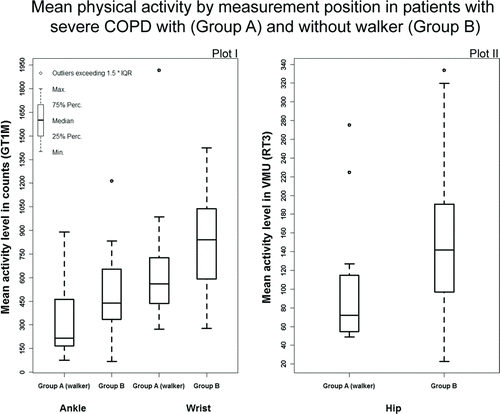
Table 2. Measurements of activity obtained by the GT1M (wrist, ankle) and the RT3 (hip) in patients with and without walker
We observed statistically significant moderate associations between wrist and ankle and wrist and hip PA data in walker-free patients (). There was a moderate to strong correlation between the hip VMU score recorded by the RT3 and the ankle activity score from the GT1M in both patient groups.
Table 3. Correlations between activity data from different monitor positions in patients with and without walker
Relationship between PA assessments and functional capacity and disease severity
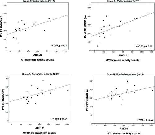
The relationships between daily mean physical activity data from hip, ankle and wrist and the 6MWD at the start and at the end of the PR were examined in both groups separately (). In this analysis, statistically significant positive associations were observed only for mean ankle activity counts and both 6MWD examinations. Mean ankle PA data was moderately related to the 6MWD assessed at the start of PR in COPD patients with (r = 0.55, p < 0.05) and without walkers (r = 0.60, p < 0.01) and to the 6MWD assessed at the end of PR (r = 0.63, p < 0.01; r = 0.53, p < 0.05, respectively; ). Wrist and hip activity measurements were not significantly associated with the 6MWD.
Figure 2. Relationship between the mean PA values assessed at the ankle and the 6MWD assessed at the beginning and end of the PR program in both patient groups.
To assess the individual contribution of ankle, wrist and hip recordings on disease severity as the dependent variable, a multivariate regression model was used including group (walker vs. non-walker) and gender information as independent variables (adjusted R2 = 0.406, p < 0.01; ). The model revealed that only ankle activity was significantly associated with the BODE score (p < 0.01), explaining 16% of the variation. Hip PA only explained 3% (p = 0.227), wrist recordings 8% (p = 0.054) of the variation in the model.
Table 4. Standard multivariate regression model to assess the impact of group and gender and all simultaneously measured PA positions, on the disease severity as described by the BODE score
Discussion
Motion sensors can be attached at different positions, but recordings at one location of the body might not be representative for overall activity (Citation21, Citation23). It remains unclear whether the sampling location affects the relationship to clinical data. Our study aimed at clarifying this issue. We therefore examined the associations between objective data gathered at different body positions and assessed their relationship to the 6MWD and disease severity in in-clinic patients suffering from COPD and undergoing LTOT. Recently, Casaburi et al. (Citation42) assessed PA patterns of LTOT patients similar to the present study, but detected no difference in PA. To our knowledge, no previous study simultaneously assessed upper, lower and trunk activity by means of three accelerometers in COPD during an in-patient rehabilitation setting.
Louvaris et al. (Citation41), in a short report, compared the output of 6 different sensors in patients with COPD, also with different body locations, but only assessing correlations among sensor models, not considering location as a possible influence. They found that if similar constructs are assessed by different sensors the outcome is moderately correlated. Although using different body locations in our study, moderate correlations between activity counts (GT1M) and VMU (RT3) were only found in patients without walker (0.44 < r < 0.65, p < 0.05). With increasing limitation of mobility, especially in patients using a walker, the activities of ankle and wrist appeared to become less associated. In line with the results reported by Walker et al. (Citation21) we found a good agreement between data from the uniaxial ankle (Actigraph) and triaxial hip worn accelerometer (Stayhealthy) in patients.
Lower limb activity is the key determinant of whole body activity (Citation37), but our results also indicate that patients were limited in their arm movements, although to a lesser extent compared to leg and hip. Wrist accelerometry has recently been shown to be a valid and sensitive instrument to measure upper-extremity movement during PR (Citation38) and is responsive to differences between days. Ankle and wrist assessments are lacking complete agreement with the hip sensor in our study and cannot be fully substituted when determining whole body energy expenditure (Citation21, Citation23).
We observed a moderate-to-strong association between 6MWD and the mean ankle activity data. This was not found for the hip or wrist recording. Although the 6MWD is related to mortality and morbidity in COPD and can indicate changes in the functional exercise capacity (Citation40), it does not necessarily track daily activities, at least not under the setting of PR and in the presence of heterogeneous handicaps. Daily activity is certainly not only determined by exercise capacity but also highly influenced by habits, modulated by psychosocial and environmental factors particularly in a rehabilitation setting. Irrespective of these considerations, our findings support the notion that the placement of the motion sensor affects the association of activity recordings with the 6MWD. This is also underlined by the finding that an association between disease severity and PA only exists for the assessment at the ankle. PA is probably an important outcome measure for PR, and, compared to self-reported information, quantitative PA data is better suited for the analysis of quantitative relationships. Currently, only few devices have been validated in COPD and data comparison among devices is hampered by manufacturer-dependent peculiarities. Nevertheless, accelerometry in principle can track changes in PA before and after PR programs, even in slow walking patients (Citation30, Citation39).
A limitation of our study is the comparison of two different types of accelerometers at the different locations having different responses to similar activities. The presented results might therefore not only reflect the influence of the body placement but also include differences between the accelerometers (i.e., accuracy, sensitivity) themselves. However, in a very recent report from Vanhelst et al. (Citation43), no significant difference between the output of GT1M and RT3 sensors was found in a group of adolescents when both devices were worn at the same location.
Given the inpatient PR setting of this study, the results might differ from those acquired in free-living, because the training sessions recorded by the accelerometers have also been included in the analysis, and therefore they probably do not reflect what a patient does normally in daily living. Furthermore, self-exercises and training sessions were individualized and for this reason may differ in intensity and amount in these severely ill patients. Nevertheless, the overall time of prescribed therapy for each group was nearly the same. The higher age and frailty in patients with a walker might have been a confounding factor in activity decline.Despite these limitations, our results underline the importance of sensor position for accelerometry in COPD patients. In patients with very severe COPD, recording at the ankle seems to best reflect the clinical state of the patients.
Acknowledgments
The authors thank the medical team in the rehabilitation clinic Bad Reichenhall, and the colleagues at IMSE, Dr. Tibor Schuster and Alexander Hapfelmeier, for their statistical support.
Declaration of Interest
None of the authors has any conflicts of interest to disclose. The authors are responsible for the content and the writing of this paper. This research was funded/supported by the Graduate School of Information Science in Health (GSISH) and the Technische Universität München Graduate School. Lukas Gorzelniak received university grant monies as a PhD scholarship from the 3/1/2009 to 6/30/2012. The total grant for 3.5 years is 80,000 € ∼ 112,288 US $. André Dias is supported by the Portuguese Foundation for Science and Technology (FCT), by scholarship SFRH/BD/39867/2007 and Research Council of Norway Grant No. 174934.
References
- Polkey MI, Rabe KF. Chicken or egg: physical activity in COPD revisited. Eur Respir J 2009;33(2):227–229. Editorial.
- Garcia-Aymerich J, Lange P, Benet M, Schnohr P, Anto JM. Regular physical activity modifies smoking-related lung function decline and reduces risk of chronic obstructive pulmonary disease: a population-based cohort study. Am J Respir Crit Care Med 2007;175:458e63.
- Garcia-Aymerich J, Lange P, Benet M, Schnohr P, Anto JM. Regular physical activity reduces hospital admission and mortality in chronic obstructive pulmonary disease: a population based cohort study. Thorax 2006;61:772e8.
- Garcia-Aymerich J, Farrero E, Félez MA, Izquierdo J, Marrades RM, Antó JM; Estudi del Factors de Risc d'Agudització de la MPOC investigators. Risk factors of readmission to hospital for a COPD exacerbation: a prospective study. Thorax 2003;58(2):100–105.
- Pitta F, Troosters T, Probst VS, Spruit MA, Decramer M, Gosselink R. Quantifying physical activity in daily life with questionnaires and motion sensors in COPD. Eur Respir J 2006;27(5):1040–1055.
- Pitta F, Troosters T, Spruit MA, Probst VS, Decramer M, Gosselink R. Characteristics of physical activities in daily life in chronic obstructive pulmonary disease. Am J Respir Crit Care Med 2005;171: 972–977.
- Troosters T, Sciurba F, Battaglia S, Physical inactivity in patients with COPD, a controlled multi-center pilot-study. Respir Med. 2010;104(7):1005–11. Epub 2010 Feb 18.
- Watz H, Waschki B, Meyer T, Magnussen H. Physical activity in patients with COPD. Eur Respir J. 2009 Feb;33(2):262–72. Epub 2008 Nov 14. Erratum in: Eur Respir J 2010;36(2):462.
- Global Initiative for Chronic Obstructive Lung Disease. Global strategy for the diagnosis, management and prevention of chronic obstructive pulmonary disease. NHLBI/WHO workshop report, National Heart, Lung and Blood Institute, Bethesda, April 2001; GOLD website: /www.goldcopd.com S. Update 2009.
- Garcia-Aymerich J, Serra I, Gómez FP, Phenotype and course of COPD study group. Physical activity and clinical and functional status in COPD. Chest 2009;136(1):62–70. Epub 2009 Mar 2.
- Hecht A, Ma S, Porszasz J, Casaburi R. Methodology for using long-term accelerometry monitoring to describe daily activity patterns in COPD. COPD. 2009 Apr;6(2):121–9.
- Singh S, Morgan MD. Activity monitors can detect brisk walking in patients with chronic obstructive pulmonary disease. J Cardiopulm Rehabil 2001;21:143e8.
- Coronado M, Janssens JP, de Muralt B, Terrier P, Schutz Y, Fitting JW. Walking activity measured by accelerometry during respiratory rehabilitation. J Cardiopulm Rehab 2003;23(5):357–64.
- Garcia-Aymerich J, Félez MA, Escarrabill J, Physical activity and its determinants in severe chronic obstructive pulmonary disease. Med Sci Sports Exer 2004;36(10):1667–73.
- Dallas MI, McCusker C, Haggerty MC, Rochester CL, Zuwallack R. Using pedometers to monitor walking activity in outcome assessment for pulmonary rehabilitation. Chron Respir Dis 2009;6(4):217–224.
- Belza B, Steele BG, Hunziker J, Lakshminaryan S, Holt L, Buchner DM. Correlates of physical activity in chronic obstructive pulmonary disease. Nurs Res 2001;50(4):195–202.
- Pitta F, Troosters T, Spruit MA, Decramer M, Gosselink R. Activity monitoring for assessment of physical activities in daily life in patients with chronic obstructive pulmonary disease. Arch Phys Med Rehabil. 2005;86(10):1979–85.
- Pitta F, Takaki MY, Oliveira NH, Relationship between pulmonary function and physical activity in daily life in patients with COPD. Respir Med 2008;102(8):1203–1207. Epub 2008 Jun 24.
- Schönhofer B, Ardes P, Geibel M, Köhler D, Jones PW. Evaluation of a movement detector to measure daily activity in patients with chronic lung disease. Eur Respir J 1997;10(12):2814–2819.
- Steele BG, Holt L, Belza B, Quantitating physical activity in COPD using a triaxial accelerometer. Chest 2000;117(5):1359–1367.
- Walker PP, Burnett A, Flavahan PW, Calverley PM. Lower limb activity and its determinants in COPD. Thorax. 2008 Aug;63(8):683–9. Epub 2008 May 16.
- Swartz AM, Strath SJ, Bassett DR Jr, O'Brien WL, King GA. Estimation of energy expenditure using CSA accelerometers at hip and wrist sites. Med Sci Sports Exerc. 2000 Sep;32(9 Suppl):S450–6.
- Cohen MD, Cutaia M. A novel approach to measuring activity in chronic obstructive pulmonary disease: using 2 activity monitors to classify daily activity. J Cardiopulm Rehabil Prev. 2010 May-Jun;30(3):186–94.
- GOLD. Global strategy for diagnosis, management, and prevention of COPD, www.goldcopd.com; 2010 accessed 09.08.10.
- Magnussen H, Kirsten AM, Köhler D, Morr H, Sitter H, Worth H. Leitlinien zur Langzeit-Sauerstofftherapie. Pneumologie 2008; 62:748–756.
- Redelmeier DA, Bayoumi AM, Goldstein RS, Guyatt GH. Interpreting small differences in functional status: the six minute walk test in chronic lung disease patients. Am J Respir Crit Care Med 1997; 155:1278–1282.
- Miller MR, Hankinson J, Brusasco V, ATS/ERS Task Force. Standardisation of spirometry. Eur Respir J 2005;26:319e38.
- Borg GAV. Psychophysical basis of perceived exertion. Med Sci Sports Exerc 1982;14:377–387.
- Mahler D, Wells C. Evaluation of clinical methods for rating dyspnea. Chest 1988; 93:580–586.
- Le Masurier GC, Tudor-Locke C. Comparison of pedometer and accelerometer accuracy under controlled conditions. Med Sci Sports Exer 2003;35(5):867–871.
- John D, Tyo B, Bassett DR. Comparison of four ActiGraph accelerometers during walking and running. Med Sci Sports Exerc 2010 Feb;42(2):368–374.
- vanHees T, Pias M, Thaerian S, Ekelund U, Brage S. A method to compare new and traditional accelerometry data in physical activity monitoring. 2010 IEEE 11th International Symposium on a World of Wireless, Mobile and Multimedia Networks (WoWMoM). Montreal, QC, Canada, 14–17 June, 2010.
- Nichols JF, Morgan CG, Chabot LE, Sallis JF, Calfas KJ. Assessment of physical activity with the Computer Science and Applications, Inc., accelerometer: laboratory versus field validation. Res Q Exerc Sport 2000, 71(1):36–43.
- Powell SM, Jones DI, Rowlands AV. Technical variability of the RT3 accelerometer. Med Sci Sports Exer 2003;35(10):1773–1778.
- R Development Core Team. R: A language and environment for statistical computing. R Foundation for Statistical Computing, Vienna, Austria. ISBN 3–900051-07–0, URL http: //www.R-project.org, 2010.
- Wood M. Statistical inference using bootstrap confidence intervals. Significance 2004; 1(4):180–182.
- Morgan M. Life in slow motion: quantifying physical activity in COPD. Thorax 2008;63(8):663–664. Editorial.
- Bauldoff GS, Ryan-Wenger NA, Diaz PT. Wrist actigraphy validation of exercise movement in COPD. West J Nurs Res 2007;29(7):789–802. Epub 2007 Jul 17.
- Mador MJ, Patel AN, Nadler J. Effects of pulmonary rehabilitation on activity levels in patients with chronic obstructive pulmonary disease. J Cardiopulm Rehab Prev 2011;31(1):52–59.
- Cote CG, Casanova C, Marín JM, Validation and comparison of reference equations for the 6-min walk distance test. Eur Respir J 2008 Mar;31(3):571–578. Epub 2007 Nov 7.
- Louvaris Z, Langer D, Giavedoni S, Raste Y, Van Remoortel H, Gatti Regueiro E. M, Wilson F, Vogiatzis I, Hopkinson N, Rabinovich R, Peterson B, Troosters B. Activity monitor outcomes in COPD –Assessment of variability of 6 monitors as part of the IMI PROactive project. Thematic Poster Session : Clinical physiology for clinical problem. P2157. ERS Annual Congress, Amsterdam 2011.
- Casaburi R, Porszasz J, Hecht A, Tiep B, Albert RK, Anthonisen NR, Bailey WC, Connett JE, Cooper JA Jr, Criner GJ, Curtis J, Dransfield M, Lazarus SC, Make B, Martinez FJ, McEvoy C, Niewoehner DE, Reilly JJ, Scanlon P, Scharf SM, Sciurba FC, Woodruff P; COPD Clinical Research Network. Influence of lightweight ambulatory oxygen on oxygen use and activity patterns of COPD patients receiving long-term oxygen therapy. COPD 2012 Feb;9(1):3–11.
- Vanhelst J, Beghin L, Duhamel A, Bergman P, Sjotrom M, Gottrand F. Comparison of uniaxial and triaxial accelerometry in the assessment of physical activity among adolescents under free-living conditions: the HELENA study. BMC Med Res Methodol 2012;12(1):26. (Epub ahead of print).
Appendix
Sensitivity analysis for imputation of missing values and re-assessment of correlations.
Based on the variables Age, Sex, FEV1%pred., Ankle, Hip and Wrist PA data, Group (Walker vs Walker-free), 6MWD at the start and end of the PR program, missing values for the accelerometer data and both 6MWD were imputed by means of linear regression.
Correlations for Non-Walker Patients before Imputation (Group B).
Correlations for Walker Patients after Imputation (Group A).
Correlations for Walker Patients before Imputation (Group A).